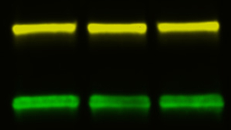
Western Blot Example: Demonstrating Antibody Specificity
Overview
A simple way to demonstrate that the band seen on a Western blot is specific to the interaction between a target protein and a properly matched antibody is by blocking the antibody from binding to the target epitope through competition with a peptide that matches the sequence of the epitope . Bio-Rad offers a range of blocking peptides for this purpose .
The experiment is carried out during the antibody incubation stage and compares antibody alone to antibody treated with the blocking peptide . If the peptide sequence correctly corresponds to the epitope then the antibody will bind to the peptide and will not be available to bind to the target protein on the blot . This technique would only be used with monoclonal antibodies, since they are directed against a single epitope, and when the epitope is known . In the example below, lane 2 has no signal because the peptide has effectively blocked the antibody from binding, whereas in lane 1 the band is visible as expected.
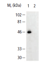
Fig.17. Peptide Blocks Binding of Anti-CX3CR1 Antibody (AHP566). Lane 1: THP-1 cell lysate (LYS008) . Lane 2: THP-1 cell lysate + peptide to aa175-189 of human CX3Cr1.
Popular Resources
Generate Western Blot Data You Can Trust
Best practices for image documentation, integrity, and verification.
TidyBlot Western Blot Detection Reagent:HRP
TidyBlot Reagent:HRP is a western blotting detection reagent that specifically binds to native (non-reduced) antibodies.