s
TidyBlot Western Blot Detection Reagent:HRP
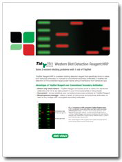
TidyBlot Reagent:HRP is a western blotting detection reagent that specifically binds to native (non-reduced) antibodies.
s

TidyBlot FAQs
Find out more about TidyBlot's mode of action and how to benefit from using the product.
Overview
This protocol provides a procedure when using TidyBlot HRP Conjugated Western Blot Detection Reagent. TidyBlot Reagent is very versatile because it is compatible with a variety of both monoclonal and polyclonal primary antibodies.
TidyBlot preferentially binds to the native antibody used during the western blotting procedure rather than the SDS-denatured/reduced form present in lysates/immunoprecipitates. Best results are therefore achieved by denaturing the immunoprecipitate/lysate fully. If your target protein is phosphorylated, we recommend using our protocol for detection of phosphorylated proteins by western blotting.
Please note that a certain level of technical skill and immunological knowledge is required for the successful design and implementation of these techniques - these are guidelines only and may need to be adjusted for particular applications.
View TidyBlot’s compatibility chart
Protocol
Reagents
Blocking Buffer
Block Ace (BUF029) dissolved in ddH2O (100 ml of ddH2O per sachet of Block Ace).
TGS Running Buffer
100 ml 10x TGS Running Buffer (Bio-Rad, 1610772) diluted in 900 ml deionized water (DI H2O)
Washing buffer
Block Ace (BUF029) dissolved in PBS (1 liter of PBS per sachet of Block Ace) + 0.1% Tween 20
Ponceau S
Acetic acid, 5 ml
Distilled water, 95 ml
Ponceau S (Sigma P3504), 0.1 g
*Note: Ponceau S is light sensitive.
Method
- Prepare samples of your protein of interest in sample buffer such as Bio-Rad’s 2x Laemmli Sample Buffer (1610737) with reducing agent for example, Dithiothreitol (DTT) (1610610). Boil the samples for 5 min at 95 ⁰C and allow to cool to room temperature (RT). Please note that this step is vital as complete reduction and denaturation of proteins in your samples is required to prevent TidyBlot Reagent from detecting antibodies present in your samples.
- Place an SDS page gel into a suitable gel tank. The size and type of gel selected should be optimized for the system you are using and the size of your target protein. In general, gels with a higher percentage of acrylamide are more suitable for targets of a lower molecular weight.
- Fill the inner chamber and outer chamber of the gel tank with running buffer according to manufacturer’s instructions.
- Load equal amounts of prepared lysate into the wells of an SDS page gel. Remember to load a suitable molecular weight marker such as Bio-Rad’s Precision Plus All Blue Standards (1610373) alongside your samples to determine the molecular weight of any proteins you visualize at the end of the blotting process.
- Perform gel electrophoresis using a suitable protocol. For Bio-Rad’s Criterion Gel System, we recommend running the gel at 300 V for approximately 25 min at RT.
- Following SDS-PAGE, transfer proteins onto blotting membrane using a suitable transfer system, such as Bio-Rad’s Trans-Blot Turbo Transfer System, according to the manufacturer’s instructions.
- Once transfer is complete, check that proteins have transferred from the gel to the membrane efficiently by staining the membrane with a total protein stain. Our recommendation is to stain the membrane post transfer with Ponceau S for 1 min, then completely destain the blot by washing with DI H2O with agitation.
- Place the membrane into blocking solution and incubate for 30 min at RT with agitation.
- Remove the blocking buffer and wash the blot in wash buffer for 5 min with agitation.
- Remove the wash buffer and add the primary antibody diluted in the wash buffer. Incubate overnight at 4°C with agitation. Please consult the product specific datasheet for the primary antibody you are using for recommended dilutions, bearing in mind that titration experiments may need to be performed in order to find the optimal primary antibody dilution for your experimental set up.
- Remove the primary antibody. Wash the blot in wash buffer 3x for 10 min with agitation.
- Remove wash buffer and add TidyBlot diluted in wash buffer. We recommend diluting TidyBlot between 1/40 and 1/400 in wash buffer. However, it is important that titration experiments are performed to optimize the dilution of TidyBlot for the system you are using and your target protein. Incubate for 1 hr at RT with agitation.
- Wash the membrane 4x for 5 min in wash buffer with agitation.
- Wash the membrane 2x for 5 min in PBS.
- Add an appropriate enzyme substrate solution to the membrane, such as Bio-Rad’s Clarity ECL (1705061), and incubate as recommended by the manufacturer.
- Develop the blot using an appropriate developing system for example Bio-Rad’s ChemiDoc MP Imager according to manufacturer’s instructions.
Appropriate controls should always be carried out. It may be useful to include a sample in which no primary antibody is used at all, in order to determine any nonspecific binding of the secondary reagent to the target tissue. Please contact Bio-Rad’s Technical Services Department to learn about recommended secondary reagents for specific applications.
View Our Detection of Phosphorylated Proteins by Western Blotting Protocol
Explore Our Full List of Recommended Antibody Protocols
See Our General Western Blotting Protocol


