MIP-2 antibody
Rabbit anti Mouse MIP-2
- Product Type
- Polyclonal Antibody
- Isotype
- Polyclonal IgG
- Specificity
- MIP-2

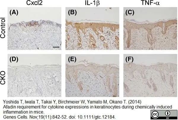
Rabbit anti Mouse MIP-2 antibody (AAM48) used for the identification of MIP-2 positive cells in mouse skin by immunohistochemistry on formailn fixed paraffin embedded tissue sections.
Image caption:
Immunohistochemical analysis of 12-O-tetradecanoylphorbol 13-acetate (TPA)-treated skin. TPA-treated skin from control (A–C) and afadin conditional knockout (CKO) (D–F) mice was stained with the antibodies shown at the top of each picture. Scale bar 50μm.
From: Yoshida T, Iwata T, Takai Y, Birchmeier W, Yamato M, Okano T.
Afadin requirement for cytokine expressions in keratinocytes during chemically induced inflammation in mice.
Genes Cells. 2014 Nov;19(11):842-52.
10.1111/gtc.12184.
This image is from an open access article distributed under terms of a Creative Commons Attribution License.
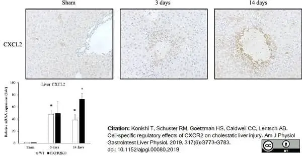
Rabbit anti Mouse MIP-2 antibody (AAM48) used to assess MIP-2 (CXCL2) expression in mouse hepatic tissue by immunohistochemistry on formalin fixed, paraffin embedded tissue sections.
Image caption:
CXCL2 expression after bile duct ligation (BDL). B: CXCL2 immunohistochemical staining. CXCL2 protein was found to be expressed around areas of hepatocyte necrosis after BDL in wild-type mice. Original magnification: ×400. Original magnification: ×400.
From: Konishi T, Schuster RM, Goetzman HS, Caldwell CC, Lentsch AB.
Cell-specific regulatory effects of CXCR2 on cholestatic liver injury.
Am J Physiol Gastrointest Liver Physiol. 2019 Dec 1;317(6):G773-G783.
doi: 10.1152/ajpgi.00080.2019
This image is from an open access article distributed under terms of a Creative Commons Attribution License.
Filter by Application:
P Reset| Rabbit anti Mouse MIP-2 antibody recognizes mouse macrophage inflammatory protein-2 (MIP-2), also known as CXCL2. MIP-2 is expressed by macrophages and epidermal langerhans cells and is chemotactic for neutrophils. |
- Target Species
- Mouse
- Species Cross-Reactivity
-
Target Species Cross Reactivity Rat - N.B. Antibody reactivity and working conditions may vary between species.
- Product Form
- Purified IgG - lyophilized
- Reconstitution
- Reconstitute with 1.0 ml distilled water
Care should be taken during reconstitution as the protein may appear as a film at the bottom of the vial. Bio-Rad recommend that the vial is gently mixed after reconstitution. For long term storage the addition of 0.09% sodium azide is recommended.
N.B. For functional studies do not add sodium azide - Antiserum Preparation
- Antisera to mouse MIP-2 were raised by repeated immunisations of rabbits with highly purified antigen. Purified IgG was prepared from whole serum by affinity chromatography.
- Buffer Solution
- Phosphate buffered saline
- Preservative Stabilisers
- None present
- Carrier Free
- Yes
- Immunogen
- Recombinant mouse MIP-2
- Approx. Protein Concentrations
- IgG concentration 0.1 mg/ml after reconstitution.
- Regulatory
- For research purposes only
- Guarantee
- 12 months from date of despatch
This product is shipped at ambient temperature.
Prior to reconstitution store at -20°C.
After reconstitution store at -20°C.
This product should be stored undiluted. Storage in frost-free freezers is not recommended. Avoid repeated freezing and thawing as this may denature the antibody. Should this product contain a precipitate we recommend microcentrifugation before use.
This product has been reported to work in the following applications. This information is derived from testing within our laboratories, peer-reviewed publications or personal communications from the originators. Please refer to references indicated for further information. For general protocol recommendations, please visit the antibody protocols page.
| Application Name | Verified | Min Dilution | Max Dilution |
|---|---|---|---|
| ELISA |  |
0.5ug/ml | |
| Functional Assays | |
0.50ug/ml | 0.90ug/ml |
| Immunohistology - Frozen | |
||
| Immunohistology - Paraffin 1 | |
||
| Western Blotting | |
0.1ug/ml | 0.2ug/ml |
- 1This product requires antigen retrieval using heat treatment prior to staining of paraffin sections.
Sodium citrate buffer pH 6.0 is recommended for this purpose.
Where this product has not been tested for use in a particular technique this does not necessarily exclude its use in such procedures. Suggested working dilutions are given as a guide only. It is recommended that the user titrates the product for use in their own system using appropriate negative/positive controls.
- ELISA
- Rabbit anti Mouse MIP-2 antibody may be used in an indirect ELISA or as the capture reagent in a sandwich ELISA with biotinylated Rabbit anti mouse MIP-2 antibody (AAM48B) as the detection antibody and recombinant Mouse MIP-2 protein (PMP55) as a standard.
- Western Blotting
- Rabbit anti Mouse MIP-2 antibody may be used under either reducing or non-reducing conditions with recombinant Mouse MIP-2 (PMP55) protein as a positive control.
| Description | Product Code | Applications | Pack Size | List Price | Your Price | Quantity | |
|---|---|---|---|---|---|---|---|
| Goat anti Rabbit IgG (Fc):Biotin | STAR121B | E WB | 1 mg |

|
Log in | ||
| List Price | Your Price | ||||||
|
|
Log in | ||||||
| Description | Goat anti Rabbit IgG (Fc):Biotin | ||||||
| Goat anti Rabbit IgG (Fc):FITC | STAR121F | F | 1 mg |
|
Log in | ||
| List Price | Your Price | ||||||
|
|
Log in | ||||||
| Description | Goat anti Rabbit IgG (Fc):FITC | ||||||
| Goat anti Rabbit IgG (Fc):HRP | STAR121P | E WB | 1 mg |
|
Log in | ||
| List Price | Your Price | ||||||
|
|
Log in | ||||||
| Description | Goat anti Rabbit IgG (Fc):HRP | ||||||
| Goat anti Rabbit IgG (H/L):HRP | STAR124P | C E WB | 1 mg |
|
Log in | ||
| List Price | Your Price | ||||||
|
|
Log in | ||||||
| Description | Goat anti Rabbit IgG (H/L):HRP | ||||||
| Sheep anti Rabbit IgG:RPE | STAR35A | F | 1 ml |
|
Log in | ||
| List Price | Your Price | ||||||
|
|
Log in | ||||||
| Description | Sheep anti Rabbit IgG:RPE | ||||||
| Description | Product Code | Applications | Pack Size | List Price | Your Price | Quantity | |
|---|---|---|---|---|---|---|---|
| Antigen Retrieval Buffer, pH8.0 | BUF025A | P | 500 ml | Log in | |||
| List Price | Your Price | ||||||
| Log in | |||||||
| Description | Antigen Retrieval Buffer, pH8.0 | ||||||
| TidyBlot Western Blot Detection Reagent:HRP | STAR209P | WB * | 0.5 ml | Log in | |||
| List Price | Your Price | ||||||
| Log in | |||||||
| Description | TidyBlot Western Blot Detection Reagent:HRP | ||||||
| Description | Product Code | Applications | Pack Size | List Price | Your Price | Quantity | |
|---|---|---|---|---|---|---|---|
| Recombinant Mouse MIP-2 | PMP55 | E FN WB | 20 µg |
|
Log in | ||
| List Price | Your Price | ||||||
|
|
Log in | ||||||
| Description | Recombinant Mouse MIP-2 | ||||||
References for MIP-2 antibody
-
Di Carlo, E. et al. (2000) The combined action of IL-15 and IL-12 gene transfer can induce tumor cell rejection without T and NK cell involvement.
J Immunol. 165: 3111-8. -
Cavallo, F. et al (2001) Interleukin 12-activated lymphocytes influence tumor genetic programs.
Cancer Res. 61: 3518-23. -
Roche, J.K. et al. (2007) CXCL1/KC and CXCL2/MIP-2 are critical effectors and potential targets for therapy of Escherichia coli O157:H7-associated renal inflammation.
Am J Pathol. 170: 526-37. -
González-López, A. et al. (2011) Inflammation and matrix remodeling during repair of ventilator-induced lung injury.
Am J Physiol Lung Cell Mol Physiol. 301: L500-9. -
Tittel, A.P. et al. (2011) Kidney dendritic cells induce innate immunity against bacterial pyelonephritis.
J Am Soc Nephrol. 22 (8): 1435-41. -
Yoshida, T. et al. (2014) Afadin requirement for cytokine expressions in keratinocytes during chemically induced inflammation in mice.
Genes Cells. 19: 842-52. -
Lazic, M. et al. (2014) Differential regulation of inflammation and apoptosis in Fas-resistant hepatocyte-specific Bid-deficient mice.
J Hepatol. 61: 107-15. -
Nicholas, J. et al. (2015) Time course of chemokine expression and leukocyte infiltration after acute skeletal muscle injury in mice.
Innate Immun. 21 (3): 266-74.
View The Latest Product References
-
Lasarte, S. et al. (2016) Sex Hormones Coordinate Neutrophil Immunity in the Vagina by Controlling Chemokine Gradients.
J Infect Dis. 213 (3): 476-84. -
Salinas-Muñoz, L. et al. (2019) Estradiol impairs epithelial CXCL1 gradient in the cervix to delay neutrophil transepithelial migration during insemination.
J Reprod Immunol. 132: 9-15. -
Konishi, T. et al. (2019) Cell-specific regulatory effects of CXCR2 on cholestatic liver injury.
Am J Physiol Gastrointest Liver Physiol. 317 (6): G773-G783. -
Shimada, A. & Hasegawa-Ishii, S. (2021) Increased cytokine expression in the choroid plexus stroma and epithelium in response to endotoxin-induced systemic inflammation in mice.
Toxicol Rep. 8: 520-8.
- Synonyms
- CXCL2
- RRID
- AB_2086327
- UniProt
- P10889
- Entrez Gene
- Cxcl2
- GO Terms
- GO:0005615 extracellular space
- GO:0006954 inflammatory response
- GO:0006955 immune response
- GO:0008009 chemokine activity
AAM48
If you cannot find the batch/lot you are looking for please contact our technical support team for assistance.
View more products with MIP-2 specificity
Please Note: All Products are "FOR RESEARCH PURPOSES ONLY"
View all Anti-Mouse ProductsAlways be the first to know.
When we launch new products and resources to help you achieve more in the lab.
Yes, sign me up