Collagen IV antibody
Rabbit anti Mouse Collagen IV
- Product Type
- Polyclonal Antibody
- Isotype
- Polyclonal IgG
- Specificity
- Collagen IV

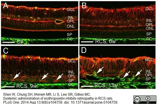
Rabbit anti Mouse collagen 4 antibody (2150-1470) used for detection of collagen 4 in rat retina by immunofluorescence on cryostat sections.
Image caption:
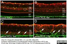
Retinal gliosis and subretinal neovascularization in RCS rats. Double label immunostaining was performed using antibodies against vimentin (red) and collagen type IV (green) on frozen sections. (A) Non-dystrophic rat at 21 weeks (w) of age showed a normal distribution of retinal vessels confined to the inner retina and absence of glial cells in the subretinal space. (B, C) RCS rats at 6 w (B) and 9 w (C) of age showed activation of Muller cells but without subretinal neovascularization. Muller cell invasion into the subretinal space was more obvious at 9 w compared with 6 w of age (arrows in C). (D) Subretinal neovascularization (arrows) in RCS rats at 13 w of age. GCL = ganglion cell layer, INL = inner nuclear layer, OPL = outer plexiform layer, ONL = outer nuclear layer, SP = subretinal space, CC = choroidal capillaries. Scale bars: 50 μm.
From: Shen W, Chung SH, Irhimeh MR, Li S, Lee S-R, et al. (2014)
Systemic Administration of Erythropoietin Inhibits Retinopathy in RCS Rats.
PLoS ONE 9(8): e104759.
doi: 10.1371/journal.pone.0104759.
This image is from an open access article distributed under the terms of the Creative Commons Attribution License.
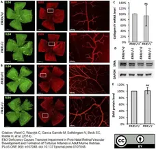
Image caption:
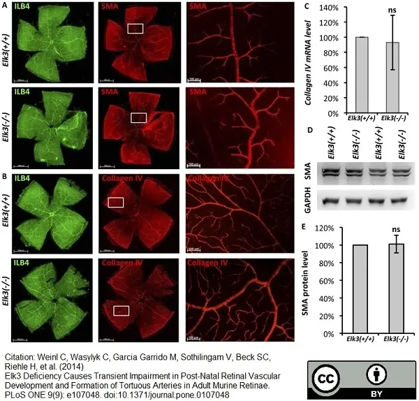
Smooth muscle actin and CollagenIV levels are not altered in Elk3(−/−) knockout retinae. (A) IsolectinB4 (ILB4) (left) and smooth muscle actin (SMA) (middle and right) staining of retinal flat-mounts of control (upper panel) and Elk3(−/−) knockout animals. (B) IsolectinB4 (left) and collagenIV (middle and right) staining of retinal flat-mounts of control (upper panel) and Elk3(−/−) knockout animals. (C) Quantitation of collagenIV RNA levels in control and Elk3(−/−) knockout retinae (n = 5 independent experiments). (D) Representative pairs of control and Elk3(−/−) knockout retinal lysates tested for smooth muscle actin expression in Western Blot analysis. (E) Quantitation of control and Elk3(−/−) knockout retinal levels of SMA (n = 5 independent experiments). Scale bar in (A, B left and middle) 1000 μm, in (A, B right) 100 μm.
From: Weinl C, Wasylyk C, Garcia Garrido M, Sothilingam V, Beck SC, et al. (2014)
Elk3 Deficiency Causes Transient Impairment in Post-Natal Retinal Vascular Development and Formation of Tortuous Arteries in Adult Murine Retinae.
PLoS ONE 9(9): e107048.
doi: 10.1371/journal.pone.0107048
This image is from an open access article distributed under the terms of the Creative Commons Attribution License.

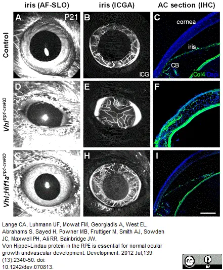
Rabbit anti Mouse collagen IV antibody (2150-1470) used for the evaluation of collagen IV expression in murine pupillary membranes by immunofluorescence on cryosections.
Image caption:
Vhltrp1-creKO mice demonstrate a Hif1a-dependent persistence of the pupillary membrane. (A-I) Autofluorescence scanning laser ophthalmoscope (AF-SLO) imaging (A,D,G), indocyanin-green angiography (ICGA; B,E,H) and collagen IV-stained cryosections (C,F,I) of Vhltrp1-creKO, Vhl;Hif1atrp1-creKO and control littermates at P21 show a persistent pupillary membrane in Vhltrp1-creKO mice that is absent in Vhl;Hif1atrp1-creKO and control mice. CB, ciliary body. Scale bar: 75 μm.
From: Lange CA, Luhmann UF, Mowat FM, Georgiadis A, West EL, Abrahams S, Sayed H, Powner MB, Fruttiger M, Smith AJ, Sowden JC, Maxwell PH, Ali RR, Bainbridge JW.
Von Hippel-Lindau protein in the RPE is essential for normal ocular growth and vascular development.
Development. 2012 Jul;139(13):2340-50.
doi: 10.1242/dev.070813.
This image is from an open access article distributed under the terms of the Creative Commons Attribution License.
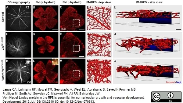
Rabbit anti Mouse collagen IV antibody (2150-1470) used for the evaluation of collagen IV expression in retinal flat-mounts by immunofluorescence.
Image caption:
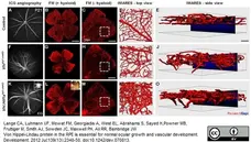
Vhltrp1-creKO mice show a persistence of the hyaloid vasculature and a disorganised retinal vasculature that is independent of Hif1a. (A,B,F,G,K,L) Indocyanin-green angiography (A,F,K) and corresponding collagen 4-stained retinal flatmounts (B,G,L) show a persistent hyaloid vasculature in Vhltrp1-creKO and Vhl;Hif1atrp1-creKO mice that is absent in control littermates at P21. Inset in K shows angiography focussed on the persistent hyaloid vasculature. Inset in L shows a light microscopy image of a retinal flat mount with haemorrhage. (C,H,M) Vhltrp1-creKO and Vhl;Hif1atrp1-creKO mice demonstrate a disorganised retinal vasculature with areas of intra-retinal vascular proliferation compared with age-matched controls (hyaloid vessels were removed). Outlined regions indicate the areas selected for IMARES imaging. (D,E,I,J,N,O) IMARES reconstruction reveals a disorganised retinal vasculature with abrogated vascular layering and retinal vessels growing into the outer retina in Vhltrp1-creKO and Vhl;Hif1atrp1-creKO mice. Scale bars: 1 mm for B; 500 μm for C,M; 700 μm for H; 50 μm for D,E,I,J,N,O.
From: Lange CA, Luhmann UF, Mowat FM, Georgiadis A, West EL, Abrahams S, Sayed H, Powner MB, Fruttiger M, Smith AJ, Sowden JC, Maxwell PH, Ali RR, Bainbridge JW.
Von Hippel-Lindau protein in the RPE is essential for normal ocular growth and vascular development.
Development. 2012 Jul;139(13):2340-50.
doi: 10.1242/dev.070813.
This image is from an open access article distributed under the terms of the Creative Commons Attribution License.

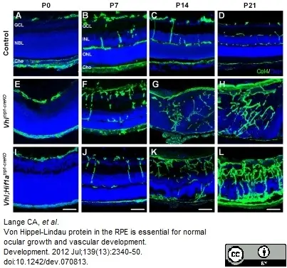
Rabbit anti Mouse collagen IV antibody (2150-1470) used for the detection of collagen IV in mouse retina by immunofluorescence.
Image caption:
Deletion of Vhl in the RPE results in disruption of the retinal vascular architecture and formation of chorioretinal anastomoses. (A-L) Representative collagen IV-stained cryosections of control (A-D), Vhltrp1-creKO mice (E-H) and Vhl;Hif1atrp1-creKO (I-L) and at different time points during development show a disorganised retinal vasculature and the formation of chorioretinal anastomoses in Vhl;Hif1atrp1-creKO and Vhltrp1-creKO mice (G,K). GCL, ganglion cell layer; NBL, neuroblast layer; Cho, choroid; INL, inner nuclear layer, ONL, outer nuclear layer. Scale bars: 25 μm.
From: Lange CA, Luhmann UF, Mowat FM, Georgiadis A, West EL, Abrahams S, Sayed H, Powner MB, Fruttiger M, Smith AJ, Sowden JC, Maxwell PH, Ali RR, Bainbridge JW.
Von Hippel-Lindau protein in the RPE is essential for normal ocular growth and vascular development.
Development. 2012 Jul;139(13):2340-50.
doi: 10.1242/dev.070813.
This image is from an open access article distributed under the terms of the Creative Commons Attribution License.
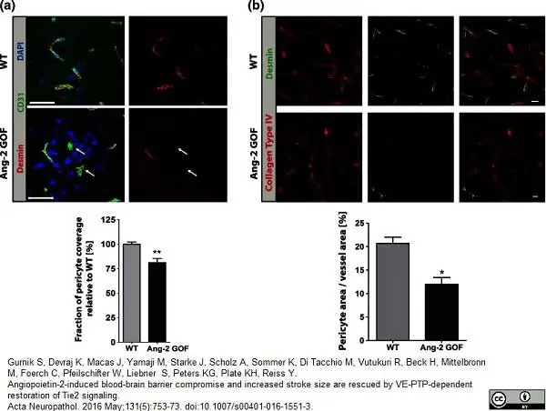
Rabbit anti Murine cytokeratin 4 antibody (2150-1470) used for the identification of collagen IV expressing vasculature in mouse brains by immunofluorescence on vibratome sections.
Image caption:
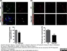
Immunohistochemistry analysis of pericytes in Ang-2 GOF mice. a Pericytes (desmin+) were decreased in GOF mice utilizing 10 μm cryosections for analysis (n = 8, scale bars 25μm). b 50 μm vibratome sections of Ang-2 GOF and WT mice revealed decrease in pericyte area/vessel area [%] (n = 3, scale bars 10 μm) indicating decreased pericyte coverage of the vessels in GOF mice
From: Gurnik S, Devraj K, Macas J, Yamaji M, Starke J, Scholz A, Sommer K, Di Tacchio M, Vutukuri R, Beck H, Mittelbronn M, Foerch C, Pfeilschifter W, Liebner S, Peters KG, Plate KH, Reiss Y.
Angiopoietin-2-induced blood-brain barrier compromise and increased stroke size are rescued by VE-PTP-dependent restoration of Tie2 signaling.
Acta Neuropathol. 2016 May;131(5):753-73.
doi: 10.1007/s00401-016-1551-3.
This image is from an open access article distributed under the terms of the Creative Commons Attribution License.
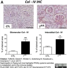
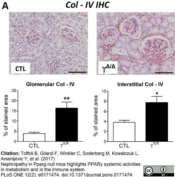
Rabbit anti Human collagen IV antibody (2150-1470) showing increased renal collagen IV deposition in aged PpargΔ/Δ mice over controls by immunohistochemistry on formalin fixed, paraffin embedded tissue sections.
Image caption:
Immunohistochemistry and gene expression of fibrosis markers in kidney of ageing PpargΔ/Δ mice, and plasmatic levels of anti-β2-glycoprotein 1 antibodies.
52 weeks old control (CTL) and PparΔ/Δ (γΔ/Δ) animals were analyzed for the following parameters: (A) Top panels: Kidney Immunohistochemistry for collagen IV (Col-IV). Positive staining is in brown. Scale bar represents 100μm.
From: Toffoli B, Gilardi F, Winkler C, Soderberg M, Kowalczuk L, Arsenijevic Y, et al. (2017)
Nephropathy in Pparg-null mice highlights PPARγ systemic activities in metabolism and in the immune system.
PLoS ONE 12(2): e0171474.
doi: 10.1371/journal.pone.0171474.
This image is from an open access article distributed under the terms of the Creative Commons Attribution License.
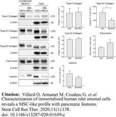
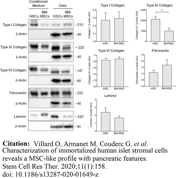
Rabbit anti Human collagen IV antibody (2150-4140) used for the evaluation of collagen IV expression in human stem cell preparations by western blotting.
Image caption:
hISCs secrete pancreatic ECM proteins. Extracted proteins from hISCs and BM-MSCs and corresponding conditioned media were analyzed by western blotting. Type I, IV, and VI collagens as well as fibronectin and laminin were detected in both cell types but at different levels of expression. β-actin was used as loading control. The right panels represent the quantification of the Western blot, n = 4, *p <0.05, significant differences
From: Villard O, Armanet M, Couderc G, et al.
Characterization of immortalized human islet stromal cells reveals a MSC-like profile with pancreatic features.
Stem Cell Res Ther. 2020;11(1):158.
doi:10.1186/s13287-020-01649-z
This image is from an open access article distributed under the terms of the Creative Commons Attribution License.
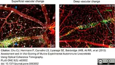
Rabbit anti Mouse collagen IV antibody (2150-1470) used to demonstrate blood vessels by immunofluorescence.
Image caption:
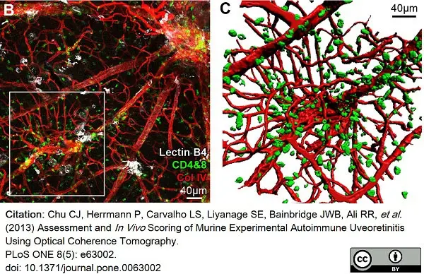
OCT identifies vascular changes in EAU and appearances are altered by depth and degree of infiltration.
The vessel (collagen IV in red) with altered OCT signal in the perivascular tissue (upward pointing arrowheads) is densely infiltrated with myeloid - lectin B4 binding (white) and T cell - CD4+ & CD8+ (green) cells as shown by immunohistochemistry on retinal flat mount (H). This is in contrast to the other vessel (downward pointing arrowheads), which lacks any localised infiltrate. Diffuse hyper-reflective signal on OCT (I) corresponds to a deep retinal vein, with associated cellular infiltrate (J). The blue line indicates alignment with the OCT scan.
From: Chu CJ, Herrmann P, Carvalho LS, Liyanage SE, Bainbridge JWB, Ali RR, et al. (2013)
Assessment and In Vivo Scoring of Murine Experimental Autoimmune Uveoretinitis Using Optical Coherence Tomography.
PLoS ONE 8(5): e63002.
doi: 10.1371/journal.pone.0063002
This image is from an open access article distributed under terms of a Creative Commons Attribution License.
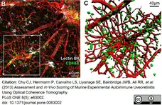
Rabbit anti Mouse collagen IV antibody (2150-1470) used to demonstrate blood vessels by immunofluorescence.
Image caption:
OCT imaging can guide identification and characterisation of novel features within the EAU model.
Retinal flat-mount revealed an atypical vascular lesion arising along the course of a large vessel (B). 3D reconstruction of the region enclosed by the white box, with CD4+ & CD8+ cells (green) displayed, collagen IV (red) (C).
From: Chu CJ, Herrmann P, Carvalho LS, Liyanage SE, Bainbridge JWB, Ali RR, et al. (2013)
Assessment and In Vivo Scoring of Murine Experimental Autoimmune Uveoretinitis Using Optical Coherence Tomography.
PLoS ONE 8(5): e63002.
doi: 10.1371/journal.pone.0063002
This image is from an open access article distributed under terms of a Creative Commons Attribution License.
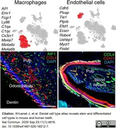
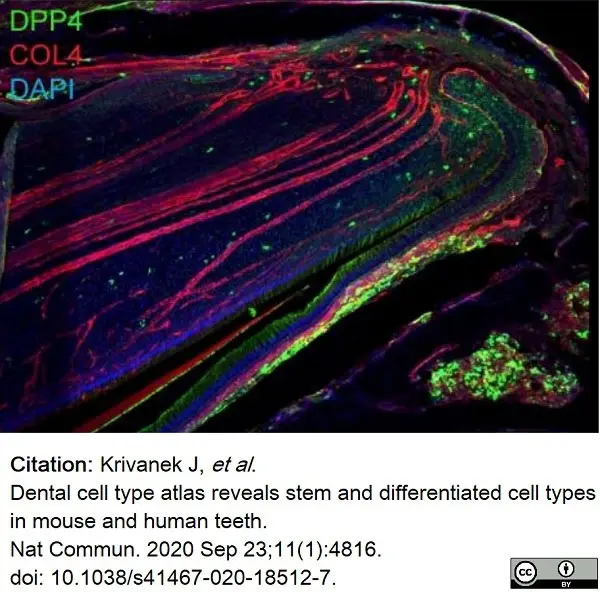
Rabbit anti Mouse collagen IV antibody (2150-1470) used to label blood vessels in mouse dental tissue by immunofluorescence.
Image caption:
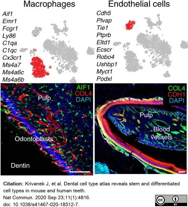
Unbiased identification, validation, and spatial mapping of major dental cell types and subpopulations.
Validations and mapping of unbiasedly identified populations based on the expression of selected marker genes. Note. COL4 is a well known marker for blood vessels, this marker genes is highly and specifically expressed in corresponding clusters (Supplementary Table 1), but do not belong to top10 genes shown in plots above the images. (LiCL Lingual Cervical Loop, LaCL Labial Cervical Loop, SI Stratum Intermedium, SR Stellate reticulum, OEE Outer Enamel Epithelium). Scare bars: 50 μm.
From: Krivanek J, et al.
Dental cell type atlas reveals stem and differentiated cell types in mouse and human teeth.
Nat Commun. 2020 Sep 23;11(1):4816.
doi: 10.1038/s41467-020-18512-7.
This image is from an open access article distributed under terms of a Creative Commons Attribution License.
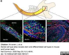
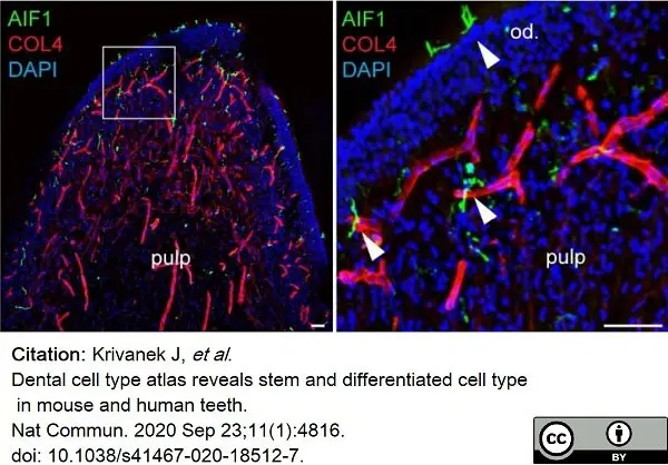
Rabbit anti Mouse collagen IV antibody (2150-1470) used to label blood vessels in mouse dental tissue by immunofluorescence.
Image caption:
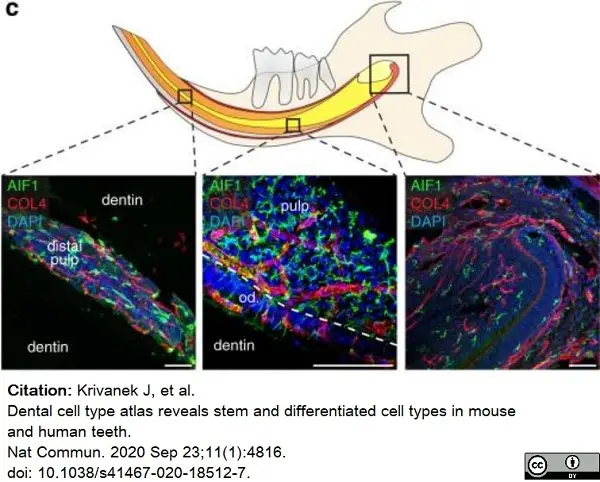
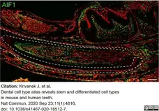
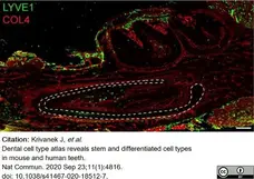
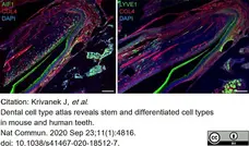
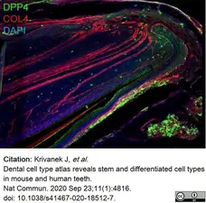
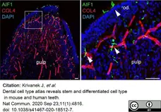
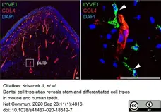
Heterogeneity of immune cells in mouse incisor.
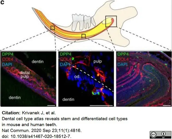
c Location of tissue-residential immune cells in the different parts of mouse incisor. AIF1+ macrophages are located in the whole incisor including apical pulp, cervical loop, odontoblast layer, and distal pulp in contrast to LYVE+ macrophages which mostly reside in the middle part of the pulp, but not inside the odontoblast layer. DPP4+ immune cells are sporadically located in the apical part of the tooth and odontoblast layer. COL4 immunohistochemical staining visualize the blood vessels. (Od. Odontoblasts), Scale bars: 50 μm.
From: Krivanek J, et al.
Dental cell type atlas reveals stem and differentiated cell types in mouse and human teeth.
Nat Commun. 2020 Sep 23;11(1):4816.
doi: 10.1038/s41467-020-18512-7.
This image is from an open access article distributed under terms of a Creative Commons Attribution License.
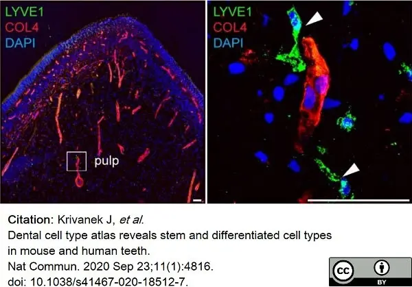
Rabbit anti Mouse collagen IV antibody (2150-1470) used to label blood vessels in mouse dental tissue by immunofluorescence.
Image caption:
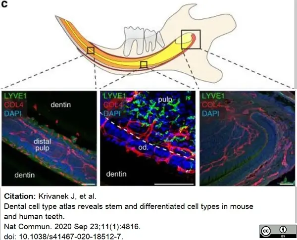
Heterogeneity of immune cells in mouse incisor.
c Location of tissue-residential immune cells in the different parts of mouse incisor. AIF1+ macrophages are located in the whole incisor including apical pulp, cervical loop, odontoblast layer, and distal pulp in contrast to LYVE+ macrophages which mostly reside in the middle part of the pulp, but not inside the odontoblast layer. DPP4+ immune cells are sporadically located in the apical part of the tooth and odontoblast layer. COL4 immunohistochemical staining visualize the blood vessels. (Od. Odontoblasts), Scale bars: 50 μm.
From: Krivanek J, et al.
Dental cell type atlas reveals stem and differentiated cell types in mouse and human teeth.
Nat Commun. 2020 Sep 23;11(1):4816.
doi: 10.1038/s41467-020-18512-7.
This image is from an open access article distributed under terms of a Creative Commons Attribution License.
Rabbit anti Mouse collagen IV antibody (2150-1470) used to label blood vessels in mouse dental tissue by immunofluorescence.
Image caption:
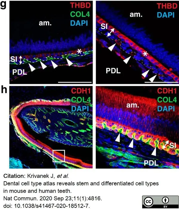
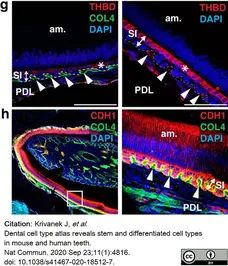
Identification of previously unrecognized cell types in dental epithelium and stem cells.
g, h Immunohistochemistry identification of the Cuboidal layer of stratum intermedium (expressing THBD) and spatial relation to the neighboring blood vessels submerged into the papillary structure of stratum intermedium. COL4 expression characterizes the blood vessels on left panel. h Papillary structure of stratum intermedium with submerged blood vessels (COL4) and CDH1 expressing ameloblasts and cells from stratum intermedium. Note. Cuboidal layer characterized by THBD expression (g) forms subpopulation of stratum intermedium cells (h). Immunohistochemistry. (am. ameloblasts, od. odontoblasts, LaCL Labial Cervical Loop, SI stratum intermedium, OEE Outer Enamel Epithelium, am. Ameloblasts, PDL periodontal ligamentum). Scale bars: 50 μm.
From: Krivanek J, et al.
Dental cell type atlas reveals stem and differentiated cell types in mouse and human teeth.
Nat Commun. 2020 Sep 23;11(1):4816.
doi: 10.1038/s41467-020-18512-7.
This image is from an open access article distributed under terms of a Creative Commons Attribution License.
Rabbit anti Mouse collagen IV antibody (2150-1470) used to label blood vessels in mouse dental tissue by immunofluorescence.
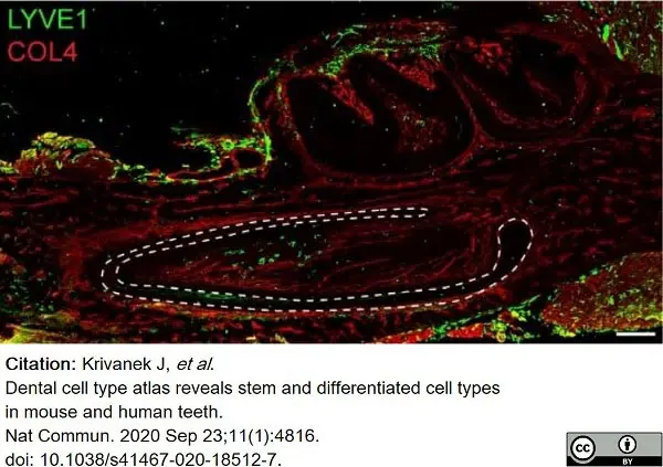
Image caption:
Cross-section through P3 mandible showing the differential spatial distribution of AIF1+ and LYVE1+ macrophages (immunohistochemistry). COL4 immunohistochemical staining to visualize the blood vessels.
From: Krivanek J, et al.
Dental cell type atlas reveals stem and differentiated cell types in mouse and human teeth.
Nat Commun. 2020 Sep 23;11(1):4816.
doi: 10.1038/s41467-020-18512-7.
This image is from an open access article distributed under terms of a Creative Commons Attribution License.
Rabbit anti Mouse collagen IV antibody (2150-1470) used to label blood vessels in mouse dental tissue by immunofluorescence.
Image caption:
Cross-section through P3 mandible showing the differential spatial distribution of AIF1+ and LYVE1+ macrophages (immunohistochemistry). COL4 immunohistochemical staining to visualize the blood vessels.
From: Krivanek J, et al.
Dental cell type atlas reveals stem and differentiated cell types in mouse and human teeth.
Nat Commun. 2020 Sep 23;11(1):4816.
doi: 10.1038/s41467-020-18512-7.
This image is from an open access article distributed under terms of a Creative Commons Attribution License.
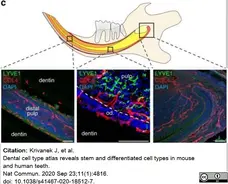
Rabbit anti Mouse collagen IV antibody (2150-1470) used to label blood vessels in mouse dental tissue by immunofluorescence.
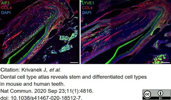
Image caption:
Immunohistochemistry-based validation of AIF1+, LYVE1+ macrophages inside themouse incisor. Together with the figure 5c these panels prove the different histological positioning of two different kind of macrophages (LYVE1+ and LYVE1-). AIF1+ macrophages are located in the whole incisor including apical pulp, cervical loop, odontoblast layer and distal pulp in contrast to LYVE+ macrophages which mostly resides in the middle part of the pulp, but not inside the odontoblast layer. COL4 immunohistochemical staining to visualize the blood vessels.
From: Krivanek J, et al.
Dental cell type atlas reveals stem and differentiated cell types in mouse and human teeth.
Nat Commun. 2020 Sep 23;11(1):4816.
doi: 10.1038/s41467-020-18512-7.
This image is from an open access article distributed under terms of a Creative Commons Attribution License.
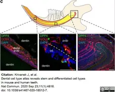
Rabbit anti Mouse collagen IV antibody (2150-1470) used to label blood vessels in mouse dental tissue by immunofluorescence.
Image caption:
Immunohistochemistry-based validation of the NK-cells cluster using DPP4 marker. Position of DPP4+ cells outside the blood vessels in the tooth was proved in 5 independent animals. COL4 immunohistochemical staining visualize the blood vessels.
From: Krivanek J, et al.
Dental cell type atlas reveals stem and differentiated cell types in mouse and human teeth.
Nat Commun. 2020 Sep 23;11(1):4816.
doi: 10.1038/s41467-020-18512-7.
This image is from an open access article distributed under terms of a Creative Commons Attribution License.
Rabbit anti Mouse collagen IV antibody (2150-1470) used to label blood vessels in human dental tissue by immunofluorescence.
Image caption:
Two different types of macrophages (LYVE1+ and LYVE1-) demonstrate spatially restricted distribution also in human molar (immunohistochemistry). COL4 immunohistochemical staining visualize the blood vessels. Scale bars: 50 μm.
From: Krivanek J, et al.
Dental cell type atlas reveals stem and differentiated cell types in mouse and human teeth.
Nat Commun. 2020 Sep 23;11(1):4816.
doi: 10.1038/s41467-020-18512-7.
This image is from an open access article distributed under terms of a Creative Commons Attribution License.
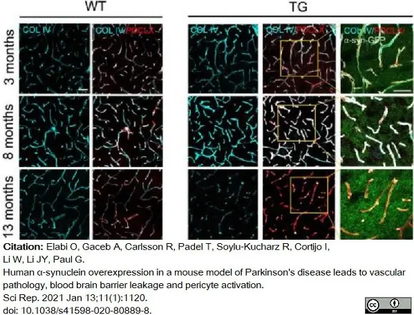
Rabbit anti Mouse collagen IV antibody (2150-1470) used to highlight collagen in relation to microvessels in mouse brain by immunofluorescence.
Image caption:
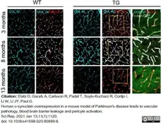
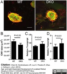
Reduction of Col IV in TG mice at late age.
Confocal images illustrating Col IV density in relation to the PDCLX+ vessels in WT (left panel) and TG mice (right panel).Scale bar: 50 μm. PDCLX = Podocalyxin, Col IV = Collagen IV.
From: Elabi O, Gaceb A, Carlsson R, Padel T, Soylu-Kucharz R, Cortijo I, Li W, Li JY, Paul G.
Human α-synuclein overexpression in a mouse model of Parkinson's disease leads to vascular pathology, blood brain barrier leakage and pericyte activation.
Sci Rep. 2021 Jan 13;11(1):1120.
doi: 10.1038/s41598-020-80889-8..
This image is from an open access article distributed under terms of a Creative Commons Attribution License.
Rabbit anti Murine cytokeratin 4 antibody (2150-1470) used for the identification of collagen IV expressing vasculature in mouse brains by immunofluorescence on vibratome sections.
Image caption:
Immunohistochemistry analysis of pericytes in Ang-2 GOF mice. a Pericytes (desmin+) were decreased in GOF mice utilizing 10 μm cryosections for analysis (n = 8, scale bars 25μm). b 50 μm vibratome sections of Ang-2 GOF and WT mice revealed decrease in pericyte area/vessel area [%] (n = 3, scale bars 10 μm) indicating decreased pericyte coverage of the vessels in GOF mice
From: Gurnik S, Devraj K, Macas J, Yamaji M, Starke J, Scholz A, Sommer K, Di Tacchio M, Vutukuri R, Beck H, Mittelbronn M, Foerch C, Pfeilschifter W, Liebner S, Peters KG, Plate KH, Reiss Y.
Angiopoietin-2-induced blood-brain barrier compromise and increased stroke size are rescued by VE-PTP-dependent restoration of Tie2 signaling.
Acta Neuropathol. 2016 May;131(5):753-73.
doi: 10.1007/s00401-016-1551-3.
This image is from an open access article distributed under the terms of the Creative Commons Attribution License.
Rabbit anti Mouse collagen IV antibody (2150-1470) used to visualize collagen IV expression in murine retina by immunofluorecence.
Image caption:
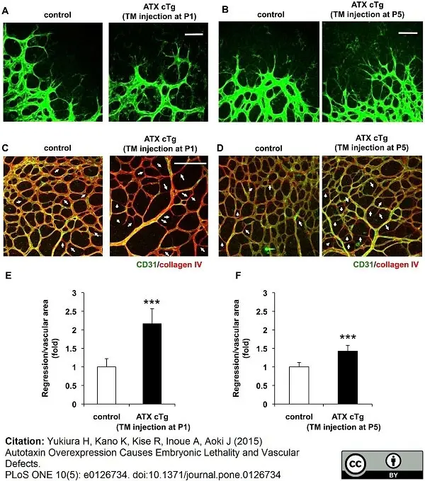
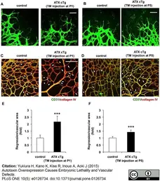
Overexpression of ATX causes abnormal vessel morphology and vessel regression. (A and B) Magnification view of angiogenic front in retina from ATX cTg mice at P6. Control and ATX cTg retinas had similar filopodia protrusion. Scale bar, 50 μm. TM, tamoxifen. (C and D) ATX cTg retinas displayed vessel regression at vascular plexus. Control (wild type) and ATX cTg retinas labeled for CD31 (green) and collagen IV (red). Arrows highlight empty collagen IV sleeves, indicating vessel regression. Scale bar, 100 μm. (E and F) Vessel regression was evaluated quantitatively. Error bars indicate s.d. (control; n = 7, ATX cTg; n = 5). P-values were estimated by student’s t-test, ***P < 0.001. Data were pooled from three independent experiments.
From: Yukiura H, Kano K, Kise R, Inoue A, Aoki J (2015)
Autotaxin Overexpression Causes Embryonic Lethality and Vascular Defects.
PLoS ONE 10(5): e0126734.
doi: 10.1371/journal.pone.0126734
This image is from an open access article distributed under the terms of the Creative Commons Attribution License.
Rabbit anti Mouse collagen IV antibody (2150-1470) used to label collagen IV+ vessels in mouse retina by immunofluorescence.
Image caption:
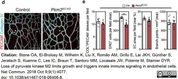
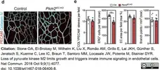
Endothelial specific deletion of Pkm2 in the post-natal mouse retina.
(d) Representative confocal projections of central plexus vessels in PECAM and Collagen IV stained retinas from control and Pkm2iEC-KO mice. (e) Quantification of Collagen IV+/PECAM+ sleeves, ERG+ ECs, EC area per field and branch point density at the angiogenic front of control and Pkm2iEC-KO mice (n=6). e data represent means ± s.e.m. (**P <0.01 by two tailed students t-test) Scale bars in d = 100μm.
From: Stone OA, El-Brolosy M, Wilhelm K, Liu X, Romão AM, Grillo E, Lai JKH, Günther S, Jeratsch S, Kuenne C, Lee IC, Braun T, Santoro MM, Locasale JW, Potente M, Stainier DYR.
Loss of pyruvate kinase M2 limits growth and triggers innate immune signaling in endothelial cells.
Nat Commun. 2018 Oct 9;9(1):4077.
doi: 10.1038/s41467-018-06406-8.
This image is from an open access article distributed under terms of a Creative Commons Attribution License.
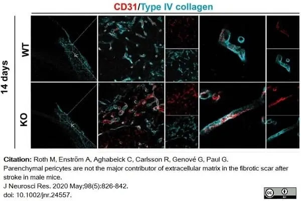
Rabbit anti Mouse collagen IV antibody (2150-1470) used to highlight collagen IV staining in mouse brain by immunofluorescence.
Image caption:
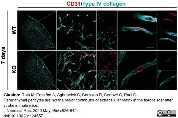
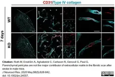
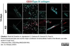
RGS5‐KO mice have reduced thickness of type IV collagen+ vascular basement membrane after stroke. (a) Confocal images of type IV collagen (cyan) and the blood vessel marker CD31 (red) at 7 and 14 days. Left column shows type IV collagen distribution in wild‐type (WT) and RGS5‐KO mice at both time points. The box indicates where the higher magnification picture in middle column has been taken from. Right column shows a high magnification of single z‐stack through a blood vessel to illustrate the thickness of the vascular basement membrane that is reduced in RGS5‐KO mice.
From:Roth M, Enström A, Aghabeick C, Carlsson R, Genové G, Paul G.
Parenchymal pericytes are not the major contributor of extracellular matrix in the fibrotic scar after stroke in male mice
J Neurosci Res. 2020 May;98(5):826-842.
doi: 10.1002/jnr.24557.
This image is from an open access article distributed under terms of a Creative Commons Attribution License.
Rabbit anti Mouse collagen IV antibody (2150-1470) used to highlight collagen IV staining in mouse brain by immunofluorescence.
Image caption:
RGS5‐KO mice have reduced thickness of type IV collagen+ vascular basement membrane after stroke. (a) Confocal images of type IV collagen (cyan) and the blood vessel marker CD31 (red) at 7 and 14 days. Left column shows type IV collagen distribution in wild‐type (WT) and RGS5‐KO mice at both time points. The box indicates where the higher magnification picture in middle column has been taken from. Right column shows a high magnification of single z‐stack through a blood vessel to illustrate the thickness of the vascular basement membrane that is reduced in RGS5‐KO mice.
From:Roth M, Enström A, Aghabeick C, Carlsson R, Genové G, Paul G.
Parenchymal pericytes are not the major contributor of extracellular matrix in the fibrotic scar after stroke in male mice
J Neurosci Res. 2020 May;98(5):826-842.
doi: 10.1002/jnr.24557.
This image is from an open access article distributed under terms of a Creative Commons Attribution License.
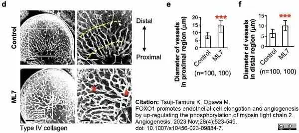
Rabbit anti Mouse collagen IV antibody (2150-1470) used to stain vasculature in mouse embryo wholemounts by immunofluorescence.
Image caption:
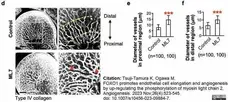
MLCK-inhibition causes an abnormal vascular structure in mouse embryos. d The forelimb vessels of E11.5 mouse embryos were investigated by whole-mount immunostaining for Type IV collagen. Right panels show a higher magnification of the boxed area in each left panel. Yellow dot lines indicate the position of vessels (proximal and distal) measured in (e) and (f). Red arrowheads indicate abnormally thick and dilated vessels. Scale bars indicate 100 μm (left panels) or 50 μm (right panels). Similar results were obtained from three independent experiments using embryos of different litters. e, f Diameters of the vessels in the proximal region (e) and distal region (f). The total number of vessels examined in 20 embryos in three independent experiments is indicated in the brackets. Data were analyzed using an F-test, followed by an unpaired two-tailed t test (***P <0.001)
From: Tsuji-Tamura K, Ogawa M.
FOXO1 promotes endothelial cell elongation and angiogenesis by up-regulating the phosphorylation of myosin light chain 2.
Angiogenesis. 2023 Nov;26(4):523-45.
doi: 10.1007/s10456-023-09884-7.
This image is from an open access article distributed under terms of a Creative Commons Attribution License.
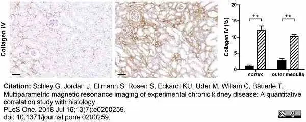
Rabbit anti Mouse collagen IV antibody (2150-1470) used to demonstrate collagen IV deposition in murine kidneys by immunohistochemistry on formalin fixed, paraffin embedded tissue sections.
Image caption:
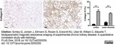
Interstitial fibrosis in CKD kidneys.
Kidney sections of control and CKD mice were stained for collagen IV. Shown are representative immunohistological stainings of control and CKD kidneys and quantitative analyses of staining categorized for renal cortex and outer medulla. Black bars represent control (n = 15) and dashed bars CKD kidneys (n = 16). Scale bars, 50 μm. ** p <0.01.
From: Schley G, Jordan J, Ellmann S, Rosen S, Eckardt KU, Uder M, Willam C, Bäuerle T.
Multiparametric magnetic resonance imaging of experimental chronic kidney disease: A quantitative correlation study with histology.
PLoS One. 2018 Jul 16;13(7):e0200259.
doi: 10.1371/journal.pone.0200259.
This image is from an open access article distributed under terms of a Creative Commons Attribution License.
Rabbit anti mouse collagen IV antibody (2150-1470) used to label collagen IV in murine tissues by immunofluorescence.
Image caption:
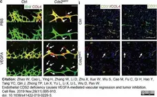
Vessel regression in CDS2 endothelium-specific knockout mice. a Model of tamoxifen treatment on mouse pups. Confocal images of IB4/Collagen IV (COL4) (c) double stained retinal vessels at postnatal day (P) 7 in Cds2iΔEC or control mice with or w/o VEGFA injection. Asterisks display the blunt angiogenic front. Arrows indicate COL4+/IB4− empty sleeves. Anti-COL4 and anti-CD31 co-immunostaining (i) Scale bars, 70 μm (c) and 100 μm (i).
From: Zhao W, Cao L, Ying H, Zhang W, Li D, Zhu X, Xue W, Wu S, Cao M, Fu C, Qi H, Hao Y, Tang YC, Qin J, Zhong TP, Lin X, Yu L, Li X, Li L, Wu D, Pan W.
Endothelial CDS2 deficiency causes VEGFA-mediated vascular regression and tumor inhibition.
Cell Res. 2019 Nov;29(11):895-910.
doi: 10.1038/s41422-019-0229-5.
This image is from an open access article distributed under terms of a Creative Commons Attribution License.
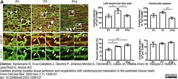
Rabbit anti Human collagen IV antibody (2150-1470) used to label basemant membrane in mouse heart by immunofluorescence.
Image caption:
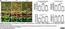
Vascular remodeling occurs by capillary pruning in the mouse postnatal cardiac microvasculature and correlates with changes in tissue oxygenation. (A), Confocal microscopy maximum intensity projections (MIP) of ICAM2 (green) and collagen IV (red) staining (left) and quantification of empty sleeves, vascular density and endothelial cells in the LVFW myocardium and of empty sleeves in the septum (right) in P1, P7 and P14 mouse hearts. Arrows indicate ‘empty sleeves’. Scale bar, 100 μm and 25 μm in the magnifications.
From: Santamaría R, Cruz-Caballero J, Gkontra P, Jiménez-Montiel A, Clemente C, López JA, Villalba-Orero M, Vázquez J, Hutloff A, Lara-Pezzi E, Arroyo AG.
Capillary pruning couples tissue perfusion and oxygenation with cardiomyocyte maturation in the postnatal mouse heart.
Front Cell Dev Biol. 2023 Nov 7;11:1256127.
doi: 10.3389/fcell.2023.1256127.
This image is from an open access article distributed under terms of a Creative Commons Attribution License.
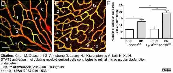
Rabbit anti Mouse collagen IV antibody (2150-1470) used to label mouse retinal capillaries by immunofluorescence.
Image caption:
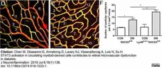
Acellular capillary was examined by confocal microscopy of collagen IV (red) and Isolectin B4 (green) stained retinal flatmounts from in SOCS3fl/fl and LysMCre/+SOCS3fl/fl mice with/without 6-month diabetes. Representative confocal images of retina flatmount stained with collagen IV (Red) and Isolectin B4 (green) in diabetic SOCS3fl/fl (d) and LysMCre/+SOCS3fl/fl (e) mice. Acellular capillaries are positive for collagen IV but negative for Isolectin B4 (numbered). f Bar figure showing the quantification of acellular capillaries. Data were presented as mean ± SEM. N > 6 per group. Two-way ANOVA followed by Bonferroni test. CON: non-diabetes, DM: diabetes. *p < 0.05; ***p < 0.001
From: Chen M, Obasanmi G, Armstrong D, Lavery NJ, Kissenpfennig A, Lois N, Xu H.
STAT3 activation in circulating myeloid-derived cells contributes to retinal microvascular dysfunction in diabetes.
J Neuroinflammation. 2019 Jul 8;16(1):138.
doi: 10.1186/s12974-019-1533-1.
This image is from an open access article distributed under terms of a Creative Commons Attribution License.
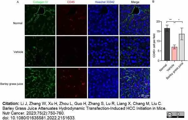
Rabbit anti Mouse collagen IV antibody (2150-1470) used to stain the vasculature in murine liver by immunofluorescence.
Image caption:
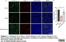
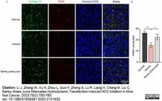
Immune cell and Kupffer cell numbers were raised by barley grass juice treatment of hepatocellular carcinoma (HCC) in the liver. A: Representative images of the CD45 immune staining in the normal, barley grass juice, and vehicle groups. B: Static images of (A). Collagen IV was used to stain the vascular structure and Hoechst 33342 was used to stain the nuclei. Scale bar, 25 μm. Data are representative of three independent experiments. Each value represents the mean ± SD (n = 3). *p<05, **p<01, as measured by Student’s t-test.
From: Li J, Zhang W, Xu H, Zhou L, Guo H, Zhang S, Lu R, Liang X, Chang M, Liu C.
Barley Grass Juice Attenuates Hydrodynamic Transfection-Induced HCC Initiation in Mice.
Nutr Cancer. 2023;75(2):750-60.
doi: 10.1080/01635581.2022.2151633.
This image is from an open access article distributed under terms of a Creative Commons Attribution License.
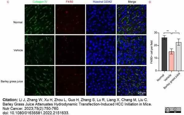
Rabbit anti Mouse collagen IV antibody (2150-1470) used to stain the vasculature in murine liver by immunofluorescence.
Image caption:
Immune cell and Kupffer cell numbers were raised by barley grass juice treatment of hepatocellular carcinoma (HCC) in the liver. C: Representative images of the F4/80 immune staining in the normal, barley grass juice, and vehicle groups. D: Static images of (C). Collagen IV was used to stain the vascular structure and Hoechst 33342 was used to stain the nuclei. Scale bar, 25 μm. Data are representative of three independent experiments. Each value represents the mean ± SD (n = 3). *p<05, **p<01, as measured by Student’s t-test.
From: Li J, Zhang W, Xu H, Zhou L, Guo H, Zhang S, Lu R, Liang X, Chang M, Liu C.
Barley Grass Juice Attenuates Hydrodynamic Transfection-Induced HCC Initiation in Mice.
Nutr Cancer. 2023;75(2):750-60.
doi: 10.1080/01635581.2022.2151633.
This image is from an open access article distributed under terms of a Creative Commons Attribution License.
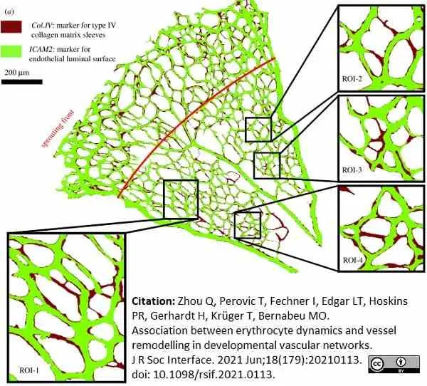
Rabbit anti Mouse collagen IV antibody (2150-1470) used to label collagen IV expressing cells in mouse retina by immunofluorescence.
Image caption:
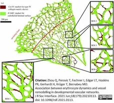
Simulated RBC velocity and cell flux in the primitive vasculature of developing mouse retina. (a) A vascular plexus of postnatal day 5 (P5) mouse retina, with vessel lumen and collagen matrix sleeves labelled by ICAM2 (light green) and Col.IV (dark red), respectively. The insets show four regions of interest (ROI-1, ROI-2, ROI-3 and ROI-4) selected from the remodelling region of the plexus. The red line indicates the transitional border between the sprouting and remodelling regions
From: Zhou Q, Perovic T, Fechner I, Edgar LT, Hoskins PR, Gerhardt H, Krüger T, Bernabeu MO.
Association between erythrocyte dynamics and vessel remodelling in developmental vascular networks.
J R Soc Interface. 2021 Jun;18(179):20210113.
doi: 10.1098/rsif.2021.0113.
This image is from an open access article distributed under terms of a Creative Commons Attribution License.

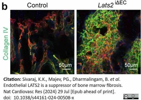
Rabbit anti Mouse collagen IV antibody (2150-1470) used for the evaluation of collagen IV expression in mouse bone sections by immunofluorescence.
Image caption:
Confocal images showing increased staining for collagen IV (COL IV) in Lats2iΔEC mutant relative to control.
From: Sivaraj, K.K., Majev, PG., Dharmalingam, B. et al.
Endothelial LATS2 is a suppressor of bone marrow fibrosis.
Nat Cardiovasc Res (2024).3(8):951-969.
10.1038/s44161-024-00508-x.
This image is from an open access article distributed under terms of a Creative Commons Attribution License.

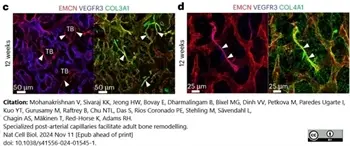
Rabbit anti Mouse collagen IV antibody (2150-1470) used to label collagen IV fibers in murine femurs by immunofluorescence.
Image caption:
c,d, Type III collagen (COL3A1) (c) and type IV collagen (COL4A1) (d) are tightly associated with EMCN+VEGFR3− type R capillaries (arrowheads) in 12-week-old wild-type femur, whereas the surrounding sinusoidal vessels show a loose reticular fibre network.
From: Mohanakrishnan V, Sivaraj KK, Jeong HW, Bovay E, Dharmalingam B, Bixel MG, Dinh VV, Petkova M, Paredes Ugarte I, Kuo YT, Gurusamy M, Raftrey B, Chu NTL, Das S, Rios Coronado PE, Stehling M, Sävendahl L, Chagin AS, Mäkinen T, Red-Horse K, Adams RH.
Specialized post-arterial capillaries facilitate adult bone remodelling.
Nat Cell Biol. 2024 26 (12): 2020-2034.
doi: 10.1038/s41556-024-01545-1.
This image is from an open access article distributed under terms of a Creative Commons Attribution License.
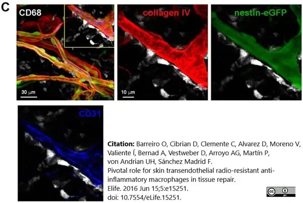
Rabbit anti Mouse collagen IV antibody (2150-1470) used to label collagen IV in mouse ear by immunofluorescence.
Image caption:
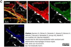
(C) Maximal projection of a z-stack of skin dermis from a nestin-eGFP mouse ear showing macrophages (CD68+, white) on the outer surface of vessel walls, which are defined by basal lamina staining (collagen IV, red), pericytes (eGFP+ cells covered with collagen) and endothelium (CD31, blue). The boxed area is shown as a magnified view with split channels.
From: Barreiro O, Cibrian D, Clemente C, Alvarez D, Moreno V, Valiente Í, Bernad A, Vestweber D, Arroyo AG, Martín P, von Andrian UH, Sánchez Madrid F.
Pivotal role for skin transendothelial radio-resistant anti-inflammatory macrophages in tissue repair.
Elife. 2016 Jun 15;5:e15251.
doi: 10.7554/eLife.15251.
This image is from an open access article distributed under terms of a Creative Commons Attribution License.
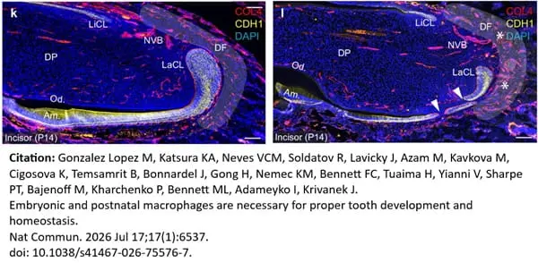
Rabbit anti Mouse collagen IV antibody (2150-1470) used to identify colllagen IV expression in mice during tooth development by immunofluorescence.
Image caption:
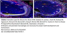
Wnt1Cre/Csf1fl/fl mice display an altered cervical loop shape, disruption of the epithelial layer (white arrowheads), and increased vascularization (asterisks) in the dental follicle space (k, l). Scale bars ( k, l) = 100 μm.
From: Gonzalez Lopez M, Katsura KA, Neves VCM, Soldatov R, Lavicky J, Azam M, Kavkova M, Cigosova K, Temsamrit B, Bonnardel J, Gong H, Nemec KM, Bennett FC, Tuaima H, Yianni V, Sharpe PT, Bajenoff M, Kharchenko P, Bennett ML, Adameyko I, Krivanek J.
Embryonic and postnatal macrophages are necessary for proper tooth development and homeostasis.
Nat Commun. 2026 Jul 17;17(1):6537.
doi: 10.1038/s41467-026-75576-7.
This image is from an open access article distributed under terms of a Creative Commons Attribution License.
Filter by Application:
IF P WB C Reset| Rabbit anti Mouse Collagen IV antibody recognizes mouse collagen type IV. Collagen IV is a 1682 amino acid ~160 kDa (predicted) matrx protein and major component of glomerular basement membranes. Multiple isoforms exist each capable of forming triple helical structures with two other chains to form the type IV collagen network. The collagen IV alpha chain can be cleaved between residues 1444-1445 to yield the c-terminal 225 amino acid, ~28 kDa arresten fragment, collagen α2(IV) yields a c-terminal canstatin fragment while Collagen α3(IV) yeilds a tumstatin fragment. Collagen IV bears a single collagen IV NC1 (C-terminal non-collagenous) domain (UniProt: Q9QZR9). Mutations in collagen IV genes have been implicated in inherited nephropathies and potentially in cystic kidney disease and intracranial aneurysms (Plaisier et al. 2007). Rabbit amti Mouse Collagen IV antibody has been successfully employed for the detection of collagen IV by immunofluorescence and immunohistochemistry in mice (Tang et al. 2010), rats (Shen et al. 2014) and oragutan (Bredies et al. 2013). The following cross reactivities have been observed:
|
- Target Species
- Mouse
- Species Cross-Reactivity
-
Target Species Cross Reactivity Orangutan Rat Human - N.B. Antibody reactivity and working conditions may vary between species.
- Product Form
- Purified Ig - liquid
- Preparation
- Purified IgG prepared by affinity chromatography on Protein A
- Buffer Solution
- Phosphate buffered saline
0.1M citrate - Preservative Stabilisers
- <0.1% Sodium Azide (NaN3)
Antibiotic antimycotic mixture 1% - Immunogen
- Collagen IV purified from mouse EHS tumor.
- Regulatory
- For research purposes only
- Guarantee
- 12 months from date of despatch
This product is shipped at ambient temperature. It is recommended to aliquot and store at -20°C on receipt. When thawed, aliquot the sample as needed. Keep aliquots at 2-8°C for short term use (up to 4 weeks) and store the remaining aliquots at -20°C.
Avoid repeated freezing and thawing as this may denature the antibody. Storage in frost-free freezers is not recommended.
Avoid repeated freezing and thawing as this may denature the antibody. Storage in frost-free freezers is not recommended.
This product has been reported to work in the following applications. This information is derived from testing within our laboratories, peer-reviewed publications or personal communications from the originators. Please refer to references indicated for further information. For general protocol recommendations, please visit the antibody protocols page.
| Application Name | Verified | Min Dilution | Max Dilution |
|---|---|---|---|
| ELISA |  |
1/2000 | |
| Immunofluorescence | |
1/40 | |
| Immunohistology - Frozen | |
1/500 | |
| Immunohistology - Paraffin | |
1/500 |
Where this product has not been tested for use in a particular technique this does not necessarily exclude its use in such procedures. Suggested working dilutions are given as a guide only. It is recommended that the user titrates the product for use in their own system using appropriate negative/positive controls.
| Description | Product Code | Applications | Pack Size | List Price | Your Price | Quantity | |
|---|---|---|---|---|---|---|---|
| Goat anti Rabbit IgG (Fc):Biotin | STAR121B | E WB | 1 mg |

|
Log in | ||
| List Price | Your Price | ||||||
|
|
Log in | ||||||
| Description | Goat anti Rabbit IgG (Fc):Biotin | ||||||
| Goat anti Rabbit IgG (Fc):FITC | STAR121F | F | 1 mg |
|
Log in | ||
| List Price | Your Price | ||||||
|
|
Log in | ||||||
| Description | Goat anti Rabbit IgG (Fc):FITC | ||||||
| Goat anti Rabbit IgG (Fc):HRP | STAR121P | E WB | 1 mg |
|
Log in | ||
| List Price | Your Price | ||||||
|
|
Log in | ||||||
| Description | Goat anti Rabbit IgG (Fc):HRP | ||||||
| Goat anti Rabbit IgG (H/L):HRP | STAR124P | C E WB | 1 mg |
|
Log in | ||
| List Price | Your Price | ||||||
|
|
Log in | ||||||
| Description | Goat anti Rabbit IgG (H/L):HRP | ||||||
| Sheep anti Rabbit IgG:RPE | STAR35A | F | 1 ml |
|
Log in | ||
| List Price | Your Price | ||||||
|
|
Log in | ||||||
| Description | Sheep anti Rabbit IgG:RPE | ||||||
| Description | Product Code | Applications | Pack Size | List Price | Your Price | Quantity | |
|---|---|---|---|---|---|---|---|
| Antigen Retrieval Buffer, pH8.0 | BUF025A | P | 500 ml | Log in | |||
| List Price | Your Price | ||||||
| Log in | |||||||
| Description | Antigen Retrieval Buffer, pH8.0 | ||||||
References for Collagen IV antibody
-
Xu Q. et al. (2004) Vascular development in the retina and inner ear: control by Norrin and Frizzled-4, a high-affinity ligand-receptor pair.
Cell. 116: 883-95. -
Kojima, T. et al. (2007) Proangiogenic role of ephrinB1/EphB1 in basic fibroblast growth factor-induced corneal angiogenesis.
Am J Pathol. 170: 764-73. -
Fantin, A. et al. (2010) Tissue macrophages act as cellular chaperones for vascular anastomosis downstream of VEGF-mediated endothelial tip cell induction.
Blood.116: 829-40. -
Chen, M. et al. (2010) Immune activation in retinal aging: a gene expression study.
Invest Ophthalmol Vis Sci. 51: 5888-96. -
Rubin, A.N. et al. (2010) The germinal zones of the basal ganglia but not the septum generate GABAergic interneurons for the cortex.
J Neurosci. 30: 12050-62. -
Scott, A. et al. (2010) Astrocyte-derived vascular endothelial growth factor stabilizes vessels in the developing retinal vasculature.
PLoS One. 5: e11863. -
Armulik, A. et al. (2010) Pericytes regulate the blood-brain barrier.
Nature. 468: 557-61. -
Tang, Z. et al. (2010) Survival effect of PDGF-CC rescues neurons from apoptosis in both brain and retina by regulating GSK3beta phosphorylation.
J Exp Med. 207: 867-80.
View The Latest Product References
-
Stenzel, D. et al. (2011) Integrin-dependent and -independent functions of astrocytic fibronectin in retinal angiogenesis.
Development. 138: 4451-63. -
Li, W. and Mukouyama, Y.S. (2011) Whole-mount immunohistochemical analysis for embryonic limb skin vasculature: a model system to study vascular branching morphogenesis in embryo.
J Vis Exp. 51: pii: 2620. -
Dulauroy, S. et al. (2012) Lineage tracing and genetic ablation of ADAM12(+) perivascular cells identify a major source of profibrotic cells during acute tissue injury.
Nat Med. 18: 1262-70. -
Powner, M.B. et al. (2012) Visualization of gene expression in whole mouse retina by in situ hybridization.
Nat Protoc. 7: 1086-96. -
Zuercher, J. et al. (2012) Norrin stimulates cell proliferation in the superficial retinal vascular plexus and is pivotal for the recruitment of mural cells.
Hum Mol Genet. 21: 2619-30. -
Arnold, T.D. et al. (2012) Defective retinal vascular endothelial cell development as a consequence of impaired integrin αVβ8-mediated activation of transforming growth factor-β.
J Neurosci. 32: 1197-206. -
Takagi, N. et al. (2012) Mineralocorticoid Receptor Blocker Protects against Podocyte-Dependent Glomerulosclerosis.
Nephron Extra. 2: 17-26. -
Ma, S. et al. (2012) Ric-8a, a Guanine Nucleotide Exchange Factor for Heterotrimeric G Proteins, Regulates Bergmann Glia-Basement Membrane Adhesion during Cerebellar Foliation.
J Neurosci. 32: 14979-93. -
McKenzie, J.A. et al. (2012) Apelin is required for non-neovascular remodeling in the retina.
Am J Pathol. 180: 399-409. -
Schulz, C. et al. (2012) A lineage of myeloid cells independent of Myb and hematopoietic stem cells.
Science. 336: 86-90. -
Edgar, K. et al. (2012) eNOS Overexpression Exacerbates Vascular Closure in the Obliterative Phase of OIR and Increases Angiogenic Drive in the Subsequent Proliferative Stage.
Invest Ophthalmol Vis Sci. 53: 6833-50. -
Lange, C.A. et al. (2012) Von Hippel-Lindau protein in the RPE is essential for normal ocular growth and vascular development.
Development. 139: 2340-50. -
Lutter, S. et al. (2012) Smooth muscle-endothelial cell communication activates Reelin signaling and regulates lymphatic vessel formation.
J Cell Biol. 197: 837-49. -
Bredies, K. et al. (2013) Computer-assisted counting of retinal cells by automatic segmentation after TV denoising.
BMC Ophthalmol. 13: 59. -
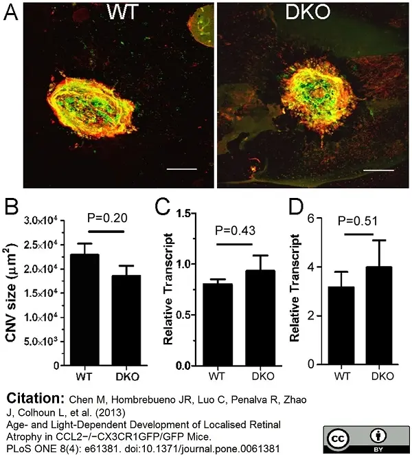
Chen, M. et al. (2013) Age- and light-dependent development of localised retinal atrophy in CCL2(-/-)CX3CR1(GFP/GFP) mice.
PLoS One. 8: e61381. -
Chu, C.J. et al. (2013) Assessment and in vivo scoring of murine experimental autoimmune uveoretinitis using optical coherence tomography.
PLoS One. 8 (5): e63002. -
Weinl, C. et al. (2014) Elk3 deficiency causes transient impairment in post-natal retinal vascular development and formation of tortuous arteries in adult murine retinae.
PLoS One. 9: e107048. -
Scott, A. et al. (2014) Quantification of vascular tortuosity as an early outcome measure in oxygen induced retinopathy (OIR)
Exp Eye Res. 120: 55-60. -
Shen, W. et al. (2014) Systemic Administration of Erythropoietin Inhibits Retinopathy in RCS Rats.
PLoS One. 9: e104759. -
Yukiura H et al. (2015) Autotaxin overexpression causes embryonic lethality and vascular defects.
PLoS One. 10 (5): e0126734. -
Wu, W.K. et al. (2015) IL-4 regulates specific Arg-1(+) macrophage sFlt-1-mediated inhibition of angiogenesis.
Am J Pathol. 185 (8): 2324-35. -
Williams, J.A. et al. (2016) Regulation of C3 Activation by the Alternative Complement Pathway in the Mouse Retina.
PLoS One. 11 (8): e0161898. -
Piñero, G. et al. (2017) Lithium Reversibly Inhibits Schwann Cell Proliferation and Differentiation Without Inducing Myelin Loss.
Mol Neurobiol. 54 (10): 8287-307. -
Gurnik, S. et al. (2016) Angiopoietin-2-induced blood-brain barrier compromise and increased stroke size are rescued by VE-PTP-dependent restoration of Tie2 signaling.
Acta Neuropathol. 131 (5): 753-73. -
Misra, A. et al. (2016) Integrin β3 inhibition is a therapeutic strategy for supravalvular aortic stenosis.
J Exp Med. 213 (3): 451-63. -
Yanagida, K. et al. (2017) Size-selective opening of the blood-brain barrier by targeting endothelial sphingosine 1-phosphate receptor 1.
Proc Natl Acad Sci U S A. 114 (17): 4531-6. -
Toffoli, B. et al. (2017) Nephropathy in Pparg-null mice highlights PPARγ systemic activities in metabolism and in the immune system.
PLoS One. 12 (2): e0171474. -
Fernández-robredo, P. et al. (2017) Neuropilin 1 Involvement in Choroidal and Retinal Neovascularisation.
PLoS One. 12 (1): e0169865. -
Kim, B. et al. (2018) Endothelial pyruvate kinase M2 maintains vascular integrity.
J Clin Invest. 128 (10): 4543-56. -
Stone, O.A. et al. (2018) Loss of pyruvate kinase M2 limits growth and triggers innate immune signaling in endothelial cells.
Nat Commun. 9 (1): 4077. -
Niaudet, C. et al. (2019) Adgrf5 contributes to patterning of the endothelial deep layer in retina.
Angiogenesis. 22 (4): 491-505. -
Villard, O. et al. (2020) Characterization of immortalized human islet stromal cells reveals a MSC-like profile with pancreatic features.
Stem Cell Res Ther. 11 (1): 158. -
Krivanek, J. et al. (2020) Dental cell type atlas reveals stem and differentiated cell types in mouse and human teeth.
Nat Commun. 11 (1): 4816. -
Roth, M. et al. (2020) Parenchymal pericytes are not the major contributor of extracellular matrix in the fibrotic scar after stroke in male mice.
J Neurosci Res. 98 (5): 826-842. -
Elabi, O. et al. (2021) Human α-synuclein overexpression in a mouse model of Parkinson's disease leads to vascular pathology, blood brain barrier leakage and pericyte activation.
Sci Rep. 11 (1): 1120. -
Månberg, A. et al. (2021) Altered perivascular fibroblast activity precedes ALS disease onset.
Nat Med. 27 (4): 640-6. -
Mohanta, S.K. et al. (2022) Neuroimmune cardiovascular interfaces control atherosclerosis.
Nature. 605 (7908): 152-9. -
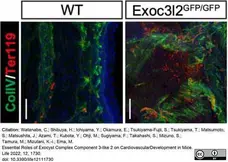
Watanabe, C. et al. (2022) Essential Roles of Exocyst Complex Component 3-like 2 on Cardiovascular Development in Mice.
Life (Basel). 12 (11): 1730. -
Tsuji-Tamura, K. & Ogawa, M. (2023) FOXO1 promotes endothelial cell elongation and angiogenesis by up-regulating the phosphorylation of myosin light chain 2.
Angiogenesis. 26 (4): 523-45. -
Yin, C. et al. (2019) ApoE attenuates unresolvable inflammation by complex formation with activated C1q.
Nat Med. 25 (3): 496-506. -
Chen, M. et al. (2018) Cytokine Signaling Protein 3 Deficiency in Myeloid Cells Promotes Retinal Degeneration and Angiogenesis through Arginase-1 Up-Regulation in Experimental Autoimmune Uveoretinitis.
Am J Pathol. 188 (4): 1007-20. -
Schley, G. et al. (2018) Multiparametric magnetic resonance imaging of experimental chronic kidney disease: A quantitative correlation study with histology.
PLoS One. 13 (7): e0200259. -
Zhao, W. et al. (2019) Endothelial CDS2 deficiency causes VEGFA-mediated vascular regression and tumor inhibition.
Cell Res. 29 (11): 895-910. -
Kool, H.M. et al. (2019) Inhibition of retinoic acid signaling induces aberrant pericyte coverage and differentiation resulting in vascular defects in congenital diaphragmatic hernia.
Am J Physiol Lung Cell Mol Physiol. 317 (3): L317-L331. -
Cottarelli, A. et al. (2020) Fgfbp1 promotes blood-brain barrier development by regulating collagen IV deposition and maintaining Wnt/β-catenin signaling.
Development. 147 (16): dev185140. -
Santamaría, R. et al. (2023) Capillary pruning couples tissue perfusion and oxygenation with cardiomyocyte maturation in the postnatal mouse heart.
Front Cell Dev Biol. 11: 1256127. -
Chen, M. et al. (2019) STAT3 activation in circulating myeloid-derived cells contributes to retinal microvascular dysfunction in diabetes.
J Neuroinflammation. 16 (1): 138. -
Kautzman, A.G. et al. (2018) Sox2 regulates astrocytic and vascular development in the retina.
Glia. 66 (3): 623-36. -
Li, J. et al. (2023) Barley Grass Juice Attenuates Hydrodynamic Transfection-Induced HCC Initiation in Mice.
Nutr Cancer. 75 (2): 750-60. -
Wang, F. et al. (2021) Neutralization of Hv1/HVCN1 With Antibody Enhances Microglia/Macrophages Myelin Clearance by Promoting Their Migration in the Brain.
Front Cell Neurosci. 15: 768059. -
Zhou, Q. et al. (2021) Association between erythrocyte dynamics and vessel remodelling in developmental vascular networks.
J R Soc Interface. 18 (179): 20210113. -
Sivaraj, K.K. et al. (2024) Endothelial LATS2 is a suppressor of bone marrow fibrosis
Nat Cardiovasc Res. 29 Jul. [Epub ahead of print]. -
Wang, D. et al. (2024) Differences in Acute Expression of Matrix Metalloproteinases-9, 3, and 2 Related to the Duration of Brain Ischemia and Tissue Plasminogen Activator Treatment in Experimental Stroke.
Int J Mol Sci. 25 (17): 9442. -
Mohanakrishnan, V. et al. (2024) Specialized post-arterial capillaries facilitate adult bone remodelling.
Nat Cell Biol. 26 (12): 2020-34. -
Barreiro, O. et al. (2016) Pivotal role for skin transendothelial radio-resistant anti-inflammatory macrophages in tissue repair.
Elife. 5:e15251. -
Teng, Q. et al. (2025) TGF-β3 promotes vascular normalization of prostate cancer to potentiate immunotherapy and chemotherapy.
Cancer Immunol Immunother. 74 (8): 268. -
Zhao, W. et al. (2026) NAD-dependent redox control enables endothelial quiescence and vascular stabilization during angiogenesis.
Cell Metab. S1550-4131(26)00142-7. -
Gonzalez, M.L. et al. (2026) Embryonic and postnatal macrophages are necessary for proper tooth development and homeostasis.
Nat Commun. 17 (1): 6537.
- RRID
- AB_2082660
- UniProt
- P02463
- P08122
- Q9QZS0
- Q9QZR9
- Q80V57
- Q6PFB1
- Entrez Gene
- Col4a1
- Col4a2
- Col4a4
- Col4a3
- GO Terms
- GO:0001525 angiogenesis
- GO:0005201 extracellular matrix structural constituent
- GO:0005587 collagen type IV
- GO:0005488 binding
- GO:0007155 cell adhesion
- GO:0005178 integrin binding
- GO:0006917 induction of apoptosis
- GO:0006919 activation of caspase activity
- GO:0007166 cell surface receptor linked signaling pathway
- View More GO Terms
- GO:0008283 cell proliferation
- GO:0016525 negative regulation of angiogenesis
- GO:0007528 neuromuscular junction development
- GO:0032836 glomerular basement membrane development
View more products with COLLAGEN specificity
Please Note: All Products are "FOR RESEARCH PURPOSES ONLY"
View all Anti-Mouse ProductsAlways be the first to know.
When we launch new products and resources to help you achieve more in the lab.
Yes, sign me up