CD5 antibody | YKIX322.3
Rat anti Dog CD5
- Product Type
- Monoclonal Antibody
- Clone
- YKIX322.3
- Isotype
- IgG2a
- Specificity
- CD5


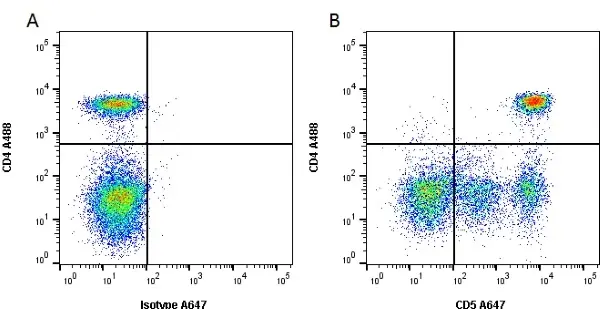
Figure B. Alexa Fluor® 488 conjugated Rat anti Canine CD4 antibody, clone YKIX302.9 (MCA1038A488) and Alexa Fluor® 647 conjugated Rat anti Canine CD5 antibody, clone YKIX322.3 (MCA1037A647). All experiments performed on red cell lysed canine blood gated on lymphocytes in the presence of 10% canine serum.
Data acquired on the ZE5 Cell Analyzer.

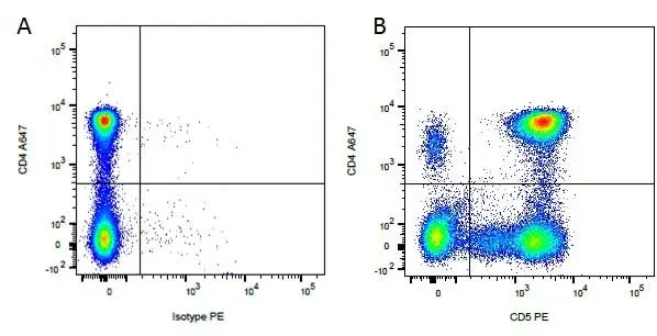
Figure B. Alexa Fluor® 647 conjugated Rat anti Canine CD4 antibody, clone YKIX302.9 (MCA1038A647) and RPE conjugated Rat anti Canine CD5 antibody, clone YKIX322.3 (MCA1037PE). All experiments performed on red cell lysed canine blood gated on mononuclear cells.
Data acquired on the ZE5 Cell Analyzer.

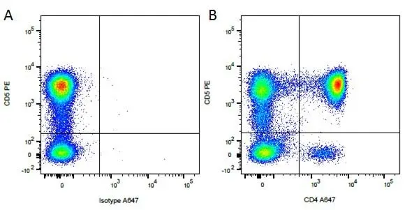
Figure B. RPE conjugated Rat anti Canine CD5 antibody, clone YKIX322.3 (MCA1037PE) and Alexa Fluor® 647 conjugated Rat anti Canine CD4 antibody, clone YKIX302.9 (MCA1038A647). All experiments performed on red cell lysed canine blood gated on mononuclear cells.
Data acquired on the ZE5 Cell Analyzer.
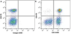
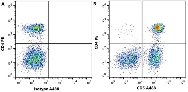
Figure B. PE conjugated Rat anti Dog CD4 antibody, clone YKIX302.9 (MCA1038PE) and Alexa Fluor®488 conjugated Rat anti Dog CD5 antibody, clone YKIX322.3 (MCA1037A488).
All experiments performed on red cell lysed canine blood gated on live single lymphocytes, in the presence of 10% canine serum. Data acquired on the ZE5 Cell analyser.

Fitc conjugated Rat anti Dog CD5 antibody, clone YKIX322.3 (MCA1037F) used to label canine lymphocytes by flow cytometry.
Image caption:
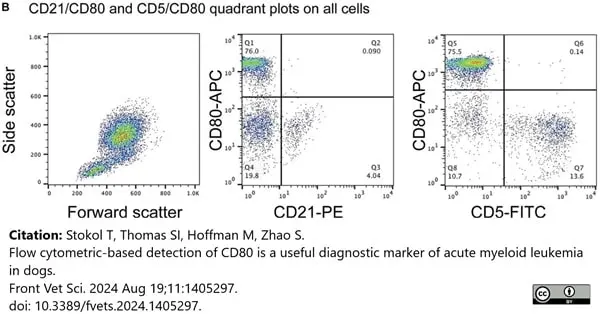
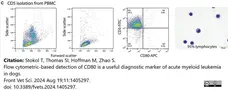
Flow cytometric dot plots of anti-CD80 antibody labeling of leukocytes in blood from healthy dogs. (B) Triple-labeling of dog leukocytes with CD80-APC, CD21-PE and CD5-FITC shows that CD21+ B cells and CD5+ T cells are CD80− (representative result from 1 of 3 dogs). All leukocyte events were combined for analysis vs. splitting the events into different gates based on forward and side scatter.
From: Stokol T, Thomas SI, Hoffman M, Zhao S.
Flow cytometric-based detection of CD80 is a useful diagnostic marker of acute myeloid leukemia in dogs.
Front Vet Sci. 2024 Aug 19;11:1405297.
doi: 10.3389/fvets.2024.1405297.
This image is from an open access article distributed under terms of a Creative Commons Attribution License.
Fitc conjugated Rat anti Dog CD5 antibody, clone YKIX322.3 (MCA1037F) used to lable canine lymphocytes by flow cytometry.
Image caption:
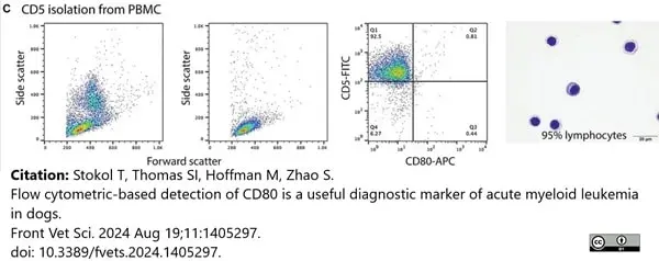
Labeling of monocytes, B cells, T cells, and neutrophils isolated from the blood of healthy dogs (representative results from 1 of 2–3 dogs for each cell type) with the anti-CD80 antibody. (C) CD5-FITC-isolated cells were mostly lymphocytes, which were CD80− (third panel). Lymphocytes were small cells with a few large or reactive forms. Several lymphocytes had a few clear cytoplasmic vacuoles, which could be due to the isolation procedure (fourth panel).
From: Stokol T, Thomas SI, Hoffman M, Zhao S.
Flow cytometric-based detection of CD80 is a useful diagnostic marker of acute myeloid leukemia in dogs.
Front Vet Sci. 2024 Aug 19;11:1405297.
doi: 10.3389/fvets.2024.1405297.
This image is from an open access article distributed under terms of a Creative Commons Attribution License.

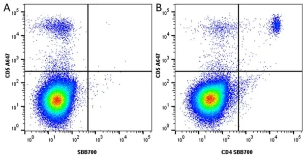
Figure B. Alexa Flour®A647 conjugated Rat anti Dog CD5 antibody, clone YKIX322.3 (MCA1037A647) and StarBright Blue 700 conjugated Rat anti Dog CD4 antibody, clone YKIX302.9 (MCA1038SBB700).
All experiments performed on red cell lysed dog blood gated on live single Lymphocytes, in the presence of 10% dog serum. Data acquired on the Data acquired on the ZE5 Cell Analyzer.
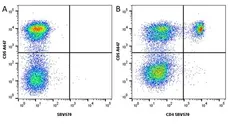
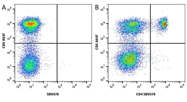
Figure B. Alexa Flour®647 conjugated Rat anti Dog CD5 antibody, clone YKIX322.3 (MCA1037A647) and StarBright Violet 570 conjugated Rat anti Dog CD4 antibody, clone YKIX302.9 (MCA1038SBV570).
All experiments performed on red cell lysed dog blood gated on live single lymphocytes, in the presence of 10% dog serum. Data acquired on the ZE5 Cell analyser.
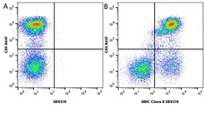
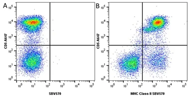
Figure B. Alexa Flour®647 conjugated Rat anti Dog CD5 antibody, clone YKIX322.3 (MCA1037A647) and StarBright Violet 570 conjugated Rat anti Dog MHC Class II monomorphic antibody, clone YKIX334.2 (MCA1044SBV570).
All experiments performed on red cell lysed dog blood gated on live single lymphocytes, in the presence of 10% dog serum. Data acquired on the ZE5 Cell analyser.
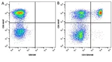
Figure B. Alexa Flour® 647 conjugated Rat anti Dog CD5 antibody, clone YKIX322.3 (MCA1037A647) and StarBright Violet 440 conjugated Rat anti Dog CD4 antibody, clone YKIX302.9 (MCA1038SBV440). All experiments performed on red blood lysed dog blood gated on live single cell lymphocytes, in the presence of 10% dog serum.
Data acquired on the ZE5 Cell analyser.

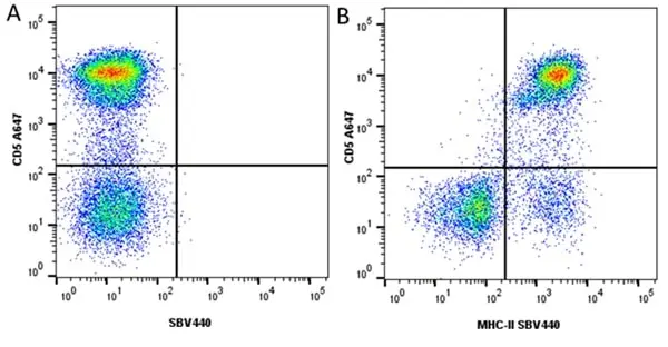
Figure B. Alexa Flour® 647 conjugated Rat anti Dog CD5 antibody, clone YKIX322.3 (MCA1037A647) and StarBright Violet 440 conjugated Rat anti Dog MHC Class II Monomorphic (MCA1044SBV440). All experiments performed on red blood lysed dog blood gated on live single cell lymphocytes, in the presence of 10% dog serum.
Data acquired on the ZE5 Cell analyser.
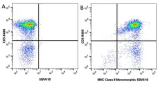
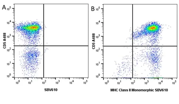
Figure B. Alexa Fluor® 488 conjugated Rat anti Dog CD5 antibody, clone YKIX322.3 (MCA1037A488) and StarBright Violet 610 conjugated Rat anti Dog MHC Class II Monomorphic antibody, clone YKIX334.2 (MCA1044SBV610). All experiments performed on red blood lysed dog blood gated on live single cell lymphocytes, in the presence of 10% dog serum.
Data acquired on the ZE5 Cell analyser
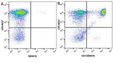
Figure B. Alexa Fluor® 647 conjugated Rat anti Dog CD5 antibody, clone YKIX322.3 (MCA1037A647) and StarBright Violet 610 conjugated Rat anti Dog CD4 antibody, clone YKIX302.9 (MCA1038SBV610). All experiments performed on red blood lysed dog blood gated on live single cell lymphocytes, in the presence of 10% dog serum.
Data acquired on the ZE5 Cell analyser
Filter by Application:
F Reset| Rat anti Dog CD5 antibody, clone YKIX322.3 recognizes canine CD5, a 67 kDa cell surface type 1 transmembrane glycoprotein also known as lymphocyte antigen T1, Ly-1 or Leu-1. CD5 is expressed on the surface of T-cells and thymocytes, CD5 is also expressed by NK cells at low levels (Huang et al. 2008). Rat anti dog CD5, cloneYKIX322.3 was clustered as canine CD5 in the First Canine Leucocyte Antigen Workshop (Cobbold et al. 1994). In a study of 73 cases of canine chronic lymphocytic leukemia (CLL) CD5 expression was absent on all cases of B-cell CLL as defined by CD21 expression and lack of CD3 or other T cell antigen expression (Vernau and Moore 1999). Rat anti dog CD5 serves as a useful marker for the discrimination of canine leukemias of differing origins (Deravi et al. 2017). |
- Target Species
- Dog
- Product Form
- Purified IgG - liquid
- Preparation
- Purified IgG prepared by affinity chromatography on Protein G from tissue culture supernatant
- Buffer Solution
- Phosphate buffered saline
- Preservative Stabilisers
- 0.09% sodium azide (NaN3)
- Carrier Free
- Yes
- Immunogen
- Concanavilin A activated canine peripheral blood cells
- Approx. Protein Concentrations
- IgG concentration 1.0 mg/ml
- Fusion Partners
- Spleen cells from an immunized DA rat were fused with cells of the rat Y3/Ag1.2.3 myeloma cell line
- Regulatory
- For research purposes only
- Guarantee
- 12 months from date of despatch
This product is shipped at ambient temperature. It is recommended to aliquot and store at -20°C on receipt. When thawed, aliquot the sample as needed. Keep aliquots at 2-8°C for short term use (up to 4 weeks) and store the remaining aliquots at -20°C.
Avoid repeated freezing and thawing as this may denature the antibody. Storage in frost-free freezers is not recommended.
Avoid repeated freezing and thawing as this may denature the antibody. Storage in frost-free freezers is not recommended.
This product has been reported to work in the following applications. This information is derived from testing within our laboratories, peer-reviewed publications or personal communications from the originators. Please refer to references indicated for further information. For general protocol recommendations, please visit the antibody protocols page.
| Application Name | Verified | Min Dilution | Max Dilution |
|---|---|---|---|
| Flow Cytometry |  |
1/50 | 1/100 |
| Immunohistology - Frozen | |
||
| Immunoprecipitation | |
Where this product has not been tested for use in a particular technique this does not necessarily exclude its use in such procedures. Suggested working dilutions are given as a guide only. It is recommended that the user titrates the product for use in their own system using appropriate negative/positive controls.
- Flow Cytometry
- Use 10μl of the suggested working dilution to label 106 cell in 100μl.
References for CD5 antibody
-
Cobbold, S/ & Metcalfe, S. (1994) Monoclonal antibodies that define canine homologues of human CD antigens: summary of the First International Canine Leukocyte Antigen Workshop (CLAW).
Tissue Antigens. 43 (3): 137-54. -
Hewicker-Trautwein, M. et al. (1999) Immunocytochemical demonstration of lymphocyte subsets and MHC class II antigen expression in synovial membranes from dogs with rheumatoid arthritis and degenerative joint disease.
Vet Immunol Immunopathol. 67 (4): 341-57. -
Vernau, W. & Moore, P.F. (1999) An immunophenotypic study of canine leukemias and preliminary assessment of clonality by polymerase chain reaction.
Vet Immunol Immunopathol. 69: 145-64. -
Guarga, J.L. et al. (2002) Evaluation of a specific immunochemotherapy for the treatment of canine visceral leishmaniasis.
Vet Immunol Immunopathol. 88: 13-20. -
Burnett, R.C. et al. (2003) Diagnosis of canine lymphoid neoplasia using clonal rearrangements of antigen receptor genes.
Vet Pathol. 40: 32-41. -
Lamerato-Kozicki, A.R. et al. (2006) Canine hemangiosarcoma originates from hematopoietic precursors with potential for endothelial differentiation.
Exp Hematol. 34 (7): 870-8. -
Fosmire, S.P. et al. (2007) Inactivation of the p16 cyclin-dependent kinase inhibitor in high-grade canine non-Hodgkin's T-cell lymphoma.
Vet Pathol. 44: 467-78. -
Huang, Y.C.
(2008) CD5-low expression lymphocytes in canine peripheral blood show characteristics of natural killer cells.
J Leukoc Biol. 84: 1501-10.
View The Latest Product References
-
Araújo, M.S. et al. (2011) Immunological changes in canine peripheral blood leukocytes triggered by immunization with first or second generation vaccines against canine visceral leishmaniasis.
Vet Immunol Immunopathol. 141: 64-75. -
GomesMde, O. et al. (2011) Old beagle dogs have lower faecal concentrations of some fermentation products and lower peripheral lymphocyte counts than young adult beagles.
Br J Nutr. 106 Suppl 1: S187-90. -
Rütgen, B.C. et al. (2012) Authentication of primordial characteristics of the CLBL-1 cell line prove the integrity of a canine B-cell lymphoma in a murine in vivo model.
PLoS One. 7 (6): e40078. -
Michael, H.T. et al. (2013) Isolation and characterization of canine natural killer cells.
Vet Immunol Immunopathol. 155 (3): 211-7. -
Aricò, A. et al. (2013) The role of vascular endothelial growth factor and matrix metalloproteinases in canine lymphoma: in vivo and in vitro study.
BMC Vet Res. 9: 94. -
Aresu, L. et al. (2014) VEGF and MMP-9: biomarkers for canine lymphoma.
Vet Comp Oncol. 12: 29-36. -
Gelain, M.E. et al. (2014) CD44 in canine leukemia: analysis of mRNA and protein expression in peripheral blood.
Vet Immunol Immunopathol. 159 (1-2): 91-6. -
Stokol, T. et al. (2015) Alkaline phosphatase is a useful cytochemical marker for the diagnosis of acute myelomonocytic and monocytic leukemia in the dog.
Vet Clin Pathol. 44 (1): 79-93. -
Ito, D. et al. (2015) A double blinded, placebo-controlled pilot study to examine reduction of CD34 +/CD117 +/CD133 + lymphoma progenitor cells and duration of remission induced by neoadjuvant valspodar in dogs with large B-cell lymphoma.
F1000Res. 4: 42. -
Bonnefont-Rebeix, C. et al. (2016) Characterization of a novel canine T-cell line established from a spontaneously occurring aggressive T-cell lymphoma with large granular cell morphology.
Immunobiology. 221 (1): 12-22. -
Gibbons, N. et al. (2017) Phenotypic heterogeneity of peripheral monocytes in healthy dogs.
Vet Immunol Immunopathol. 190: 26-30. -
Deravi, N. et al. (2017) Specific immunotypes of canine T cell lymphoma are associated with different outcomes.
Vet Immunol Immunopathol. 191: 5-13. -
Maria, A. P.J. et al. (2017) The effect of age and carbohydrate and protein sources on digestibility, fecal microbiota, fermentation products, fecal IgA, and immunological blood parameters in dogs.
J Anim Sci. 95 (6): 2452-66. -
Roatt, B.M. et al. (2017) A Vaccine Therapy for Canine Visceral Leishmaniasis Promoted Significant Improvement of Clinical and Immune Status with Reduction in Parasite Burden.
Front Immunol. 8: 217. -
Karayannopoulou, M. et al. (2017) Evaluation of blood T-lymphocyte subpopulations involved in host cellular immunity in dogs with mammary cancer.
Vet Immunol Immunopathol. 186: 45-50. -
Lin, C.S. et al. (2018) Activating natural killer (NK) cytotoxicity of canine CD5-CD21- cells requires low surface CD5 density NK cells.
Iran J Vet Res. 19 (2): 87-95. -
Graves, S.S. et al. (2019) Development and characterization of a canine-specific anti-CD94 (KLRD-1) monoclonal antibody.
Vet Immunol Immunopathol. 211: 10-8. -
Martini, V. et al. (2019) Prognostic role of non-neoplastic lymphocytes in lymph node aspirates from dogs with diffuse large B-cell lymphoma treated with chemo-immunotherapy.
Res Vet Sci. 125: 130-5. -
Wolf-Ringwall, A. et al. (2020) Prospective evaluation of flow cytometric characteristics, histopathologic diagnosis and clinical outcome in dogs with naïve B-cell lymphoma treated with a 19-week CHOP protocol.
Vet Comp Oncol. 18 (3): 342-52. -
Aguiar-Soares, R.D.O. et al. (2020) Phase I and II Clinical Trial Comparing the LBSap, Leishmune®, and Leish-Tec® Vaccines against Canine Visceral Leishmaniasis.
Vaccines (Basel). 8 (4)Nov 17 [Epub ahead of print]. -
Sayag, D. et al. (2020) Proof-of-concept study: Evaluation of plasma and urinary electrolytes as markers of response to L-asparaginase therapy in dogs with high-grade lymphoma.
Vet Clin Pathol. 49 (3): 476-83. -
Lee, J. et al. (2021) Canine Natural Killer Cell-Derived Exosomes Exhibit Antitumor Activity in a Mouse Model of Canine Mammary Tumor.
Biomed Res Int. 2021: 6690704. -
Grudzien, M. et al. (2021) A newly established canine NK-type cell line and its cytotoxic properties.
Vet Comp Oncol. 19 (3): 567-77. -
Lee, S.H. et al. (2021) Safety and immunological effects of recombinant canine IL-15 in dogs.
Cytokine. 148: 155599. -
Karayannopoulou, M. et al. (2022) Effect of major versus minor mastectomy on host immunity in canine mammary cancer
Vet Immunol Immunopathol. 24 Feb: 110403. -
Riccardo, F. et al. (2022) Antigen mimicry as an effective strategy to induce CSPG4-targeted immunity in dogs with oral melanoma: a veterinary trial.
J Immunother Cancer. 10 (5): e004007. [Epub ahead of print]. -
Jaensch, S. et al. (2022) Clinicopathologic and immunophenotypic features in dogs with presumptive large granular lymphocyte leukaemia
Australian Veterinary Journal. [Epub ahead of print]. -
Martini, V. et al. (2018) A retrospective study of flow cytometric characterization of suspected extranodal lymphomas in dogs.
J Vet Diagn Invest. 30 (6): 830-836. -
Lee, G.W. et al. (2021) Case Report: Long-Term Survival of a Dog With Chronic Lymphocytic Leukemia Treated With Chlorambucil, Prednisolone, and Imatinib.
Front Vet Sci. 8: 625527. -
Hughes, K. et al. (2024) Canine T zone lymphoma is a tumor of mature, previously activated αβ T cells
Vet Immunology Immunopathol 110725. -
Sheng, R. et al. (2023) Prognostic significance of CD25 expression in dogs with a noninvasive diagnosis of B-cell lymphoma treated with CHOP chemotherapy.
Vet Comp Oncol. 21 (1): 28-35. -
Marconato, L. et al. (2024) Multicentric aggressive unclassified hematopoietic neoplasm involving the placenta in a pregnant bitch.
Vet Clin Pathol. Oct 09 [Epub ahead of print]. -
Stokol, T. et al. (2024) Flow cytometric-based detection of CD80 is a useful diagnostic marker of acute myeloid leukemia in dogs.
Front Vet Sci. 11: 1405297. -
Rogato, F. et al. (2024) Leukemia cutis as a prominent clinical sign in a dog with acute myeloid leukemia.
Vet Clin Pathol. 53 (4): 448-457. -
De Maria, R. et al. (2025) Development and activity of canine B7-H3-CAR.CIK lymphocytes against sarcomas: preclinical evidence and perspectives for human clinical translation.
Cancer Immunol Immunother. 74 (10): 306. -
Sulce, M. et al. (2018) Utility of flow cytometry in canine primary cutaneous and matched nodal mast cell tumor.
Vet J. 242: 15-23. -
Blockeel, A. et al. (2025) CD94 as a novel marker for immunophenotyping of leukemia and lymphoma in dogs.
Front Vet Sci. 12: 1716800.
MCA1037GA
If you cannot find the batch/lot you are looking for please contact our technical support team for assistance.
View more products with CD5 specificity
Please Note: All Products are "FOR RESEARCH PURPOSES ONLY"
View all Anti-Dog ProductsAlways be the first to know.
When we launch new products and resources to help you achieve more in the lab.
Yes, sign me up