CD4 antibody | CC8
Mouse anti Bovine CD4:FITC
- Product Type
- Monoclonal Antibody
- Clone
- CC8
- Isotype
- IgG2a
- Specificity
- CD4

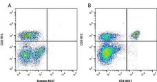
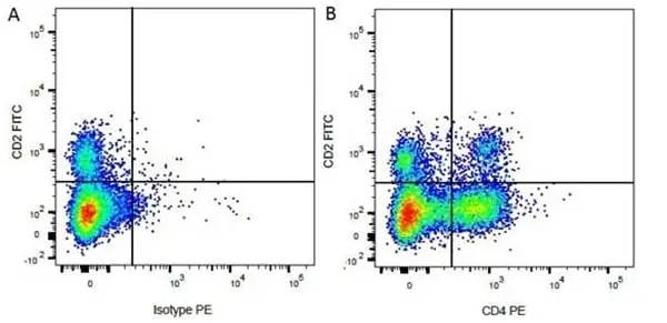
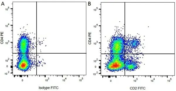
Figure B. FITC conjugated Mouse anti Bovine CD2 antibody, clone CC42 (MCA833F) and Alexa Fluor® 647 conjugated Mouse anti Bovine CD4 antibody, clone CC8 (MCA1653A647).
All experiments performed on red cell lysed bovine blood gated on lymphocytes.
Data acquired on the ZE5 Cell Analyzer.

Mouse anti Bovine CD4 antibody, clone CC8 (MCA1653) used for the evaluation of CD4 expression on proliferating bovine peripheral blood mononuclear cells by flow cytometry.
Image caption:
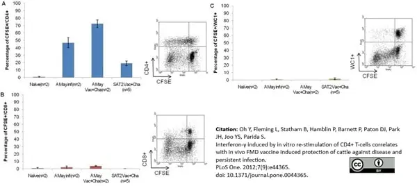
Determination of phenotypes of proliferating PBMC by FACS analysis. Determination of phenotypes (CD4+, CD8+ and WC1+) of proliferating PBMC by CFDA SE staining after re-stimulating with respective vaccine antigen and fluorescence-activated cell sorter (FACS) analysis. PBMC were separated from heparinised blood of vaccinated cattle originated from full dose vaccine group on 4 weeks after homologous FMDV challenge. Two naïve cattle without FMDV infection and two A Malaysia 97 FMDV infected cattle (4 weeks post-infection) without prior vaccination were included as control group. PBMCs from naïve animals were re-stimulated with SAT2 Eritrea vaccine antigen. FACS plots were exemplified only for one vaccinated challenged animal from SAT2 Eritrea animal experiment. Similar FACS plots were obtained for the PBMC originated from A Malaysia 97 and SAT2 Eritrea vaccinated animals. Error bars represent standard error of the mean.
From: Oh Y, Fleming L, Statham B, Hamblin P, Barnett P, Paton DJ, et al. (2012)
Interferon-γ Induced by In Vitro Re-Stimulation of CD4+ T-Cells Correlates with In Vivo FMD Vaccine Induced Protection of Cattle against Disease and Persistent Infection.
PLoS ONE 7(9): e44365.
This image is from an open access article distributed under the terms of the Creative Commons Attribution License.
Mouse anti Bovine CD4 antibody, clone CC8 (MCA1653) used for the identification of CD4 expressing lymphocytes in preipheral blood by flow cytometry.
Image caption:
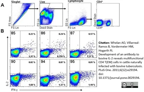
Identification of recombinant antibodies with specificity for bIL-2. PBMC from a cow naturally infected with M. bovis were cultured in the presence of PPD-B to allow screening of candidate bIL-2 by ICS flow cytometry. Panel A shows the histogram gating strategy used to interrogate responses in singlet, live CD4+ lymphocytes. Panel B shows the measurement of detectable IL-2 and/or IFN-γ within the CD4+ population for each of 6 candidate IL-2 antibody clones. The clone number is shown in the top left corner of each histogram and the percentage of CD4+ cell in which co-expression of IFN-γ and IL-2 could be detected is shown in the top right of each histogram. Data are representative of 1 of 2 independent experiments.
From: Whelan AO, Villarreal-Ramos B, Vordermeier HM, Hogarth PJ (2011)
Development of an Antibody to Bovine IL-2 Reveals Multifunctional CD4 TEM Cells in Cattle Naturally Infected with Bovine Tuberculosis.
PLoS ONE 6(12): e29194.
This image is from an open access article distributed under the terms of the Creative Commons Attribution License.
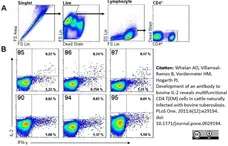
Mouse anti Bovine CD4 antibody, clone CC8 (MCA1653) used for the identification of CD4 expressing lymphocytes in preipheral blood by flow cytometry.
Image caption:
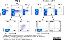
Identification of multifunctional IFN-γ, IL-2 and TNF-α CD4+ cells in natural M. bovis infection. PBMC from naturally M. bovis infected cattle were cultured in the presence of either PPD-B or medium and the co-expression of IFN-γ, IL-2 and TNF-α determined by ICS flow cytometry. Histograms were gated on singlet, live lymphocytes and then all CD4+ cells analysed for all combinations of cytokine productivity. The upper histograms show the total proportion of CD4+ cells staining for expression of IFN-γ, IL-2 or TNF-α following stimulation with PPD-B (left panel) or medium control (right panel). Subgating of the IFN-γ+ and IFN-γ− CD4+ cells provides histograms that represent all possible functionalities for the expressing of the 3 cytokines, as shown in the lower histograms. The numbers indicate percentage of CD4+ cells and data are representative of 1 of 10 naturally infected cattle.
From: Whelan AO, Villarreal-Ramos B, Vordermeier HM, Hogarth PJ (2011)
Development of an Antibody to Bovine IL-2 Reveals Multifunctional CD4 TEM Cells in Cattle Naturally Infected with Bovine Tuberculosis.
PLoS ONE 6(12): e29194.
This image is from an open access article distributed under the terms of the Creative Commons Attribution License.

APC conjugated Mouse anti Bovine CD4 antibody, clone CC8 (MCA1653APC) used for the identification of CD4 expressing T-cells in bovine peripheral blood by flow cytoometry together with RPE conjugated Mouse anti Bovine CD8 antibody, clone CC63 (MCA837PE) used to detect cytotoxic T-cells in the same samples.
Image caption:
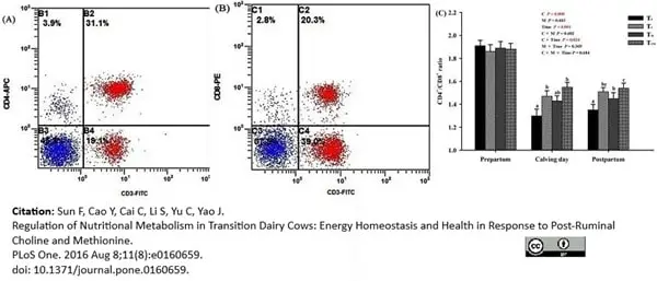
Effects of supplementation with rumen-protected choline (TC), rumen-protected methionine (TM) or both (TCM) on proportions of T lymphocyte subtypes in peripheral blood of transition dairy cows. (A) and (B) present the output images from flow cytometry. (C) displays the effects of each treatment over the three weeks before calving, on calving day, and over the three weeks after calving. a, b, c Values with different superscript letters at the same time point are significantly different (P <0.05).
From: Sun F, Cao Y, Cai C, Li S, Yu C, Yao J (2016)
Regulation of Nutritional Metabolism in Transition Dairy Cows: Energy Homeostasis and Health in Response to Post-Ruminal Choline and Methionine.
PLoS ONE 11(8): e0160659.
This image is from an open access article distributed under the terms of the Creative Commons Attribution License.
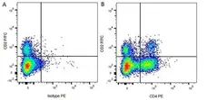
Figure B. FITC conjugated Mouse anti Bovine CD2 antibody, clone CC42 (MCA833F) and RPE conjugated Mouse anti Bovine CD4 antibody, clone CC8 (MCA1653PE).
All experiments performed on red cell lysed bovine blood gated on mononuclear cells.
Data acquired on the ZE5 Cell Analyzer.
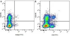
Figure B. RPE conjugated Mouse anti Bovine CD4 antibody, clone CC8 (MCA1653PE) and FITC conjugated Mouse anti Bovine CD2 antibody, clone CC42 (MCA833F).
All experiments performed on red cell lysed bovine blood gated on mononuclear cells.
Data acquired on the ZE5 Cell Analyzer.
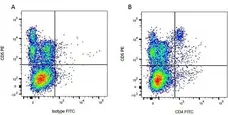
Figure B. Purified Mouse anti Bovine CD5 antibody, clone CC17 (MCA835GA) detected with RPE conjugated Goat anti Mouse IgG1 antibody (STAR132PE) and FITC conjugated Mouse anti Bovine CD4 antibody, clone CC8 (MCA1653F).
All experiments performed on red cell lysed bovine blood gated on mononuclear cells.
Data acquired on the ZE5 Cell Analyzer.
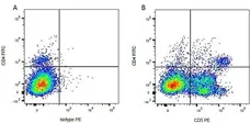
Figure B. FITC conjugated Mouse anti Bovine CD4 antibody, clone CC8 (MCA1653F) and purified Mouse anti Bovine CD5 antibody, clone CC17 (MCA835GA) detected with RPE conjugated Goat anti Mouse IgG1 antibody (STAR132PE).
All experiments performed on red cell lysed bovine blood gated on mononuclear cells.
Data acquired on the ZE5 Cell Analyzer.
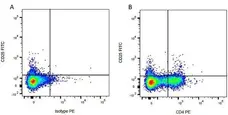
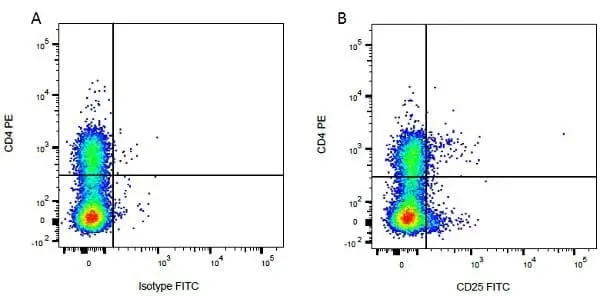
Figure B. FITC conjugated Mouse anti Bovine CD25 antibody, clone IL-A111 (MCA2430F) and RPE conjugated Mouse anti Bovine CD4 antibody, clone CC8 (MCA1653PE).
All experiments performed on red cell lysed bovine blood gated on mononuclear cells.
Data acquired on the ZE5 Cell Analyzer.
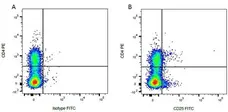
Figure B. RPE conjugated Mouse anti Bovine CD4 antibody, clone CC8 (MCA1653PE) and FITC conjugated Mouse anti Bovine CD25 antibody, clone IL-A111 (MCA2430F).
All experiments performed on red cell lysed bovine blood gated on mononuclear cells.
Data acquired on the ZE5 Cell Analyzer.

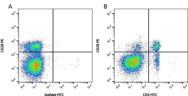
Figure B. RPE conjugated Mouse anti Bovine CD28 antibody, clone CC219 (MCA5779PE) and FITC conjugated Mouse anti Bovine CD4 antibody, clone CC8 (MCA1653F).
All experiments performed on red cell lysed bovine blood gated on lymphocytes in the presence of 10% bovine serum.
Data acquired on the ZE5 Cell Analyzer.

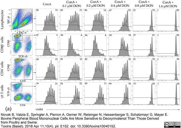
Mouse anti Bovine CD4 antibody, clone CC8 (MCA1653GA) used to evaluate CD4 expression on lymphocyte subsets from bovine peripheral blood by flow cytometry.
Image caption:
Proliferation of bovine lymphocytes and major T-cell subsets in the presence of various DON concentrations. PBMCs were stained with violet proliferation dye and cultivated in vitro in the presence of ConA and various DON concentrations for four days. After harvest, major T-cell subsets were labeled by antibodies against CD8β, CD4, and T-cell receptor (TCR)-γδ and analyzed by flow cytometry. (a) Lymphocytes were gated by light scatter properties and further subgated for the expression of CD8β, CD4, and TCR-γδ (pseudocolor plots). Histograms show the fluorescence intensities of the violet proliferation dye in lymphocytes (top) and the identified T-cell subsets under different cultivation conditions. Solid horizontal lines indicate the parental generation (on the right) and the proliferating generations (on the left). Numbers located in the two upper corners of the histograms indicate the frequency in % of the proliferating populations in comparison to the nonproliferating cells. Representative data from one animal out of six are shown. SSC-A side scatter area, FSC-A: forward scatter area.
From: Novak B, Vatzia E, Springler A, Pierron A, Gerner W, Reisinger N, Hessenberger S, Schatzmayr G, Mayer E.
Bovine Peripheral Blood Mononuclear Cells Are More Sensitive to Deoxynivalenol Than Those Derived from Poultry and Swine.
Toxins (Basel). 2018 Apr 11;10(4). pii: E152.
This image is from an open access article distributed under the terms of the Creative Commons Attribution License.
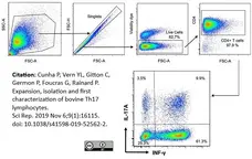
Alexa Fluor® 647 conjugated Mouse anti Bovine CD4 antibody, clone CC8 (MCA1653A647) used to assess CD4 expression on bovine lymphocytes by flow cytometry.
Image caption:
Identification of IL-17A and IFN-γ producing bovine lymphocytes. PBMC were stimulated with PMA/ionomycin, cytokine secretion blocked with Brefeldin A before flow cytometry analysis. Debris were excluded by gating according to FSC/SSC, and after gating on singlet cells, dead cells were excluded by gating on live cells. The CD4+ cells were gated by taking into account the isotype control, and the production of IL-17A and IFN-γ was measured by intracellular labelling with specific antibodies. Results shown are from a representative experiment. FSC: forward scatter: SSC: side scatter.
From: Cunha P, Vern YL, Gitton C, Germon P, Foucras G, Rainard P.
Expansion, isolation and first characterization of bovine Th17 lymphocytes.
Sci Rep. 2019 Nov 6;9(1):16115.
doi: 10.1038/s41598-019-52562-2.
This image is from an open access article distributed under the terms of the Creative Commons Attribution License.

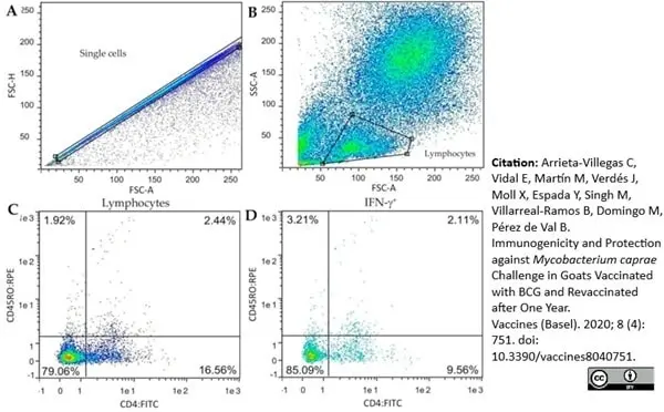
FITC conjugated Mouse anti Bovine CD4 antibody, clone CC8 (MCA1653F) used to evaluate CD4 expression on caprine PBMC by flow cytometry.
Image caption:
Gating strategy for the determination of frequency of the IFN-γ positive lymphocyte subsets. Peripheral blood mononuclear cells (PBMC) from all goats were cultured in the presence of M. bovis tuberculin (PPD-B). (A,B) Singlet lymphocytes were identified based on the degree of cellular differentiation determined by forward scatter (FSC) and side scatter (SCC). (C) Representative frequencies of the CD4/CD45RO cell populations. (D) Representative frequencies of intracellular IFN-γ staining of cells gated from prelabelled CD4+CD45RO+.
From: Arrieta-Villegas C, Vidal E, Martín M, Verdés J, Moll X, Espada Y, Singh M, Villarreal-Ramos B, Domingo M, Pérez de Val B.
Immunogenicity and Protection against Mycobacterium caprae. Challenge in Goats Vaccinated with BCG and Revaccinated after One Year.
Vaccines (Basel). 2020 Dec 10;8(4):751.
doi: 10.3390/vaccines8040751..
This image is from an open access article distributed under terms of a Creative Commons Attribution License.
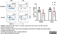
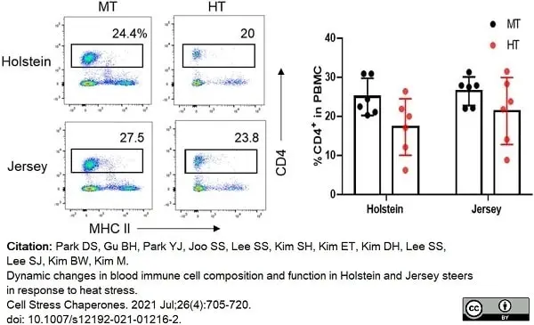
Alexa Fluor® 647 conjugated Mouse anti Bovine CD4 antibody, clone CC8 (MCA1653A647) used to identify T cell populations in bovime blood samples by flow cytometry.
Image caption:
Changes in T and B lymphocytes among PBMCs in Holstein and Jersey steers subjected to heat stress. Flow cytometry analysis to identify lymphocytes subset population. Heat stress caused reduction in lymphocyte populations in the PBMCs of both Holstein and Jersey steers. The lymphocytes comprised CD4+ T cells. MT indicates moderate THI condition and HT indicates high THI condition. * = P <0.05, ** = P <0.01
From: Park DS, Gu BH, Park YJ, Joo SS, Lee SS, Kim SH, Kim ET, Kim DH, Lee SS, Lee SJ, Kim BW, Kim M.
Dynamic changes in blood immune cell composition and function in Holstein and Jersey steers in response to heat stress.
Cell Stress Chaperones. 2021 Jul;26(4):705-20.
doi: 10.1007/s12192-021-01216-2.
This image is from an open access article distributed under terms of a Creative Commons Attribution License.
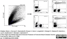
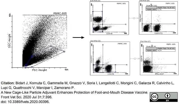
Mouse anti Bovine CD4 antibody, clone CC8 (MCA1653GA) used to evaluate CD4 expression on bovine PBMC by flow cytometry.
Image caption:
Representative dot plots, from bovine PBMCs, used for selecting the lymphocyte region based on side sideward scatter (SSC) on the y-axis and forward side scatter (FSC) on the x-axis. Then, we selected the CD8 region based on fluorescence anti-CD8 stain on the y-axis and CD4 region based on fluorescence anti-CD4 stain on the x-axis. PBMCs incubated for 18 h with iFMDV are shown.
From: Bidart J, Kornuta C, Gammella M, Gnazzo V, Soria I, Langellotti C, Mongini C, Galarza R, Calvinho L, Lupi G, Quattrocchi V, Marcipar I, Zamorano P.
A New Cage-Like Particle Adjuvant Enhances Protection of Foot-and-Mouth Disease Vaccine.
Front Vet Sci. 2020 Jul 31;7:396.
doi: 10.3389/fvets.2020.00396.
This image is from an open access article distributed under terms of a Creative Commons Attribution License.
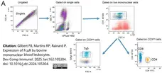
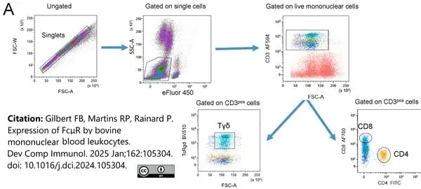
FITC conjugated Mouse anti Bovine CD4 antibody, clone CC8 (MCA1653F) used to label T lymphocytes in bovine blood by flow cytometry.
Image caption:
Expression of the FcμR by blood T lymphocytes. A) Gating strategy to distinguish viable CD3pos T cells.
From: Gilbert FB, Martins RP, Rainard P.
Expression of FcμR by bovine mononuclear blood leukocytes.
Dev Comp Immunol. 2025 Jan;162:105304.
doi: 10.1016/j.dci.2024.105304.
This image is from an open access article distributed under terms of a Creative Commons Attribution License.
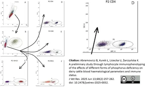
Alexa Fluor® conjugated Mouse anti Bovine CD4 antibody, clone CC8 (MCA1653A647) used for gating of bovine cell populations by flow cytometry. [ Enlargement on right].
Image caption:
Gating strategy for immunophenotyping of bovine peripheral blood lymphocytes. A – single-cell gating; B – lymphocyte gating; C–F – fluorescence cytograms of positive cells.
From: Abramowicz B, Kurek Ł, Lisiecka U, Żarczyńska K.
A preliminary study through lymphocyte immunophenotyping of the effects of different forms of phosphorus deficiency on dairy cattle blood haematological parameters and immune status.
J Vet Res. 2025 Jun 13;69(2):257-262.
doi: 10.2478/jvetres-2025-0032. PMID: 40552028; PMCID: PMC12182921.
This image is from an open access article distributed under terms of a Creative Commons Attribution License.
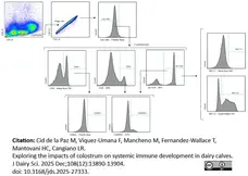
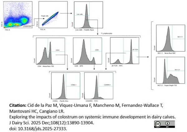
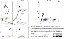
Alexa Fluor® 700 conjugated Mouse anti Bovine CD4 antibody, clone CC8 (MCA1653A700) used in the gating strategy for bovine lymphocytes by flow cytometry.
Image caption:
Gating strategy for T lymphocytes. Lymphocytes were identified by a scatter plot, followed by single cells, live cells, and then CD4+ Lymphocytes, CD8+ Lymphocytes, CD4+ Lymphocytes, γδ T Lymphocytes, WC1.1+ γδ T Lymphocytes, WC1.2+ γδ T Lymphocytes, CD62L, and CD45RO. SSC = side scatter; FSC = forward scatter; A = signal area; H = signal height.
From: Cid de la Paz M, Viquez-Umana F, Mancheno M, Fernandez-Wallace T, Mantovani HC, Cangiano LR.
Exploring the impacts of colostrum on systemic immune development in dairy calves.
J Dairy Sci. 2025 Dec;108(12):13890-13904.
doi: 10.3168/jds.2025-27333.
This image is from an open access article distributed under terms of a Creative Commons Attribution License.
Filter by Application:
F Reset| Mouse anti Bovine CD4 antibody, clone CC8 recognizes bovine CD4, the homolog of human CD4 and immunoprecipitates a ~50 kDa molecule. The phenotype, tissue distribution and function of T-cells expressing the bovine CD4 antigen are similar to those in other species. However, expression on macrophages has not yet been detected. Mouse anti Bovine CD4 antibody, clone CC8 has been reported as being suitable for use on formalin dichromate (FD5) fixed paraffin embedded tissue with amplification and antigen retrieval techniques (Eskra et al. 1991). A mutation in the bovine CD4 gene resulting in an amino acid substitution at A324 T, located in the D4 domain of the CD4 gene product can occur. This mutation results in lowered binding of Mouse anti Bovine CD4 antibody, clone CC8 to CD4 in Japanese Black (JB) cattle where this mutation has been identified (Kato-Mori, et al.. 2020). CD4 in JB cattle can be identified using clone CACT138A (MCA6081) whose binding to bovine CD4 is unaffected by the A324T mutation (Kato-Mori, et al.. 2020). |
- Target Species
- Bovine
- Product Form
- Purified IgG conjugated to Fluorescein Isothiocyanate Isomer 1 (FITC) - liquid
- Preparation
- Purified IgG prepared by affinity chromatography on Protein A from tissue culture supernatant
- Buffer Solution
- Phosphate buffered saline
- Preservative Stabilisers
- 0.09% sodium azide (NaN3)
1% bovine serum albumin - Immunogen
- Bovine lymphocytes.
- Approx. Protein Concentrations
- IgG concentration 0.1 mg/ml
- Fusion Partners
- Spleen cells from an immunized mouse were fused with cells of the mouse NS1 myeloma cell line.
- Max Ex/Em
-
Fluorophore Excitation Max (nm) Emission Max (nm) FITC 490 525 - Regulatory
- For research purposes only
- Guarantee
- 12 months from date of despatch
This product is shipped at ambient temperature. It is recommended to aliquot and store at -20°C on receipt. When thawed, aliquot the sample as needed. Keep aliquots at 2-8°C for short term use (up to 4 weeks) and store the remaining aliquots at -20°C.
Avoid repeated freezing and thawing as this may denature the antibody. Storage in frost-free freezers is not recommended. This product is photosensitive and should be protected from light.
Avoid repeated freezing and thawing as this may denature the antibody. Storage in frost-free freezers is not recommended. This product is photosensitive and should be protected from light.
This product has been reported to work in the following applications. This information is derived from testing within our laboratories, peer-reviewed publications or personal communications from the originators. Please refer to references indicated for further information. For general protocol recommendations, please visit the antibody protocols page.
| Application Name | Verified | Min Dilution | Max Dilution |
|---|---|---|---|
| Flow Cytometry |  |
Neat |
Where this product has not been tested for use in a particular technique this does not necessarily exclude its use in such procedures. Suggested working dilutions are given as a guide only. It is recommended that the user titrates the product for use in their own system using appropriate negative/positive controls.
- Flow Cytometry
- Use 10μl of the suggested working dilution to label 106 cells in 100μl
How to Use the Spectraviewer
Watch the Tool Tutorial Video ▸- Start by selecting the application you are interested in, with the option to select an instrument from the drop down menu or create a customized instrument
- Select the fluorophores or fluorescent proteins you want to include in your panel to check compatibility
- Select the lasers and filters you wish to include
- Select combined or multi-laser view to visualize the spectra
| Description | Product Code | Applications | Pack Size | List Price | Your Price | Quantity | |
|---|---|---|---|---|---|---|---|
| Mouse IgG2a Negative Control:FITC | MCA929F | F | 100 Tests |

|
Log in | ||
| List Price | Your Price | ||||||
|
|
Log in | ||||||
| Description | Mouse IgG2a Negative Control:FITC | ||||||
References for CD4 antibody
-
Bensaid, A. & Hadam, M. (1991) Individual antigens of cattle. Bovine CD4 (BoCD4).
Vet Immunol Immunopathol. 27 (1-3): 51-4. -
Eskra, L. et al. (1991) Effect of monoclonal antibodies on in vitro function of T-cell subsets.
Vet Immunol Immunopathol. 27 (1-3): 215-22. -
Howard, C.J. et al. (1991) Summary of workshop findings for leukocyte antigens of cattle.
Vet Immunol Immunopathol. 27 (1-3): 21-7. -
Gutierrez, M. et al. (1999) The detection of CD2+, CD4+, CD8+, and WC1+ T lymphocytes, B cells and macrophages in fixed and paraffin embedded bovine tissue using a range of antigen recovery and signal amplification techniques.
Vet Immunol Immunopathol. 71 (3-4): 321-34. -
Harris, J. et al. (2002) Expression of caveolin by bovine lymphocytes and antigen-presenting cells
Immunology. 105: 190-5. -
Kruger, E.F. et al. (2003) Bovine monocytes induce immunoglobulin production in peripheral blood B lymphocytes.
Dev Comp Immunol. 27 (10): 889-97. -
Buddle, B.M. et al. (2003) Revaccination of neonatal calves with Mycobacterium bovis BCG reduces the level of protection against bovine tuberculosis induced by a single vaccination.
Infect Immun. 71: 6411-9. -
Endsley, J.J. et al. (2004) Characterization of bovine homologues of granulysin and NK-lysin.
J Immunol. 173 (4): 2607-14.
View The Latest Product References
-
Brackenbury, L.S. et al. (2005) Identification of a cell population that produces alpha/beta interferon in vitro and in vivo in response to noncytopathic bovine viral diarrhea virus.
J Virol. 79: 7738-44. -
Sidders, B. et al. (2008) Screening of highly expressed mycobacterial genes identifies Rv3615c as a useful differential diagnostic antigen for the Mycobacterium tuberculosis complex.
Infect Immun. 76: 3932-9. -
Gerner, W. et al. (2009) Identification of major histocompatibility complex restriction and anchor residues of foot-and-mouth disease virus-derived bovine T-cell epitopes.
J Virol. 83: 4039-50. -
Hu, X.D. et al. (2009) Immunotherapy with combined DNA vaccines is an effective treatment for M. bovis infection in cattle
Vaccine. 27: 1317-22. -
Lynch, E.M. et al. (2010) Effect of abrupt weaning at housing on leukocyte distribution, functional activity of neutrophils, and acute phase protein response of beef calves.
BMC Vet Res. 6: 39. -
Coad, M. et al. (2010) Repeat tuberculin skin testing leads to desensitisation in naturally infected tuberculous cattle which is associated with elevated interleukin-10 and decreased interleukin-1 beta responses.
Vet Res. 41: 14. -
Kiku, Y. et al. (2010) Decrease in bovine CD14 positive cells in colostrum is associated with the incidence of mastitis after calving.
Vet Res Commun. 34: 197-203. -
Whelan, A.O. et al. (2011) Development of an Antibody to Bovine IL-2 Reveals Multifunctional CD4 T(EM) Cells in Cattle Naturally Infected with Bovine Tuberculosis.
PLoS One. 6: e29194. -
Oh, Y. et al. (2012) Interferon-γ induced by in vitro re-stimulation of CD4+ T-cells correlates with in vivo FMD vaccine induced protection of cattle against disease and persistent infection.
PLoS One. 7: e44365. -
Hine, B.C. et al. (2012) Analysis of leukocyte populations in Canadian Holsteins classified as high or low immune responders for antibody- or cell-mediated immune response.
Can J Vet Res. 76: 149-56. -
Aranday-Cortes, E. et al. (2012) Transcriptional profiling of disease-induced host responses in bovine tuberculosis and the identification of potential diagnostic biomarkers.
PLoS One. 7: e30626. -
Tenaya, I.W. et al. (2012) Flow cytometric analysis of lymphocyte subset kinetics in Bali cattle experimentally infected with Jembrana disease virus.
Vet Immunol Immunopathol. 149: 167-76. -
Wernike, K. et al. (2013) Oral exposure, reinfection and cellular immunity to Schmallenberg virus in cattle.
Vet Microbiol. pii: S0378-1135(13)00092-8. -
Dacal,. V. et al. (2013) Immunohistochemical characterization of inflammatory cells in the skin of cattle undergoing repeated infestations with Hypoderma lineatum (Diptera: Oestridae) larvae.
J Comp Pathol. 145: 282-8. -
Brodzki, P. et al. (2014) Phenotyping of leukocytes and granulocyte and monocyte phagocytic activity in the peripheral blood and uterus of cows with endometritis.
Theriogenology. 82 (3): 403-10. -
Grit, G.H. et al. (2014) Evaluation of cellular and humoral systemic immune response against Giardia duodenalis infection in cattle.
Vet Parasitol. 202 (3-4): 145-55. -
Blunt, L. et al. (2015) Phenotypic characterization of bovine memory cells responding to mycobacteria in IFN&gama; enzyme linked immunospot assays.
Vaccine. 33 (51): 7276-82. -
Okagawa, T. et al. (2016) Cooperation of PD-1 and LAG-3 Contributes to T-Cell Exhaustion in Anaplasma marginale-Infected Cattle.
Infect Immun. 84 (10): 2779-90. -
Metcalfe, H.J. et al. (2016) Protection associated with a TB vaccine is linked to increased frequency of Ag85A-specific CD4(+) T cells but no increase in avidity for Ag85A.
Vaccine. 34 (38): 4520-5. -
Diaz-San Segundo, F. et al. (2016) Combination of Adt-O1Manisa and Ad5-boIFNλ3 induces early protective immunity against foot-and-mouth disease in cattle.
Virology. 499: 340-9. -
Sun, F. et al. (2016) Regulation of Nutritional Metabolism in Transition Dairy Cows: Energy Homeostasis and Health in Response to Post-Ruminal Choline and Methionine.
PLoS One. 11 (8): e0160659. -
Wattegedera, S.R. et al. (2017) Enhancing the toolbox to study IL-17A in cattle and sheep.
Vet Res. 48 (1): 20. -
Herry, V. et al. (2017) Local immunization impacts the response of dairy cows to Escherichia coli mastitis.
Sci Rep. 7 (1): 3441. -
Denholm, S.J. et al. (2017) Estimating genetic and phenotypic parameters of cellular immune-associated traits in dairy cows.
J Dairy Sci. 100 (4): 2850-62. -
Shimizu, T. et al. (2018) Changes of leukocyte counts and expression of pro- and anti-inflammatory cytokines in peripheral leukocytes in periparturient dairy cows with retained fetal membranes.
Anim Sci J. 89 (9): 1371-8. -
Novak, B. et al. (2018) Bovine Peripheral Blood Mononuclear Cells Are More Sensitive to Deoxynivalenol Than Those Derived from Poultry and Swine.
Toxins (Basel). 10 (4):152. -
Bassi, P.B. et al. (2018) Parasitological and immunological evaluation of cattle experimentally infected with Trypanosoma vivax.
Exp Parasitol. 185: 98-106. -
de Araújo, F.F. et al. (2019) Distinct immune response profile during Rhipicephalus (Boophilus) microplus infestations of guzerat dairy herd according to the maternal lineage ancestry (mitochondrial DNA).
Vet Parasitol. 273: 36-44. -
Nakajima, N. et al. (2019) Effects of direct exposure to cold weather under grazing in winter on the physiological, immunological, and behavioral conditions of Japanese Black beef cattle in central Japan.
Anim Sci J. 90 (8): 1033-41. -
Benedictus, L. et al. (2019) Immunization of young heifers with staphylococcal immune evasion proteins before natural exposure to Staphylococcus aureus induces a humoral immune response in serum and milk.
BMC Vet Res. 15 (1): 15. -
Grandoni, F. et al. (2019) A new polymorphic epitope of bovine CD4 antigen evidenced by flow cytometry.
Vet Immunol Immunopathol. 219: 109957. -
Cunha, P. et al. (2019) Expansion, isolation and first characterization of bovine Th17 lymphocytes.
Sci Rep. 9 (1): 16115. -
Risalde, M.A. et al. (2020) BVDV permissiveness and lack of expression of co-stimulatory molecules on PBMCs from calves pre-infected with BVDV.
Comp Immunol Microbiol Infect Dis. 68: 101388. -
Okino, C.H. et al. (2020) A polymorphic CD4 epitope related to increased susceptibility to Babesia bovis. in Canchim calves.
Vet Immunol Immunopathol. 230: 110132. -
Brodzki, P. et al. (2020) Selected leukocyte subpopulations in peripheral blood and uterine washings in cows before and after intrauterine administration of cefapirin and methisoprinol.
Anim Sci J. 91 (1): e13306. -
Gondaira, S. et al. (2020) Immunosuppression in Cows following Intramammary Infusion of Mycoplasma bovis.
Infect Immun. 88 (3): e00521-19. -
Arrieta-Villegas, C. et al. (2020) Immunogenicity and Protection against Mycobacterium caprae Challenge in Goats Vaccinated with BCG and Revaccinated after One Year.
Vaccines (Basel). 8 (4): 751. -
Bidart, J. et al. (2020) A New Cage-Like Particle Adjuvant Enhances Protection of Foot-and-Mouth Disease Vaccine.
Front Vet Sci. 7: 396. -
Colombatti, M.O. et al. (2021) Evaluation of a virulent strain of Mycobacterium avium subsp. paratuberculosis used as a heat-killed vaccine.
Vaccine. 39 (51): 7401-12. -
Park, D.S. et al. (2021) Dynamic changes in blood immune cell composition and function in Holstein and Jersey steers in response to heat stress.
Cell Stress Chaperones. 26 (4): 705-20. -
Kato-Mori, Y. et al. (2021) Characterization of a variant CD4 molecule in Japanese Black cattle.
Vet Immunol Immunopathol. 232: 110167. -
Okino, C.H. et al. (2022) CD4 bovine gene: Differential polymorphisms among cattle breeds and a new tool for rapid identification.
Vet Immunol Immunopathol. 251: 110462. -
Casaro, S. et al. (2022) Flow cytometry panels for immunophenotyping dairy cattle peripheral blood leukocytes
Vet immunol Immunopathol. 248: 110417. -
Hidalgo-Ruiz, M. et al. (2022) Babesia bovis AMA-1, MSA-2c and RAP-1 contain conserved B and T-cell epitopes, which generate neutralizing antibodies and a long-lasting Th1 immune response in vaccinated cattle.
Vaccine. S0264-410X(22)00049-4. -
Elsayed, M.S.A.E. et al. (2022) Real-time PCR using atpE, conventional PCR targeting different regions of difference, and flow cytometry for confirmation of Mycobacterium bovis. in buffaloes and cattle from the Delta area of Egypt.
BMC Microbiol. 22 (1): 154. -
Tucker, N. et al. (2023) Bovine blood and milk T-cell subsets in distinct states of activation and differentiation during subclinical Staphylococcus aureus mastitis.
J Reprod Immunol. 156: 103826. -
Benedictus, L. et al. (2019) Immunization of young heifers with staphylococcal immune evasion proteins before natural exposure to Staphylococcus aureus induces a humoral immune response in serum and milk.
BMC Vet Res. 15 (1): 15. -
Yang, L. et al. (2018) Association of the expression of Th cytokines with peripheral CD4 and CD8 lymphocyte subsets after vaccination with FMD vaccine in Holstein young sires.
Res Vet Sci. 119: 79-84. -
Seemann, L. et al. (2024) Dietary L-carnitine supplementation modifies blood parameters of mid-lactating dairy cows during standardized lipopolysaccharide-induced inflammation.
Front Immunol. 15: 1390137. -
Müller, C.B.M. et al. (2024) Interactions between rumen epithelium-associated microbiota and host immunological and metabolic adaptations in response to different milk replacer feeding intensities in dairy calves
Animal Nutrition. 7 Sept [Epub ahead of print]. -
Brodzki, P. et al. (2024) The influence of probiotic administration on selected leukocyte subpopulations and the serum amyloid A concentration in the peripheral blood of dairy cows during different lactation periods
Journal of Veterinary Research. 09 Oct [Epub ahead of print]. -
Gilbert, F.B. et al. (2025) Expression of FcμR by bovine mononuclear blood leukocytes.
Dev Comp Immunol. 162: 105304. -
Díaz-Otero, F. et al. (2025) Comparative longitudinal analysis of T lymphocyte subpopulations in calves vaccinated with different doses of BCG-Phipps or with culture filtrate protein extract of Mycobacterium bovis in a natural transmission setting.
BMC Vet Res. 21 (1): 78. -
Abramowicz, B. et al. (2025) A preliminary study through lymphocyte immunophenotyping of the effects of different forms of phosphorus deficiency on dairy cattle blood haematological parameters and immune status.
J Vet Res. 69 (2): 257-262. -
Cainelli, S. et al. (2025) Dynamics of peripheral B and γδ T cells during postpartum and pregnancy in dairy cows.
Anim Reprod Sci. 282: 108012. -
Cid de la Paz, M. et al. (2025) Exploring the impacts of colostrum on systemic immune development in dairy calves.
J Dairy Sci. 108 (12): 13890-13904.
MCA1653F
If you cannot find the batch/lot you are looking for please contact our technical support team for assistance.
View more products with CD4 specificity
Please Note: All Products are "FOR RESEARCH PURPOSES ONLY"
View all Anti-Bovine ProductsAlways be the first to know.
When we launch new products and resources to help you achieve more in the lab.
Yes, sign me up