Inhibin Alpha (α) Antibodies
Our anti-inhibin alpha product range includes the highly cited R1 clone and a selection of Fab bivalent recombinant antibodies.
Inhibin and activin are part of the transforming growth factor beta (TGF-ß) family of cytokines (Bartholin et al. 2002), and are best known for their role in regulating follicle stimulating hormone (FSH) secretion, where inhibin inhibits and activin activates secretion. These protein regulators play a vital role in regulating FSH, which acts synergistically with luteinizing hormone (LH) in reproduction.
Inhibin alpha, encoded by the human INHA gene, has a negative regulatory effect upon activin. It inhibits the biosynthesis and secretion of FSH by preventing activin from binding to its receptor (Gressner, 2011). Inhibin is able to block the activity of activin by competing for the binding site on the activin receptor (ActRII) and through displacing activin from the activin-receptor complex. Inhibins were originally found in the ovary (granulosa cells) and testis (sertoli cells); they have also been found present in other tissues such as the placenta, endometrium (Mylonas et al. 2004), brain (Uccella et al. 2000), adrenal gland (McCluggage et al. 1998), testis (Bergh and Cajander, 1990) and ovary (Cuevas et al. 1987, Yamoto et al. 1992). Inhibin has a common α-subunit which is attached to either a ßA or a ßB subunit via a disulfide bond to create inhibin A (a ßA) or inhibin B (a ßB).
Did you know?
- Clone R1 is our most popular inhibin alpha antibody
- Clone R1 is trusted by researchers, with Bio-Rad antibodies highly cited (41 citations on CiteAb)
- We offer inhibin alpha antibodies generated by multiple techniques, giving more choice. You can have your antibody from mouse monoclonal, polyclonal, or recombinant fab bivalent antibodies
Inhibin alpha (R1) antibody IHC images; Ovary, testis and placental tissues
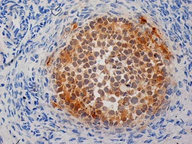
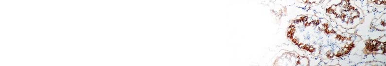
Fig 1. Cryosections of human ovary stained with mouse anti human inhibin alpha antibody, clone R1 (MCA951S) followed by HISTAR detection system. Blocking was with peroxidase blocking reagent + 10% normal goat serum. Visualization was with DAB substrate and counter-staining with haematoxylin
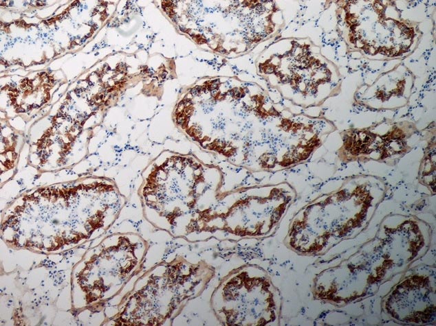
Fig 2. Cryosections of human testis stained with mouse anti human inhibin alpha antibody, clone R1 (MCA951S) followed by HISTAR detection system. Blocking was with peroxidase blocking reagent + 10% normal goat serum. Visualization was with DAB substrate and counter-staining with haematoxylin (low power).
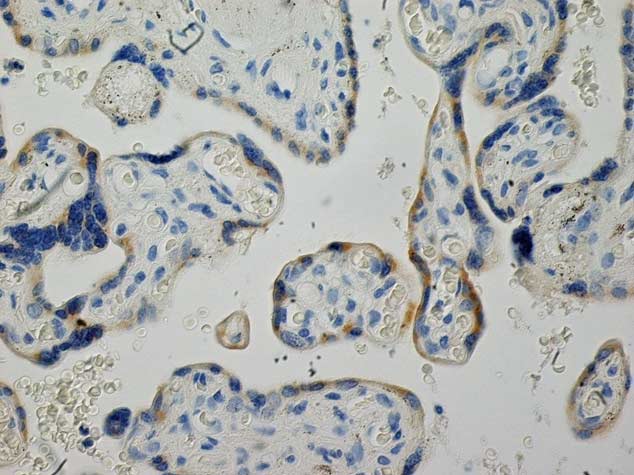
Fig 3. Formalin fixed, paraffin embedded sections of human placenta stained with mouse anti human inhibin alpha antibody, clone R1 (MCA951S) following antigen retrieval in Tris/EDTA pH 9.0. Detection is with the HISTAR detection system. Blocking was with peroxidase blocking reagent + 10% normal goat serum. Visualization was with DAB substrate and counter-staining with haematoxylin (high power).
Our Inhibin Alpha Antibody Range
| Description | Target | Format | Clone | Applications | Citations | Code |
|---|
References
-
Bartholin L et al. (2002). Transcription activation of FLRG and follistatin by activin A, through Smad proteins, participates in a negative feedback loop to modulate activin A function.
Oncogene. 21(14): 2227-2235 -
Gressner OA (2011). Chapter Four - Intracrine Signaling Mechanisms of Activin A and TGF-ß, In: Gerald Litwack, Editor(s) Vitamins & Hormones
Academic Press 85: 59-77 -
Mylonas I et al. (2004). Expression of the inhibin-alpha subunit in normal, hyperplastic and malignant endometrial tissue: an immunohistochemical analysis.
Gynecologic Oncology, 93(1):92-97 -
Uccella S et al. (2000). Localization of inhibin/activin subunits in normal pituitary adenomas.
Pituitary 3(3):131-9 -
Mc Cluggage WG et al. (1998). Immunihistochemical staining of normal, hyperplastic, and neoplastic adrenal cortex with a monoclonal antibody against alpha inhibin.
J. Clin. Pathol. 51(2):114-116 -
Bergh A and Cajander S (1990). Immunohistochemical localization of inhibin-a in the testes of normal men and in men with testicular disorders.
International Journal of Andrology, 13: 463–469. -
Cuevas P et al. (1986). Immunohistochemical detection of inhibin in the gonad.
Biochemical and Biophysical Research Communications. 142(1):23-30 -
Yamoto M et al. (1992). Immunohistochemical localization of inhibin/activin subunits in human ovarian follicles during the menstrual cycle.
The Journal of Clinical Endocrinology & Metabolism. 74(5):989-993