Actin Beta antibody
Rabbit anti Human Actin Beta (N-Terminal)
- Product Type
- Polyclonal Antibody
- Isotype
- Polyclonal IgG
- Specificity
- Actin Beta
- Region
- (N-TERMINAL)

Filter by Application:
E IC Reset| Rabbit anti Human actin beta antibody recognizes human actin beta (ACTB), one of the cytoplasmic actin isoforms, a 41.7 kDa cytoskeletal protein. Actins are highly conserved proteins that are involved in cell motility, structure and integrity and are ubiquitously expressed in all eukaryotic cells. Higher vertebrates express six actin isoforms encoded for by distinct genes. Actin isoforms are highly evolutionarily conserved. Two striated muscle isoforms, alpha skeletal and alpha cardiac, two smooth muscle isoforms, alpha smooth and gamma smooth together with two cytoplasmic isoforms, beta cytoplasmic and gamma cytoplasmic are encoded by genes on chromosomes 1, 15, 10, 2, 7 and 17 respectively. All actin isoforms have highly homologous nucleotide sequences with the heterogeneity most notably in the N terminal region. However, small differences in primary structure here and elsewhere affect the conformational state of the various isoforms and their affinity for a range of actin binding proteins (ABPs). The range of affinities for ABPs suggests that where multiple globular monomeric (G-actin) forms exist at the same site, polymerisation may result in the formation of filamentous (F-actin) enriched for one particular isoform. Defects in actin beta are a cause of juvenile dystonia, a condition that results in movement disorders due to disordered muscle tonicity, abnormal posture, development malformations and sensory hearing loss. |
- Target Species
- Human
- Species Cross-Reactivity
-
Target Species Cross Reactivity Mouse Rat - N.B. Antibody reactivity and working conditions may vary between species.
- Product Form
- Purified IgG - liquid
- Antiserum Preparation
- Antiserum to human Actin beta(N-Terminal) was raised by repeated immunisation of rabbits with highly purified antigen. Purified IgG was prepared from whole serum by affinity chromatography.
- Buffer Solution
- Phosphate buffered saline
- Preservative Stabilisers
- 0.02% Sodium Azide (NaN3)
- Immunogen
- A peptide corresponding to an 16 amino acid sequence from near the amino terminus of human Actin beta.
- Approx. Protein Concentrations
- IgG concentration 1.0mg/ml
- Regulatory
- For research purposes only
- Guarantee
- 12 months from date of despatch
This product is shipped at ambient temperature. It is recommended to aliquot and store at -20°C on receipt. When thawed, aliquot the sample as needed. Keep aliquots at 2-8°C for short term use (up to 4 weeks) and store the remaining aliquots at -20°C.
Avoid repeated freezing and thawing as this may denature the antibody. Storage in frost-free freezers is not recommended.
Avoid repeated freezing and thawing as this may denature the antibody. Storage in frost-free freezers is not recommended.
This product has been reported to work in the following applications. This information is derived from testing within our laboratories, peer-reviewed publications or personal communications from the originators. Please refer to references indicated for further information. For general protocol recommendations, please visit the antibody protocols page.
| Application Name | Verified | Min Dilution | Max Dilution |
|---|---|---|---|
| Immunohistology - Frozen |  |
1.0ug/ml | |
| Western Blotting | |
1.0 | 2.0ug/ml |
Where this product has not been tested for use in a particular technique this does not necessarily exclude its use in such procedures. Suggested working dilutions are given as a guide only. It is recommended that the user titrates the product for use in their own system using appropriate negative/positive controls.
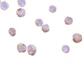
- Histology Positive Control Tissue
- HeLa cells
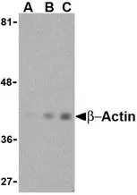
- Western Blotting
- AHP1629 detects a band of approximately 40kDa in HeLa cell lysates.
| Description | Product Code | Applications | Pack Size | List Price | Your Price | Quantity | |
|---|---|---|---|---|---|---|---|
| Goat anti Rabbit IgG (Fc):Biotin | STAR121B | E WB | 1 mg |

|
Log in | ||
| List Price | Your Price | ||||||
|
|
Log in | ||||||
| Description | Goat anti Rabbit IgG (Fc):Biotin | ||||||
| Goat anti Rabbit IgG (Fc):FITC | STAR121F | F | 1 mg |
|
Log in | ||
| List Price | Your Price | ||||||
|
|
Log in | ||||||
| Description | Goat anti Rabbit IgG (Fc):FITC | ||||||
| Goat anti Rabbit IgG (Fc):HRP | STAR121P | E WB | 1 mg |
|
Log in | ||
| List Price | Your Price | ||||||
|
|
Log in | ||||||
| Description | Goat anti Rabbit IgG (Fc):HRP | ||||||
| Goat anti Rabbit IgG (H/L):HRP | STAR124P | C E WB | 1 mg |
|
Log in | ||
| List Price | Your Price | ||||||
|
|
Log in | ||||||
| Description | Goat anti Rabbit IgG (H/L):HRP | ||||||
| Sheep anti Rabbit IgG:RPE | STAR35A | F | 1 ml |
|
Log in | ||
| List Price | Your Price | ||||||
|
|
Log in | ||||||
| Description | Sheep anti Rabbit IgG:RPE | ||||||
| Description | Product Code | Applications | Pack Size | List Price | Your Price | Quantity | |
|---|---|---|---|---|---|---|---|
| TidyBlot Western Blot Detection Reagent:HRP | STAR209P | WB * | 0.5 ml | Log in | |||
| List Price | Your Price | ||||||
| Log in | |||||||
| Description | TidyBlot Western Blot Detection Reagent:HRP | ||||||
References for Actin Beta antibody
-
Sun, L. et al. (2014) Quantitative proteomics of Xenopus laevis embryos: expression kinetics of nearly 4000 proteins during early development.
Sci Rep. 4: 4365. -
Chen, X.J. et al. (2018) Association of reduced sclerostin expression with collapse process in patients with osteonecrosis of the femoral head.
Int Orthop. 42 (7): 1675-82.
- Synonyms
- ACTB
- RRID
- AB_2223350
- UniProt
- P60709
- Entrez Gene
- ACTB
View more products with ACTIN specificity
Please Note: All Products are "FOR RESEARCH PURPOSES ONLY"
View all Anti-Human ProductsAlways be the first to know.
When we launch new products and resources to help you achieve more in the lab.
Yes, sign me up