CD8 Beta antibody | CC58
Mouse anti Bovine CD8 Beta:RPE
- Product Type
- Monoclonal Antibody
- Clone
- CC58
- Isotype
- IgG1
- Specificity
- CD8 Beta


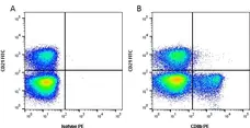
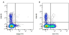
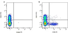
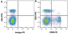
Figure B. FITC conjugated Mouse anti Bovine CD4 antibody, clone CC8 (MCA1653F) and Mouse anti Bovine CD8β antibody, clone CC58 (MCA1654GA) detected with Goat anti Mouse IgG1:RPE (STAR132PE).
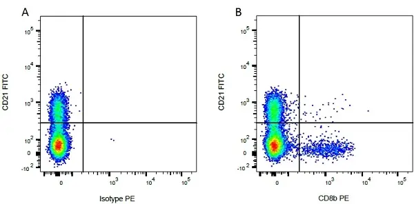
All experiments performed on bovine red blood cell lysed blood gated on live single cell lymphocytes. Data acquired on the ZE5 Cell Analyzer.
Filter by Application:
F Reset| Mouse anti Bovine CD8 beta antibody, clone CC58 recognizes an epitope associated with the bovine CD8 beta chain. CD8 is usually expressed as an α/β heterodimer. The CD8 antigen is a cell surface glycoprotein found on most cytotoxic T lymphocytes that mediates efficient cell-cell interactions within the immune system. The CD8 antigen, acting as a coreceptor, and the T-cell receptor on the T lymphocyte recognize antigens displayed by an antigen presenting cell (APC) in the context of class I MHC molecules. Mouse anti Bovine CD8 beta antibody, clone CC58 has been successfully used for the immunohistochemical detection of CD8 on formalin fixed, paraffin embedded placental tissue from water buffalo (Cantón et al. 2014). |
- Target Species
- Bovine
- Species Cross-Reactivity
-
Target Species Cross Reactivity Sheep Goat Water Buffalo - N.B. Antibody reactivity and working conditions may vary between species.
- Product Form
- Purified IgG conjugated to R. Phycoerythrin (RPE) - lyophilized
- Reconstitution
- Reconstitute with 1 ml distilled water
- Preparation
- Purified IgG prepared by affinity chromatography on Protein A from tissue culture supernatant
- Buffer Solution
- Phosphate buffered saline
- Preservative Stabilisers
- 0.09% sodium azide (NaN3)
1% bovine serum albumin
5% sucrose - Immunogen
- Bovine leucocytes
- Max Ex/Em
-
Fluorophore Excitation Max (nm) Emission Max (nm) RPE 488nm laser 496 578 - Regulatory
- For research purposes only
- Guarantee
- 12 months from date of despatch
This product is shipped at ambient temperature.
Prior to reconstitution store at +4°C. Following reconstitution store at +4°C.
DO NOT FREEZE.
This product should be stored undiluted. This product is photosensitive and should be protected from light. Should this product contain a precipitate we recommend microcentrifugation before use.
Prior to reconstitution store at +4°C. Following reconstitution store at +4°C.
DO NOT FREEZE.
This product should be stored undiluted. This product is photosensitive and should be protected from light. Should this product contain a precipitate we recommend microcentrifugation before use.
This product has been reported to work in the following applications. This information is derived from testing within our laboratories, peer-reviewed publications or personal communications from the originators. Please refer to references indicated for further information. For general protocol recommendations, please visit the antibody protocols page.
| Application Name | Verified | Min Dilution | Max Dilution |
|---|---|---|---|
| Flow Cytometry |  |
Neat | 1/10 |
Where this product has not been tested for use in a particular technique this does not necessarily exclude its use in such procedures. Suggested working dilutions are given as a guide only. It is recommended that the user titrates the product for use in their own system using appropriate negative/positive controls.
- Flow Cytometry
- Use 10μl of the suggested working dilution to label 106 cells in 100μl
How to Use the Spectraviewer
Watch the Tool Tutorial Video ▸- Start by selecting the application you are interested in, with the option to select an instrument from the drop down menu or create a customized instrument
- Select the fluorophores or fluorescent proteins you want to include in your panel to check compatibility
- Select the lasers and filters you wish to include
- Select combined or multi-laser view to visualize the spectra
| Description | Product Code | Applications | Pack Size | List Price | Your Price | Quantity | |
|---|---|---|---|---|---|---|---|
| Mouse IgG1 Negative Control:RPE | MCA928PE | F | 100 Tests | Log in | |||
| List Price | Your Price | ||||||
| Log in | |||||||
| Description | Mouse IgG1 Negative Control:RPE | ||||||
References for CD8 Beta antibody
-
Suraud, V. et al. (2008) Acute infection by conjunctival route with Brucella melitensis induces IgG+ cells and IFN-gamma producing cells in peripheral and mucosal lymph nodes in sheep.
Microbes Infect. 10: 1370-8. -
Howard, C.J. & Naessens, J. (1993) Summary of workshop findings for cattle (tables 1 and 2).
Vet Immunol Immunopathol. 39 (1-3): 25-47. -
Naessens, J. et al. (1997) Nomenclature and characterization of leukocyte differentiation antigens in ruminants.
Immunol Today. 18 (8): 365-8. -
Hein, W.R. et al. (1991) Summary of workshop findings for leukocyte antigens of sheep.
Vet Immunol Immunopathol. 27 (1-3): 28-30. -
Gerner, W. et al. (2009) Identification of major histocompatibility complex restriction and anchor residues of foot-and-mouth disease virus-derived bovine T-cell epitopes.
J Virol. 83: 4039-50. -
Gerner, W. et al. (2010) Sensitive detection of Foxp3 expression in bovine lymphocytes by flow cytometry.
Vet Immunol Immunopathol. 138: 154-8. -
MacHugh, N.D. and Sopp, P. (1991) Individual antigens of cattle. Bovine CD8 (BoCD8).
Vet Immunol Immunopathol. 27: 65-9. -
Soltys, J. and Quinn, M.T. (1999) Selective recruitment of T-cell subsets to the udder during staphylococcal and streptococcal mastitis: analysis of lymphocyte subsets and adhesion molecule expression.
Infect Immun. 67: 6293-302.
View The Latest Product References
-
Cantón, G.J. et al. (2014) Characterization of immune cell infiltration in the placentome of water buffaloes (Bubalus bubalis) infected with neospora caninum during pregnancy.
J Comp Pathol. 150: 463-8. -
Wattegedera, S.R. et al. (2017) Enhancing the toolbox to study IL-17A in cattle and sheep.
Vet Res. 48 (1): 20. -
Hecker, Y.P. et al. (2015) Cell mediated immune responses in the placenta following challenge of vaccinated pregnant heifers with Neospora caninum.
Vet Parasitol. 214 (3-4): 247-54. -
Okino, C.H. et al. (2020) A polymorphic CD4 epitope related to increased susceptibility to Babesia bovis. in Canchim calves.
Vet Immunol Immunopathol. 230: 110132. -
Pooley, H.B. et al. (2022) Sheep vaccinated against paratuberculosis have increased levels of B cells infiltrating the intestinal tissue.
Vet Immunol Immunopathol. 252: 110482. -
Cainelli, S. et al. (2025) Dynamics of peripheral B and γδ T cells during postpartum and pregnancy in dairy cows.
Anim Reprod Sci. 282: 108012.
MCA1654PE
If you cannot find the batch/lot you are looking for please contact our technical support team for assistance.
View more products with CD8 specificity
Please Note: All Products are "FOR RESEARCH PURPOSES ONLY"
View all Anti-Bovine ProductsAlways be the first to know.
When we launch new products and resources to help you achieve more in the lab.
Yes, sign me up