-
US | en

Fluidics System
One of the fundamentals of flow cytometry is the ability to measure the properties of individual particles. When a sample enters a flow cytometer, the particles are randomly distributed in the 3-D space of the sample line, the diameter of which is significantly larger than the diameter of most cells. The sample must therefore be ordered into a stream of single particles that can be interrogated individually by the instrument’s detection system. This process is managed by the fluidics system.
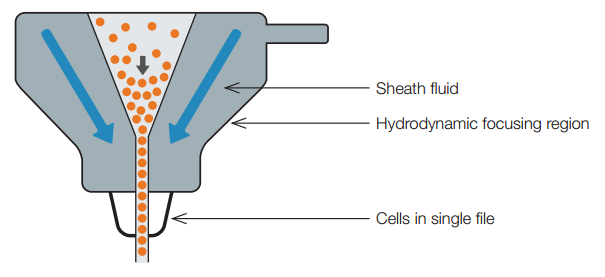
The fluidics system consists of a central core through which the sample fluid is injected, enclosed by an outer sheath fluid. Due to narrowing of the sheath (in a nozzle or cuvette) the fluid velocity is increased. The sample is introduced into the center and is focused by the Bernoulli effect (Figure 1). This allows the creation of a stream of particles in single file and is called hydrodynamic focusing. Under optimal conditions (laminar flow) there is no mixing of the central fluid stream and the sheath fluid.

Fig. 1. Hydrodynamic focusing produces a single stream of particles
Without hydrodynamic focusing, the cuvette (typically 250 x 250 μm or 180 x 480 μm), or nozzle of the instrument (typically 70-130 μm) would not create a focused stream of cells and analysis of single cells would not be possible. With hydrodynamic focusing the cells flow in single file through the illumination source, called the interrogation point, allowing single cell analysis.
