Canine Distemper Virus
- On This Page
- Canine distemper virus
- Structure
- Disease
- Canine distemper virus antibodies
- References
- Associated reading
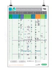
Biomarker Expression Patterns Posters
s
Canine distemper virus (CDV), along with human measles virus (HMV) and rinderpest virus (RPV) of cattle, is part of the paramyxovirinae family, genus Morbillivirus. CDV is highly infective with a worldwide distribution and apart from dogs, it can infect wild canids such as coyotes, foxes, and wolves as well as racoons, ferrets, and large wild cats. No specific treatment for CDV exists, the vaccination of dogs is the key containment strategy. CDV infection in dogs and ferrets has been used as a model to study measles virus infection.
Structure
The CDV particles are spherical, enveloped virions with a diameter of about 150 nm carrying a non-segmented single negative-stranded RNA (ssRNA) of 15,690 nucleotides coding for eight proteins (da Fontoura Budaszewski et al. 2016). The RNA codes for eight transcription units:
- N ─ nucleocapsid
- P ─ phosphoprotein, but also codes for C and V proteins
- M ─ matrix protein
- F ─ fusion protein
- H ─ hemagglutinin
- L ─ large polymerase protein
The outer layer is made up of the fusion (F) and hemagglutinin (H) transmembrane glycoproteins which are lined by the matrix (M) protein. The main function of the H and F proteins is to facilitate CDV entry into canine peripheral blood mononuclear cells via CD150 (also signaling lymphocyte activation molecule - SLAM) (von Messling et al. 2006) and to epithelial cells via nectin-4 (Pratakpiriya et al. 2012).
The RNA is encapsulated by the nucleoprotein (N) and is joined by the phosphoprotein (P) and the polymerase (L) to form the ribonucleoprotein-complex (RNP). The P gene codes for the C and V proteins through use of an alternate open reading frame and RNA editing, respectively (Whelan SP et al. 2004, da Fontoura Budaszewski and von Messling 2016). The C and V proteins modulate the infectivity of CDV (von Messling et al. 2006).
Disease
When CDV infects a dog, first signs are generally a runny nose and a nervous disposition. In the acute stage, viruses are shed in all the animal's secretions. The next phase includes cutaneous rash, copious nasal and ocular discharge, conjunctivitis and anorexia. Ensuing gastrointestinal and respiratory problems are frequently accompanied by secondary bacterial infections and neurological disorders (Beineke et al. 2009). While the immune response may lead to improvements, virus may be retained in the lymphoid system, the central nervous system (CNS), and footpads. Severe CNS pathologies tend to result in mortality; those dogs that survive will have permanent neurological damage. Demyelinating leukoencephalitis is frequently seen at this stage (Ulrich et al. 2014).
Antibodies to CDV
Bio-Rad, the leading supplier of veterinary research antibodies, supplies two monoclonal antibodies, in purified format, reactive to canine distemper virus. Mouse Anti-Canine Distemper Virus antibody, clone 7515, detects the envelop protein and is suitable for ELISA. Clone DV2-12, recognizes the CDV nucleoprotein and can be used in ELISA, western blotting, and for IHC on frozen and paraffin embedded tissue sections.
Discover an industry leading portfolio of species-specific antibodies for immune response research in our online veterinary product catalog.
Canine Distemper Virus Antibodies
| Description | Target | Format | Clone | Applications | Citations | Code |
|---|
References
- Beineke A et al. (2009). Pathogenesis and immunopathology of systemic and nervous canine distemper. Vet Immunol Immunopathol. 127, 1-18.
- da Fontoura Budaszewski R et al. (2016) Influence of vaccine strains on the evolution of canine distemper virus. Infect Genet Evol. 41, 262-269.
- da Fontoura Budaszewski R and von Messling V (2016). Morbillivirus experimental animal models: measles virus pathogenesis insights from canine distemper virus. Viruses 8(10), 274.
- Pratakpiriya W et al. (2012). Nectin4 is an epithelial cell receptor for canine distemper virus and involved in the neurovirulence. J Virol. 86(18), 10207-10210.
- Ulrich R et al. (2014). Transcriptional changes in canine distemper virus-induced demyelinating leukoencephalitis favor a biphasic mode of demyelination. PloS one. 9(4), e95917.
- von Messling V et al. (2006). Receptor (SLAM [CD150]) recognition and the V protein sustain swift lymphocyte-based invasion of mucosal tissue and lymphatic organs by a morbillivirus. J Virol. 80(12), 6084–6092.
- Whelan SP et al. (2004). Transcription and replication of non-segmented negative-strand RNA viruses. Curr Top Microbiol Immunol. 283, 61–119.