Canine Parvovirus

- On This Page
- Canine parvovirus
- Emergence
- Structure
- Disease
- Evolution
- Parvovirus antibodies
- References
- Associated reading
Biomarker Expression Patterns Posters
s
Canine parvovirus, also called parvo (CVPC, CPV-2, CPV-2a, CPV-2b, CPV-2c), is among the top three most common infections of dogs. Caused by viral infection from external parasites, it infects the intestinal epithelium and spreads to lymphoid organs. This disease can be fatal for young dogs that have not been vaccinated or don't have maternal antibodies.
Emergence
Parvo, short for canine parvovirus (CPV), is caused by canine parvovirus type 2 (CPV-2), one of the main enteric viruses affecting dogs. CPV-2 emerged in 1978 and spread out globally within two years. A series of new outbreaks followed over the years, caused by mutation of the initial type, CPV-2, to CPV-2a and then minor further changes in the capsid proteins, leading to the CPV-2b strain (Parrish et al. 1991). In 2000, a third variant (CPV-2c) was discovered (Buonavoglia et al. 2001).
Structure

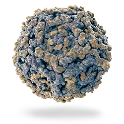
Parvoviruses are small non-enveloped viruses made up of a spherical capsid that carries a single-strand DNA chain. The approx. 5000-nucleotide long DNA molecule has two large open reading frames (ORFs) which use alternative mRNA splicing to code for overlapping genes. This makes transcription of two non-structural (NS1 and NS2) and two structural (VP1 and VP2) proteins possible. The capsid is constructed with 60 copies of a mix of VP1, VP2, and VP3 proteins. VP2 makes up around 90% of the capsid; VP3 is generated through cleavage of VP2 by host cell proteases. The structural proteins have a conserved central beta sheet formed by eight anti-parallel strands, the resulting end loops interact with other chains to form the capsid surface (Reed et al. 1988, Parrish 1999, Hoelzer et al. 2008).
Disease
CPV-2 is highly contagious with high morbidity, especially in locations like kennels. Death can occur a few days after onset of disease, young dogs are particularly susceptible between the age of 6 weeks and 6 months. If left untreated mortality is very high, however with treatment survival rates can be up to 90%. All breeds can be infected, but mixed breeds are more resistant, while pure bred rottweilers, doberman pinschers, English springer spaniels, American pit bull terriers, and German shepherds are at increased risk for infection (Houston et al. 1996, Parrish et al. 1995).
Infection is via the oral route with CPV-2 replicating in tissues with high cell turnover, such as the intestinal epithelium, bone marrow, and lymphoid tissues. One key cellular symptom is lymphocytopenia. Later, the virus accumulates in the gut-associated lymphoid tissues before it is shed via the intestines. This is the enteric form of parvo characterized by vomiting, hemorrhagic diarrhea, fever, and dehydration. For a while a second form existed in pups which led to myocarditis and death; this is no longer seen, as vaccination and immunity in older animal protects the newly born dogs (Greene and Decaro 2011).
Evolution
CPV is closely related to feline panleukopenia virus (FPV), mink enteritis virus (MEV) and raccoon parvovirus (RPV), raccoon dog parvovirus (RDPV), and blue fox parvovirus (BFPV) hinting that all CPVs derived from a common ancestor (Allison et al. 2012, Allison et al. 2013). CPV and FPV are 98 % identical, but vary in host specificity, antigenic determinants, and hemagglutination (HA) characteristics which are controlled by the capsid protein gene (Shackelton 2005). However, a few amino acid changes among isolates mean that:
- In vitro, FPV will only replicate in feline cell lines while CPV can infect canine and feline cells
- In vivo, FPV can replicate in the canine thymus and bone marrow but will not be released from the intestine
- The original canine virus, CPV-2, cannot replicate in felines
- Yet the type 2a and 2b variants can again replicate in vivo in the feline host (Truyen 2006)
- Whereas CPV-2 and its variants can bind the feline and canine transferrin receptor, FPV can only binds the feline receptor (Palermo et al. 2006)
This makes CPV an interesting virus to study from an evolutionary and receptor specificity angle. Bio-Rad supplies Mouse Anti-Canine/Feline Parvovirus, clone PV1-2A1 which will detect canine parvovirus and feline panleucopenia virus in ELISA and in immunohistology on paraffin imbedded tissue samples.
Discover an industry leading portfolio of species-specific antibodies for immune response research in our online veterinary product catalog.
Canine Parvovirus Antibodies
| Description | Target | Format | Clone | Applications | Citations | Code |
|---|
References
- Allison AB et al. (2012). Role of multiple hosts in the cross-species transmission and emergence of a pandemic parvovirus. J Virol. 86, 865–872.
- Allison AB et al. (2013). Frequent cross-species transmission of parvoviruses among diverse carnivore hosts. J Virol. 87, 2342–2347.
- Buonavoglia C et al. (2001). Evidence for evolution of canine parvovirus type 2 in Italy. J Gen Virol. 82(12), 3021–3025.
- Greene CE and Decaro N (2011). Canine Viral Enteritis. In: Sykes J and Greene CE (Ed.), Infectious Diseases of the Dog and Cat, 4th ed. WB Saunders, Philadelphia, PA.
- Hoelzer K et al. (2008). Phylogenetic analysis reveals the emergence, evolution and dispersal of carnivore parvoviruses. J Gen Virol. 89, 2280–2289.
- Houston DM et al. (1996). Risk factors associated with parvovirus enteritis in dogs: 283 cases (1982-1991). J Am Vet Med Assoc. 208, 542–546.
- Palermo LM et al. (2006). Purified feline and canine transferrin receptors reveal complex interactions with the capsids of canine and feline parvoviruses that correspond to their host ranges. J Virol. 80(17), 8482-8492.
- Parrish CR et al. (1991). Rapid antigenic-type replacement and DNA sequence evolution of canine parvovirus. J. Virol. 65 (12), 6544-6552.
- Parrish CR (1995). Pathogenesis of feline panleukopenia virus and canine parvovirus. Baillieres Clin Haematol. 8, 57–71.
- Parrish CR (1999). Host range relationships and the evolution of canine parvovirus. Vet Microbiol. 69, 29–40.
- Reed AP et al. (1988). Nucleotide sequence and genome organization of canine parvovirus. J Virol. 62, 266–276.
- Shackelton LA (2005). High rate of viral evolution associated with the emergence of carnivore parvovirus. Proc Natl Acad Sci U S A 102, 379–384.
- Truyen U (2006). Evolution of canine parvovirus-a need for new vaccines? Vet Microbiol. 117(1), 9-13.


