Chapter 2: Samples, Gels and Blotting
Overview
A typical Western blot, or immunoblot, relies upon a purified, semi-purified, or crude extract of cellular proteins containing a target protein that can be detected by antibodies. Several key steps are required to take the sample from the cellular starting point to a detectible band on a Western blot.
This chapter focuses on the preparative stages that are accomplished prior to immunodetection by antibodies. Throughout these processes it is essential that the cellular protein is prepared and stored carefully, since this will significantly impact the experimental results.
The three key preparative stages are:
- Sample production by lysis or homogenization to solubilize and release cellular proteins.
- Separation of protein mixtures using gel electrophoresis.
- Transfer of separated proteins to a blotting membrane which can be manipulated more easily than a gel.
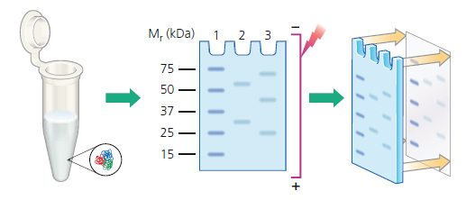
Fig.5. Sample Preparation, Electrophoresis and Transfer
Western blots are effective in detecting low nanogram to low picogram amounts of target protein, depending on the antibodies used and the detection substrate chosen.
If the target is suspected to be of very low abundance, or if there is no detectible signal on the blot, then it may be necessary to concentrate, immunoprecipitate, or fractionate the starting material.
Popular Resources
Generate Western Blot Data You Can Trust
Best practices for image documentation, integrity, and verification.
TidyBlot Western Blot Detection Reagent:HRP
TidyBlot Reagent:HRP is a western blotting detection reagent that specifically binds to native (non-reduced) antibodies.