Western Blotting Troubleshooting: Unusual or Unexpected Bands

Difference seen Possible Cause Action/ Solution

Band(s) at lower molecular weight than expected

  • Target protein has been cleaved or digested

  • Splice variants exist

  • Another protein bearing the same/similar epitope has been detected by antibody

  • Use a fresh sample which has been kept on ice
  • Add fresh protease inhibitors to the lysis buffer
  • Try alternate antibody

Band(s) at slightly higher molecular weight than expected, and may be blurred

Protein may be glycosylated or otherwise modified at one or more amino acid residues

  • Use enzymes to remove suspected modification returning molecular weight closer to expected
  • Check amino acid sequence and literature

Band(s) at significantly higher molecular weight than expected

Dimers, multimers, or protein-protein interactions may be occuring because samples have not been fully reduced and denatured.

  • Add fresh DTT or bME to samples and reheat before repeating experiment
  • Prepare new samples with fresh loading buffer

Multiple bands at various molecular weights

Primary antibody concentration may be too high, or there

is a cross-reactivity with similar epitopes on other proteins

  • Use an affinity-purified primary antibody
  • Optimize primary antibody concentration
  • Try another antibody
  • Check antibody specificity with blocking peptide

Secondary antibody concentration is too high leading to non-specific binding

  • Decrease/optimize the concentration of the secondary antibody
  • Use an affinity-purified secondary antibody
  • Repeat immunodetection with secondary antibody alone to check for non-specific binding

Protein exists in several different isoforms

  • Check literature

Bands are blurry

Gel was run at too high a voltage

  • Repeat gel at lower voltage

Incorrect running buffer composition

  • Prepare fresh running buffer

Trapped air bubble present during transfer

  • Carefully remove air bubbles between the gel and the membrane before protein transfer

Bands are smile shaped, not flat

Running conditions were too fast

so gel became over heated

  • Check and optimize gel electrophoresis conditions
  • Run gel at 4°C

White (negative) bands on the film when using ECL detection

Too much protein has been loaded

  • Load less sample
  • Repeat with dilution series of sample

Antibody concentration is too high

  • Reduce/optimize the antibody concentrations


Popular Resources

Generate Western Blot Data You Can Trust

Generate Western Blot Data You Can Trust

Best practices for image documentation, integrity, and verification.
TidyBlot Western Blot Detection Reagent:HRP

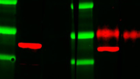
TidyBlot Western Blot Detection Reagent:HRP

TidyBlot Reagent:HRP is a western blotting detection reagent that specifically binds to native (non-reduced) antibodies.
Multiplex Fluorescent Western Blotting

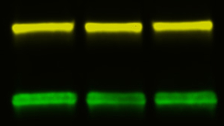
Multiplex Fluorescent Western Blotting

Multiplex fluorescent western blotting enables the simultaneous analysis of multiple proteins in a single sample on the same blot, obtaining as much information as possible in one experiment.