CD14 antibody | TÜK4
Mouse anti Human CD14:Alexa Fluor® 647
- Product Type
- Monoclonal Antibody
- Clone
- TÜK4
- Isotype
- IgG2a
- Specificity
- CD14

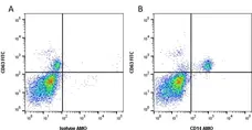
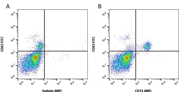
Figure B. FITC conjugated Mouse anti Human CD63 antibody, clone MEM-259 (MCA2142F) and Amethyst Orange conjugated Mouse anti Human CD14 antibody, clone TüK4 (MCA1568AMO). All experiments performed on human blood gated on live single mononuclear cells, in the presence of 10% human serum.
Data acquired on the ZE5 Cell analyser.

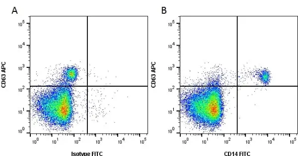
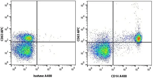
Figure B. APC conjugated Mouse anti Human CD63 antibody, clone MEM-259 (MCA2142APC) and FITC conjugated Mouse anti Human CD14 antibody, clone TüK4 (MCA1568F). All experiments performed on human blood gated on live, single mononuclear cells in the presence of 10% human serum.
Data acquired on the ZE5 Cell analyser.

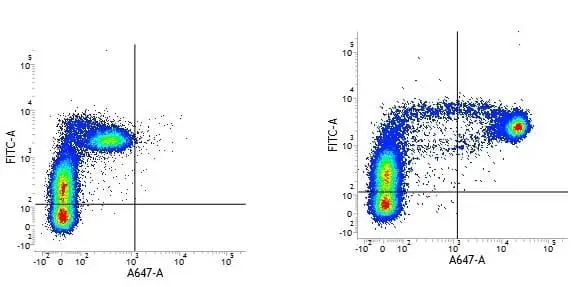
Figure B. FITC conjugated Mouse anti Human CD31 antibody, clone WM59 (MCA1738F) and Alexa Fluor® 647 conjugated Mouse anti Human CD14 antibody, clone TüK4 (MCA1568A647).
All experiments performed on human peripheral blood mononuclear cells in the presence of human SeroBlock (BUF070A).
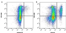
Figure B. Alexa Fluor® 488 conjugated Mouse anti Human CD9 antibody, clone MM2/57 (MCA469A488) and RPE conjugated Mouse anti Human CD14 antibody, clone TüK4 (MCA1568PE)
All experiments performed on red cell lysed human blood gated on myeloid cells in the presence of human SeroBlock (BUF070A).
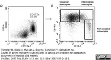
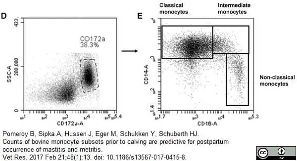
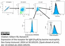
Phycoerythrin conjugated Mouse anti Human CD14 antibody, clone Tük4 (MCA1568PE) used for the evaluation of CD14 expression on bovine monocytes by flow cytometry.
Image caption:
Gating strategy utilized for flow cytometric analysis of bovine peripheral blood leukocytes showing representative data from one animal. After gating on viable (propidium-iodide-negative) cells (A), cell doublets were excluded by SSC-A and SSC-H gating (B). Bovine mononuclear cells (MNC) and granulocytes (PMN) were gated based on their forward and side scatter characteristics and their percentages were calculated (C). Three-color immunofluorescence of bovine MNC with mAbs to CD172a, CD14 and CD16 defines three monocyte subsets in peripheral blood. Viable mononuclear cells, based on forward and side scatter characteristics, were gated on CD172a-positive cells (D). Dot plots of CD14 and CD16 expression display classical monocytes (CD14+CD16−, upper left), intermediate monocytes (CD14+CD16+, upper right) and nonclassical monocytes (CD14−CD16+, lower right) (E)
From: Pomeroy B, Sipka A, Hussen J, Eger M, Schukken Y, Schuberth HJ.
Counts of bovine monocyte subsets prior to calving are predictive for postpartum occurrence of mastitis and metritis.
Vet Res. 2017 Feb 21;48(1):13.
doi: 10.1186/s13567-017-0415-8.
This is from an open access article distributed under the terms of a Creative Commons Attribution License.
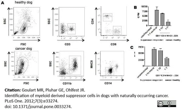
Alexa Fluor® 647 conjugated Mouse anti Human CD14 antibody Tük4 (MCA1568) used to evaluate CD14 expression on canine monocytes by flow cytometry.
Image caption:
Facs sorted CD11b+CD14−MHCII− cells isolated from a dog with osteosarcoma or healthy PBMCs were co-incubated with mitogen-stimulated CD4+ and CD8+ T cells isolated from a healthy dog for 72 hs. No stimulated cells were used as negative control. Proliferative responses were measured by 3H-thymidine incorporation from experiments performed in triplicate. CPM, counts per minute. Mean ± SEM are shown.
From: Goulart MR, Pluhar GE, Ohlfest JR (2012)
Identification of Myeloid Derived Suppressor Cells in Dogs with Naturally Occurring Cancer.
PLoS ONE 7(3): e33274.
This image is from an open access article distributed under the terms of a Creative Commons Attribution License.
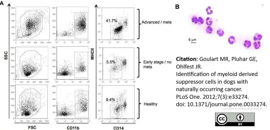
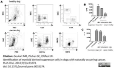
Alexa Fluor® 647 conjugated Mouse anti Human CD14 antibody Tük4 (MCA1568) used to evaluate CD14 expression on canine monocytes by flow cytometry.
Image caption:
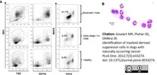
Immunophenotyping gating strategy and morphological analysis for MDSC identification in peripheral blood of dogs. PBMCs from healthy dogs and dogs with cancer were stained for the myeloid marker CD11b, monocytic marker CD14 and MHC II. (A) Representative flow cytometric analysis of forward and side scatter and gated CD11b+CD14−MHCII− cells from dogs with advanced or metastatic tumors compared to dogs with early stage non-metastatic tumors and healthy control dogs. Plots are representative of dog with advanced metastatic hemangiosarcoma (top), early stage bladder transitional cell carcinoma (middle) and a healthy dog. (B) FACS sorted CD11b+CD14−MHCII− cells were stained with diff-quick for cell morphology evaluation. A representative example of polymorphonuclear granulocyte morphology of CD11b+CD14−MHCII− cells is shown at 63× magnification.
From: Goulart MR, Pluhar GE, Ohlfest JR (2012)
Identification of Myeloid Derived Suppressor Cells in Dogs with Naturally Occurring Cancer.
PLoS ONE 7(3): e33274.
This is from an open access article distributed under the terms of a Creative Commons Attribution License.
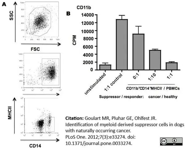
Alexa Fluor® 647 conjugated Mouse anti Human CD14 antibody clone Tük4 (MCA1568) used to evaluate CD14 expression on canine monocytes by flow cytometry.
Image caption:
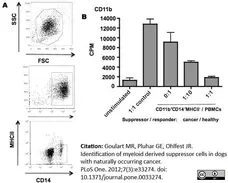
CD11b+CD14+MHCII− cells demonstrate ability to suppressive T cell proliferation. (A) CD11b+CD14+MHCII− cells were sorted from peripheral blood sample of an osteosarcoma dog (B) and co-cultured with healthy dog PBMCs in the presence of mitogen for 72 hs. Non-stimulated PBMCs were used as negative control and PBMCs co-cultured with healthy PMNs were used to control for the effect of adding cells to the assay. Proliferative responses were measured by 3H-thymidine incorporation. CPM, counts per minute. The experiment was performed in triplicate. Mean ± SEM are shown.
From: Goulart MR, Pluhar GE, Ohlfest JR (2012)
Identification of Myeloid Derived Suppressor Cells in Dogs with Naturally Occurring Cancer.
PLoS ONE 7(3): e33274.
doi: 10.1371/journal.pone.0033274.
This image is from an open access article distributed under the terms of a Creative Commons Attribution License.
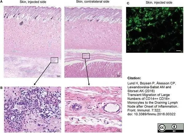
Mouse anti Human CD14 antibody, clone Tük4 (MCA1568) used to identify bovine monocytes in the skin and subcutaneous tissue of adjuvant injected calves by immunofluorescence.
Image caption:
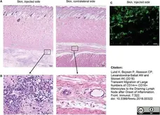
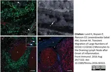
Cellular recruitment to skin and subcutaneous tissues. (A) HE stained sections of skin with subcutaneous tissue from the side injected with adjuvant and the contralateral side, at 24 h post-injection. Scale bars: 200 μm. (B) Enlargement of outlined areas in A, as indicated. Scale bars: 20 μm. (C) Immunofluorescent labeling of subcutaneous tissue on the injected side with antibody against CD14 (green). Scale bar: 20 μm.
From: Lund H, Boysen P, Åkesson CP, Lewandowska-Sabat AM and Storset AK (2016)
Transient Migration of Large Numbers of CD14++ CD16+ Monocytes to the Draining Lymph Node after Onset of Inflammation.
Front. Immunol. 7:322.
This is from an open access article distributed under the terms of a Creative Commons Attribution License.

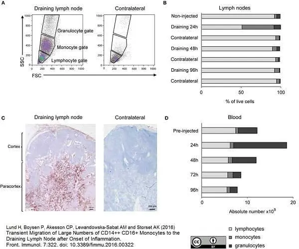
Mouse anti Human CD14 antibody, clone Tük4 (MCA1568) used to identify bovine monocytes in lymph nodes of adjuvant injected calves by immunohistochemistry.
Image caption:
Cellular recruitment to lymph nodes (LN) and peripheral blood. (A) LN cells were prepared for FCM analysis and gated on forward/side scatter (FSC/SSC) characteristics. Plots from one representative animal are presented. Panels illustrate the gating of lymphocytes, monocytes, and granulocytes as indicated and in the draining LN (left) and the contralateral LN (right), at 24 h post-injection. (B) Percentages of major immune cell populations in LNs, based on the gating strategy in A. Horizontal stacked bars show mean percentages of lymphocytes (gray), monocytes (dark gray), and granulocytes (black) of the total live cell population in non-injected animals (n = 6) and at different time points after adjuvant injection (n = 2–3). (C) IHC labeling of draining and contralateral LNs at 24 h post-injection with antibody against CD14. Different regions of the LN are indicated. (D) Cellular differential counts in peripheral blood. Horizontal stacked bars show mean absolute numbers (×109) of lymphocytes (gray), monocytes (dark gray), and granulocytes (black) at pre-injection and at different time points after adjuvant injection.
From: Lund H, Boysen P, Åkesson CP, Lewandowska-Sabat AM and Storset AK (2016)
Transient Migration of Large Numbers of CD14++ CD16+ Monocytes to the Draining Lymph Node after Onset of Inflammation.
Front. Immunol. 7:322.
doi: 10.3389/fimmu.2016.00322.
This is from an open access article distributed under the terms of a Creative Commons Attribution License.
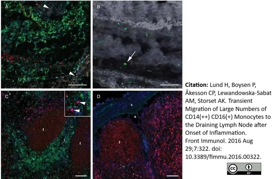
Mouse anti Human CD14 antibody, clone Tük4 (MCA1568) used to identify bovine monocytes in lymph nodes of adjuvant injected calves by immunofluorescence.
Image caption:
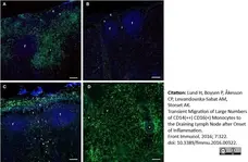
Distribution of monocytes in the LNs. Immunofluorescent labeling of LNs with antibodies against CD14 (green), CD21 (blue), and Ki67 (red). CD21 stains the LN follicles. (A) CD14+ cells were present in the capsule, subcapsular sinus, peri-trabecular sinus, and interfollicular areas of the draining LN at 24 h post-injection. (B) The contralateral LN was mainly devoid of CD14+ cells. Note the empty sub capsular sinus and trabecular sinus areas, as opposed to the infiltration in (A). (C) CD14+ cells were abundant in the capsule, but were decreased in numbers in the sinus and the cortex at 48 h post-injection. (D) CD14+ cells were present in the medulla of the LN at 48 h post-injection, and particularly around vessels. Follicle (f), interfollicular area (i), capsule (c), sinus area (s), vessel (v). Scale bars: 100 μm.
From: Lund H, Boysen P, Åkesson CP, Lewandowska-Sabat AM and Storset AK (2016)
Transient Migration of Large Numbers of CD14++ CD16+ Monocytes to the Draining Lymph Node after Onset of Inflammation.
Front. Immunol. 7:322.
doi: 10.3389/fimmu.2016.00322.
This is from an open access article distributed under the terms of a Creative Commons Attribution License.
Mouse anti Human CD14 antibody, clone Tük4 (MCA1568) used to identify bovine monocytes in lymph nodes of adjuvant injected calves by immunofluorescence.
Image caption:
Distribution of monocytes in the LNs. Immunofluorescent labeling of LNs with antibodies against CD14 (green), CD21 (blue), and Ki67 (red). CD21 stains the LN follicles. (A) CD14+ cells were present in the capsule, subcapsular sinus, peri-trabecular sinus, and interfollicular areas of the draining LN at 24 h post-injection. (B) The contralateral LN was mainly devoid of CD14+ cells. Note the empty sub capsular sinus and trabecular sinus areas, as opposed to the infiltration in (A). (C) CD14+ cells were abundant in the capsule, but were decreased in numbers in the sinus and the cortex at 48 h post-injection. (D) CD14+ cells were present in the medulla of the LN at 48 h post-injection, and particularly around vessels. Follicle (f), interfollicular area (i), capsule (c), sinus area (s), vessel (v). Scale bars: 100 μm.
From: Lund H, Boysen P, Åkesson CP, Lewandowska-Sabat AM and Storset AK (2016)
Transient Migration of Large Numbers of CD14++ CD16+ Monocytes to the Draining Lymph Node after Onset of Inflammation.
Front. Immunol. 7:322.
doi: 10.3389/fimmu.2016.00322.
This is from an open access article distributed under the terms of a Creative Commons Attribution License.
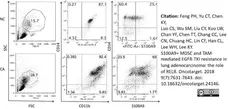
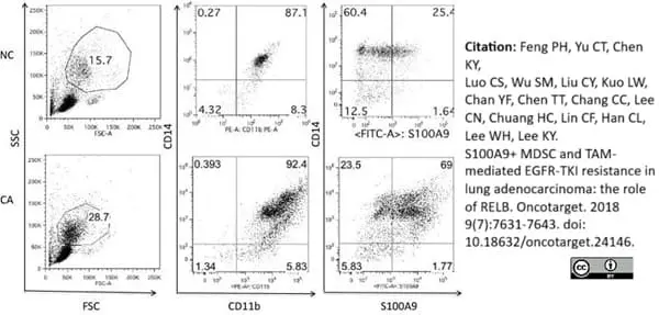
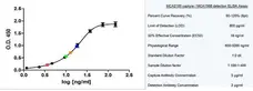
RPE-Alexa Fluor® 647 tandem conjugated Mouse anti Human CD14 antibody, clone Tük4 (MCA1568P647) used to identify CD14 expressing mononuclear cells in peripheral blood samples by flow cytometery.
Image caption:
Clinical relevance of CD11b+CD14+S100A9+ MDSCs in lung adenocarcinoma harboring activating EGFR mutation
(A) Representative dot plots of PBMC of NSCLC patients (CA) and normal health donors (NC). Non-lymphocyte mononuclear cell was gated, and CD11b+CD14+ S100A9+ cells were analyzed by flow cytometry. (B) Ratio of CD11b+CD14+ S100A9+ cells in PBMC of health donors (NC, n = 7) and NSCLC patients (CA, n = 58). (C) Clinical response of EGFR-TKI treatment and peripheral blood CD11b+CD14+ S100A9+ MDSCs, CR+PR, n = 46; SD+ PD, n = 12. (D) CD11b+CD14+ S100A9+ difference between DCB and NCB groups in training cohort. (DCB, n = 19; NCB, n = 6, p = 0.0007) (E) CD11b+CD14+ S100A9+ difference between DCB and NCB groups in validation cohort. (DCB, n = 26; NCB, n = 7, p = 0.06) (F–H) Kaplain-Meier curve of PFS according to median percentage of CD11b+CD14+S100A9+ in training cohort, validation cohort and all cases. Dark line: CD11b+CD14+S100A9+ ≤18.9% in PBMC, dashed line: CD11b+CD14+S100A9+ >18.9%. All data express as mean ±SD, * p <0.05; CR: complete response, PR: partial response, SD: stable disease, PD: progress disease, DCB: durable clinical benefit, NDB: non-durable benefit.
From: Feng PH, Yu CT, Chen KY, Luo CS, Wu SM, Liu CY, Kuo LW, Chan YF, Chen TT, Chang CC, Lee CN, Chuang HC, Lin CF, Han CL, Lee WH, Lee KY.
S100A9(+) MDSC and TAM-mediated EGFR-TKI resistance in lung adenocarcinoma: the role of RELB.
Oncotarget. 2018 Jan 10;9(7):7631-43.
doi: 10.18632/oncotarget.24146.
This image is from an open access article distributed under the terms of a Creative Commons Attribution License.
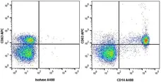
Figure B. APC conjugated Mouse anti Human CD63 antibody, clone MEM-259 (MCA2142APC) and Alexa Fluor® 488 conjugated Mouse anti Human CD14 antibody, clone TüK4 (MCA1568A488). All experiments performed on human blood gated on live single mononuclear cells, in the presence of 10% human serum.
Data acquired on the ZE5 Cell analyser.
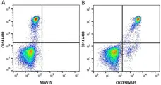
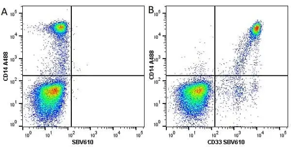
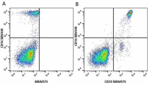
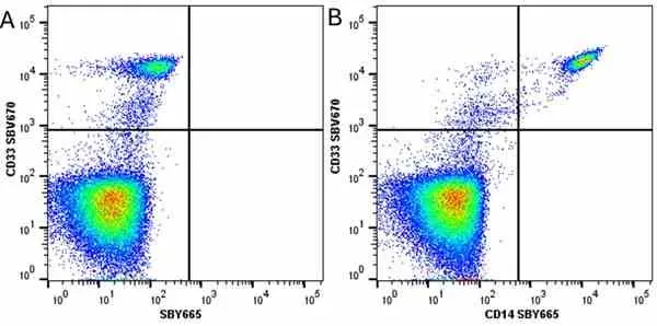
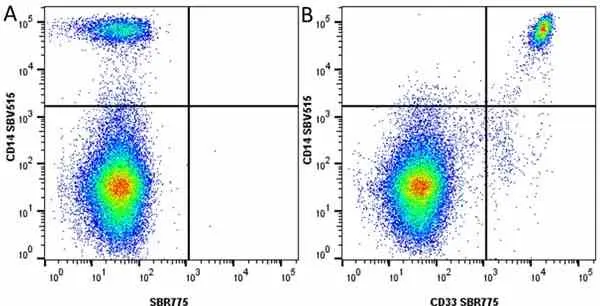
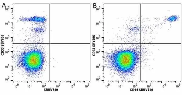
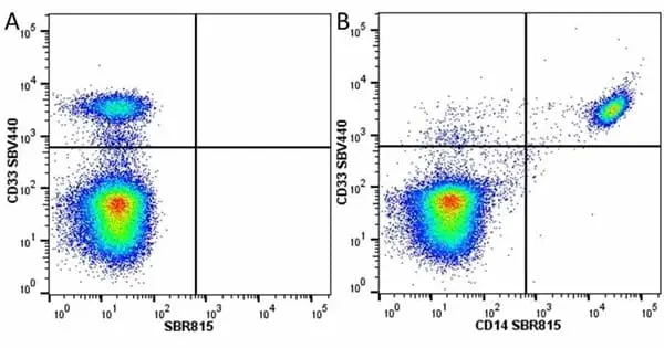
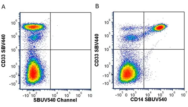
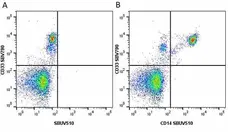
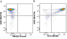
Figure B. Alexa Fluor® 488 conjugated Mouse anti Human CD14 antibody, clone TüK4 (MCA1568A647) and SBV515 conjugated Mouse anti Human CD33 antibody, clone WM53 (MCA1271SBV515). All experiments performed on red cell lysed human blood gated on live single mononuclear cells, in the presence of 10% human serum
Data acquired on the ZE5 Cell analyser.
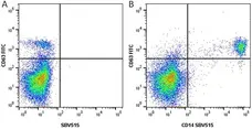
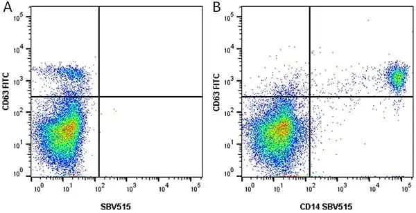
Figure B. FITC conjugated Mouse anti Human CD63 antibody, clone MEM-259 (MCA2142F) and SBV515 conjugated Mouse anti Human CD14 antibody, clone TüK4 (MCA1568SBV515). All experiments performed on red cell lysed human blood gated on live single mononuclear cells, in the presence of 10% human serum.
Data acquired on the ZE5 Cell analyser.
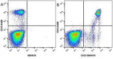
Figure B. Alexa Fluor® 488 conjugated Mouse anti Human CD14 antibody, clone TüK4 (MCA1568A488) and StarBright Violet 670 conjugated Mouse anti Human CD33 antibody, clone WM53 (MCA1271SBV670). All experiments performed on red cell lysed human blood gated on live single mononuclear cells, in the presence of 10% human serum.
Data acquired on the ZE5 Cell analyser.
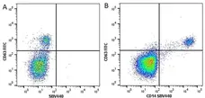
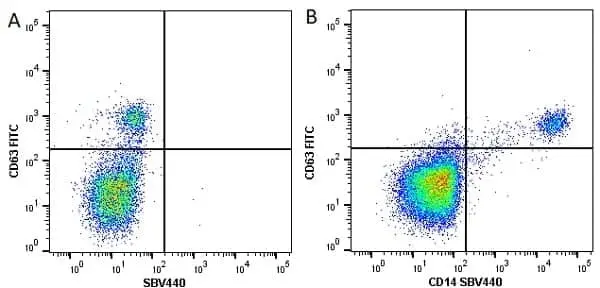
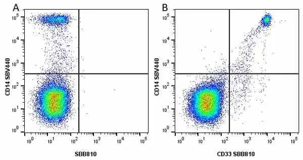
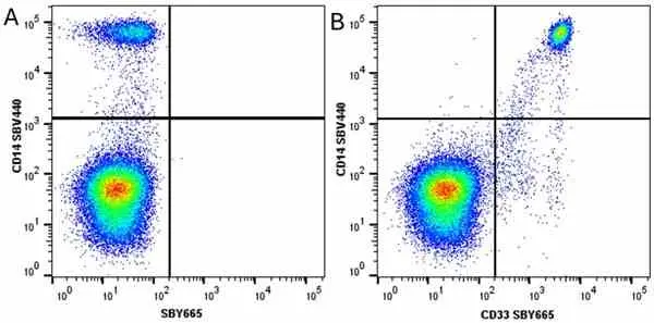
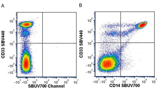
Figure B. FITC conjugated Mouse anti Human CD63 antibody, clone MEM-259 (MCA2142F) and StarBright Violet 440 conjugated Mouse anti Human CD14 antibody, clone TüK4 (MCA1568SBV440). All experiments performed on human blood gated on live single mononuclear cells, in the presence of 10% human serum.
Data acquired on the ZE5 Cell analyser.

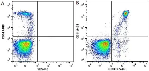
Figure B. Alexa Fluor® 488 conjugated Mouse anti Human CD14 antibody, clone TüK4 (MCA1568A488) and StarBright Violet 440 conjugated Mouse anti Human CD33 antibody, clone WM53 (MCA1271SBV440). All experiments performed on red cell lysed human blood gated on live single mononuclear cells, in the presence of 10% human serum.
Data acquired on the ZE5 Cell analyser.

Figure B. Alexa Fluor® 488 conjugated Mouse anti Human CD14 antibody, clone TüK4 (MCA1568A488) and StarBright Violet 610 conjugated Mouse anti Human CD33 antibody, clone WM53 (MCA1271SBV610) All experiments performed on red cell lysed human blood gated on live single mononuclear cells, in the presence of 10% human serum.
Data acquired on the ZE5 Cell analyser.

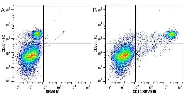
Figure B. FITC conjugated Mouse anti Human CD63 antibody, clone MEM-259 (MCA2142F) and StarBright Violet 610 conjugated Mouse anti Human CD14 antibody, clone TüK4 (MCA1568SBV610). All experiments performed on red cell lysed human blood gated on live single mononuclear cells, in the presence of 10% human serum.
Data acquired on the ZE5 Cell analyser.
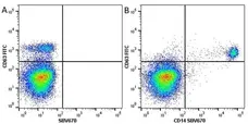
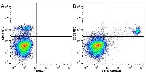
Figure B. FITC conjugated Mouse anti Human CD63 antibody, clone MEM-259 (MCA2142F) and StarBright Violet 670 conjugated Mouse anti Human CD14 antibody, clone TüK4 (MCA1568SBV670). All experiments performed on red cell lysed human blood gated on live single mononuclear cells, in the presence of 10% human serum.
Data acquired on the ZE5 Cell analyser.
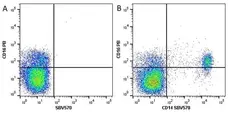
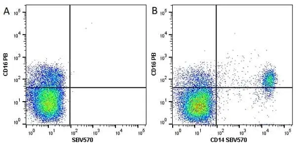
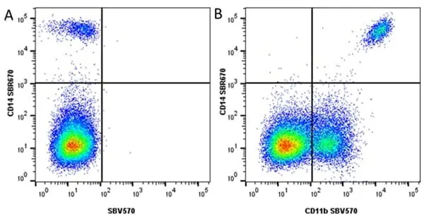
Figure B. Pacific Blue® conjugated Mouse anti Human CD16 antibody, clone DJ130c (MCA2537PB) and StarBright Violet 570 conjugated Mouse anti Human CD14 (MCA1568SBV570). All experiments performed on red cell lysed human blood gated on live single cell mononuclear cells, in the presence of 10% human serum.
Data acquired on the ZE5 Cell analyser.
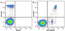
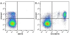
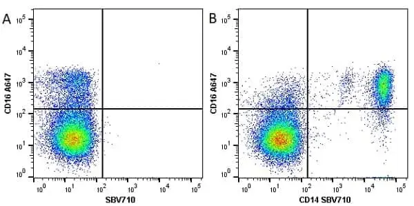
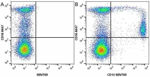
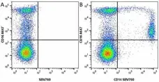
Figure B. Alexa Fluor® 647 conjugated Mouse anti Human CD16 antibody, clone DJ130c (MCA2537A647) and StarBright Violet 710 conjugated Mouse anti Human CD14 antibody, clone TüK4 (MCA1568SBV710). All experiments performed on red cell lysed human blood gated on live single mononuclear cells, in the presence of 10% human serum.
Data acquired on the ZE5 Cell analyser.
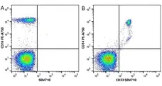
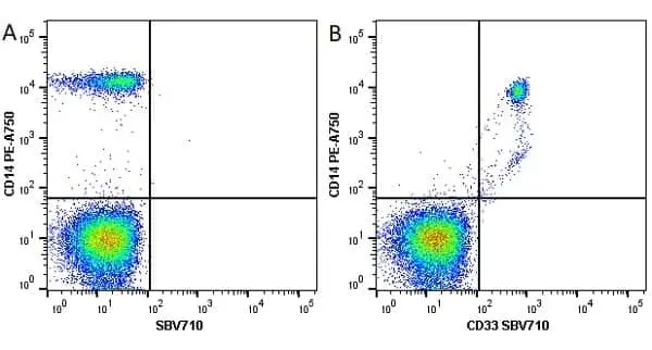
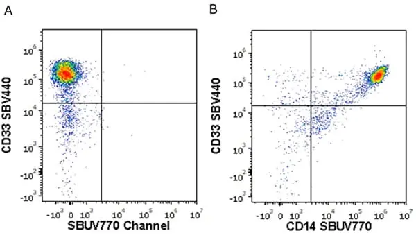
Figure B. RPE-Alexa Fluor® 750 conjugated Mouse anti Human CD14 antibody, clone TüK4 (MCA1568P750) and StarBright Violet 710 conjugated Mouse anti Human CD33 antibody, clone WM53 (MCA1271SBV710). All experiments performed on red cell lysed human blood gated on live single cell mononuclear cells, in the presence of 10% human serum.
Data acquired on the ZE5 Cell analyser.
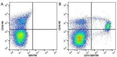
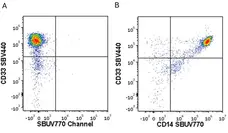
Figure B. Pacific Blue® conjugated Mouse anti Human CD16 antibody, clone DJ130c (MCA2537PB) and StarBright Violet 790 conjugated Mouse anti Human CD14 antibody, clone TüK4 (MCA1568SBV790). All experiments performed on red cell lysed human blood gated on live single cell mononuclear cells, in the presence of 10% human serum.
Data acquired on the ZE5 Cell analyser.
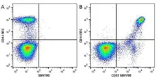
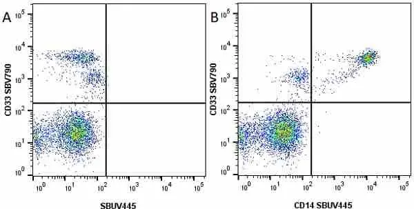
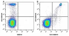
Figure B. FITC conjugated Mouse anti Human CD14 antibody, clone TüK4 (MCA1568F) and StarBright Violet 790 conjugated Mouse anti Human CD33 antibody, clone WM53 (MCA1271SBV790). All experiments performed on red blood cell lysed human blood gated on live single mononuclear cells, in the presence of 10% human serum.
Data acquired on the ZE5 Cell analyser.
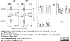
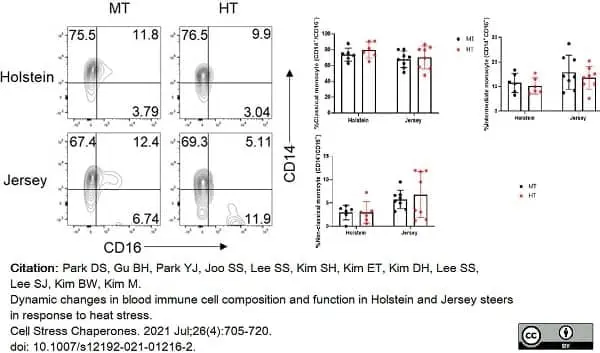
RPE conjugated Mouse anti Human CD14 antibody, clone Tük4 (MCA1568PE) used to identify monocyte populations in bovine blood samples by flow cytometry.
Image caption:
Changes in the monocyte subsets of PBMCs in Holstein and Jersey steers subjected to heat stress. Flow cytometry analysis to identify monocyte subset population. A dot plot depicting the three monocyte subsets in monocytes (CD172a+): classical CD14+CD16-, intermediate CD14+CD16+, and non-classical CD14-CD16+ monocytes. MT indicates moderate THI condition and HT indicates high THI condition
From: Park DS, Gu BH, Park YJ, Joo SS, Lee SS, Kim SH, Kim ET, Kim DH, Lee SS, Lee SJ, Kim BW, Kim M.
Dynamic changes in blood immune cell composition and function in Holstein and Jersey steers in response to heat stress.
Cell Stress Chaperones. 2021 Jul;26(4):705-20.
doi: 10.1007/s12192-021-01216-2.
This image is from an open access article distributed under terms of a Creative Commons Attribution License.

Data acquired on the ZE5 Cell analyser.
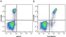
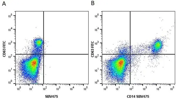
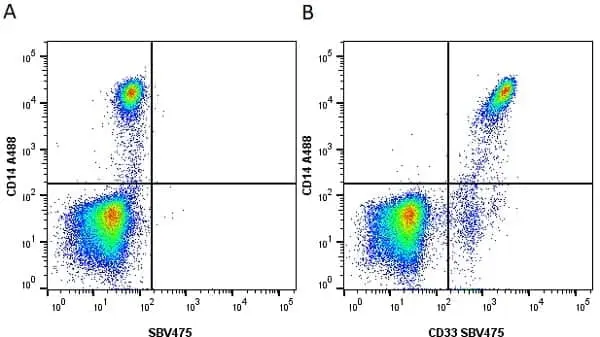
Figure B. Alexa Fluor® 488 conjugated Mouse anti Human CD14 antibody, clone TüK4 (MCA1568A488) and StarBright Violet 475 conjugated Mouse anti Human CD33 antibody, clone WM53 (MCA1271SBV475). All experiments performed on red cell lysed human blood gated on live single mononuclear cells, in the presence of 10% human serum.
Data acquired on the ZE5 Cell analyser.

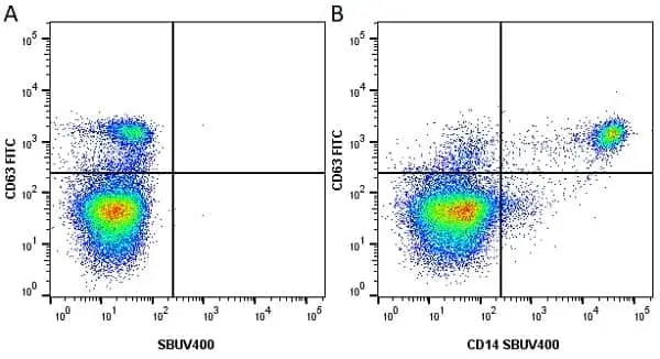
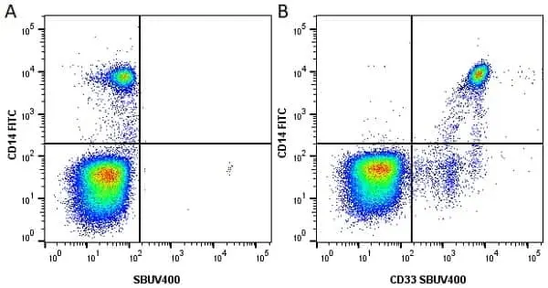
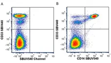
Figure B. FITC conjugated Mouse anti Human CD63 antibody, clone MEM-259 (MCA2142F) and StarBright UltraViolet 400 conjugated Mouse anti Human CD14 antibody, clone TüK4 (MCA1568SBUV400). All experiments performed on red cell lysed human blood gated on live mononuclear cells, in the presence of 10% human serum.
Data acquired on the ZE5 Cell analyser.

Data acquired on the ZE5 Cell analyser.
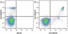
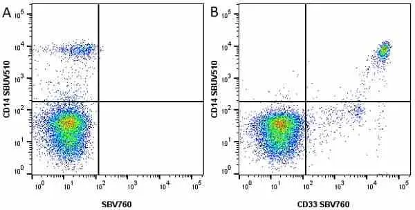
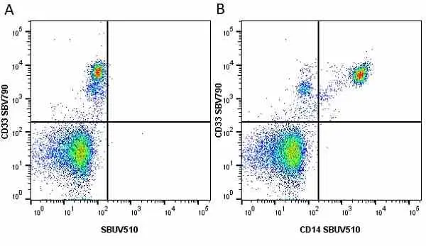
Figure B. StarBright UltraViolet 510 conjugated Mouse anti Human CD14 antibody, clone TüK4 (MCA1568SBUV510) and StarBright Violet 760 conjugated Mouse anti Human CD33 antibody, clone WM53 (MCA1271SBV760). All experiments performed on red cell lysed human blood gated on live single mononuclear cells, in the presence of 10% human serum.
Data acquired on the ZE5 Cell analyser.
Data acquired on the ZE5 Cell analyser.

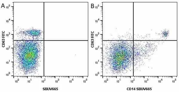
Figure B. FITC conjugated Mouse anti Human CD63 antibody, clone MEM-259 (MCA2142F) and StarBright UltraViolet 665 conjugated Mouse anti Human CD14 antibody, clone TüK4 (MCA1568SBUV665). All experiments performed on red cell lysed human blood gated on live single mononuclear cells, in the presence of 10% human serum.
Data acquired on the ZE5 Cell analyser.
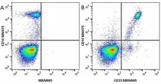
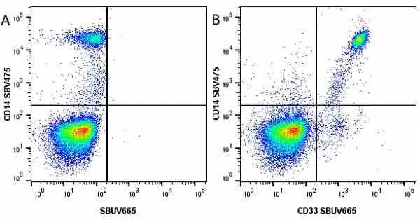
Figure B. StarBright Violet 475 conjugated Mouse anti Human CD14 antibody, clone TüK4 (MCA1568SBV475) and StarBright UltraViolet 665 conjugated Mouse anti Human CD33 antibody, clone WM53 (MCA1271SBUV665). All experiments performed on red cell lysed human blood gated on live single mononuclear cells, in the presence of 10% human serum.
Data acquired on the ZE5 Cell analyser.

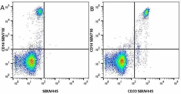
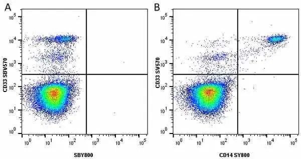
Figure B. StarBright Violet 710 conjugated Mouse anti Human CD14 antibody, clone TüK4 (MCA1568SBV710) and StarBright UltraViolet 445 conjugated Mouse anti Human CD33 antibody, clone WM57 (MCA1271SBUV445). All experiments performed on red cell lysed human blood gated on live single mononuclear cells, in the presence of 10% human serum.
Data acquired on the ZE5 Cell analyser.
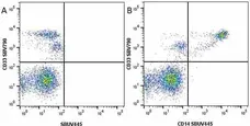
Figure B. StarBright Violet 790 conjugated Mouse anti Human CD33 antibody, clone WM53 (MCA1271SBV790) and StarBright UltraViolet 445 conjugated Mouse anti Human CD14 antibody, clone TüK4 (MCA1568SBUV445). All experiments performed on red cell lysed human blood gated on live single mononuclear cells, in the presence of 10% human serum.
Data acquired on the ZE5 Cell analyser.

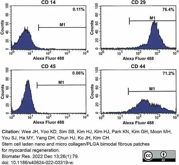
Mouse anti Human CD14 antibody, clone Tük4 (MCA1568) used for the characterization of rabbit bone marrow derived mesenchymal stem cells using flow cytometry
Image caption:
Phenotypic characterization of BMSCs using flow cytometry
From: Wee JH, Yoo KD, Sim SB, Kim HJ, Kim HJ, Park KN, Kim GH, Moon MH, You SJ, Ha MY, Yang DH, Chun HJ, Ko JH, Kim CH.
Stem cell laden nano and micro collagen/PLGA bimodal fibrous patches for myocardial regeneration.
Biomater Res. 2022 Dec 13;26(1):79.
doi: 10.1186/s40824-022-00319-w.
This image is from an open access article distributed under terms of a Creative Commons Attribution License.
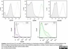
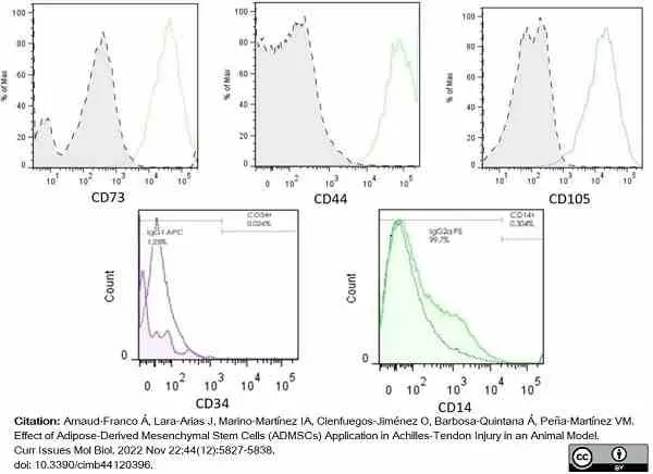
R-Phycoerythrin conjugated Mouse anti Human CD14 antibody, clone TÜK4 (MCA1568PE) used for the characterization of rabbit mesenchymal stem cells by flow cytometry.
Image caption:
Flow cytometry analysis. Adherent cells that showed MSC morphology were characterized using specific markers for stem cells according to the International Society for Cellular Therapy (ISCT). Negative controls of each antibody’s own isotype were included to rule out nonspecific signals and unlabeled cells (dotted line curves). CD73, CD44, and CD105 stained datasets were found positive after overlay, whereas CD34 and CD14 stained datasets were negative, thus discarding a hematopoietic origin of the sampled cells and suggesting an MSC phenotype.
From: Arnaud-Franco Á, Lara-Arias J, Marino-Martínez IA, Cienfuegos-Jiménez O, Barbosa-Quintana Á, Peña-Martínez VM.
Effect of Adipose-Derived Mesenchymal Stem Cells (ADMSCs) Application in Achilles-Tendon Injury in an Animal Model.
Curr Issues Mol Biol. 2022 Nov 22;44(12):5827-38.
doi: 10.3390/cimb44120396.
This image is from an open access article distributed under terms of a Creative Commons Attribution License.
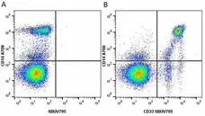
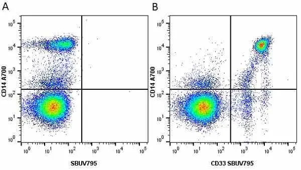
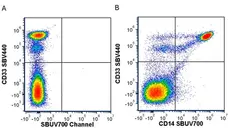
Figure B. Alexa Fluor® 700 conjugated Mouse anti Human CD14 antibody, clone TüK4 (MCA1568A700) and StarBright UltraViolet 795 conjugated Mouse anti Human CD33 antibody, clone WM53 (MCA1271SBUV795). All experiments performed on red cell lysed human blood gated on live single mononuclear cells, in the presence of 10% human serum.
Data acquired on the ZE5 Cell analyser.
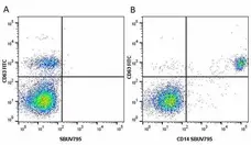
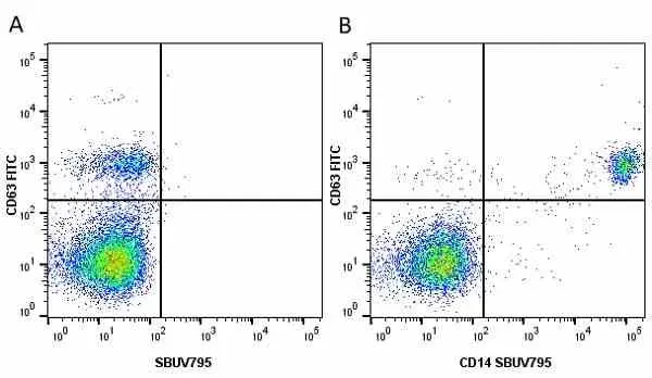
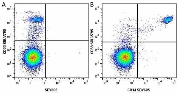
Figure B. FITC conjugated Mouse anti Human CD63 antibody, clone MEM-259 (MCA2142F) and StarBright UltraViolet 795 conjugated Mouse anti Human CD14 antibody, clone TüK4 (MCA1568SBUV795). All experiments performed on red cell lysed human blood gated on live single monocytes, in the presence of 10% human serum. Data acquired on the ZE5 Cell Analyzer.
Data acquired on the ZE5 Cell analyser.
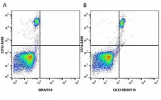
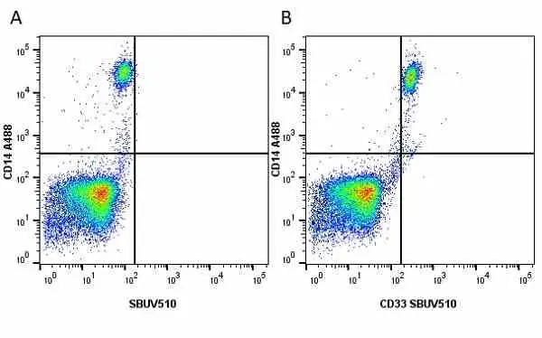
Figure B. Alexa Fluor 488 conjugated Mouse anti Human CD14 antibody, clone TüK4 (MCA1568A488) and StarBright UltraViolet 510 conjugated Mouse anti Human CD33 clone WM53 (MCA1271SBUV510). All experiments performed on red cell lysed human blood gated on live single mononuclear cells, in the presence of 10% human serum. Data acquired on the ZE5 Cell Analyzer.
Data acquired on the ZE5 Cell analyser.
Data acquired on the ZE5 Cell analyser.
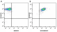
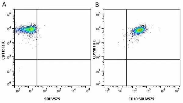
Figure B. FITC conjugated Mouse anti Human CD11b antibody, clone ICRF44 (MCA551F) and StarBright UltraViolet 575 conjugated Mouse anti Human CD14 antibody, clone TüK4 (MCA1568SBUV575). All experiments performed on red cell lysed human blood gated on live single cell granulocytes, in the presence of 10% human serum.
Data acquired on the ZE5 Cell analyser.
Data acquired on the ZE5 Cell analyser.
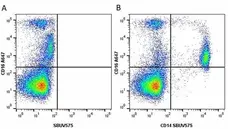
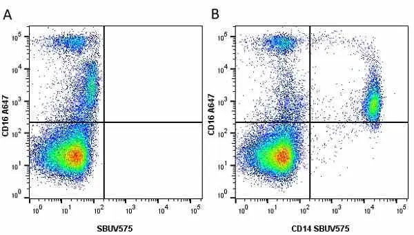
Figure B. Alexa Fluor® 647 conjugated Mouse anti Human CD16 antibody, clone KD1 (MCA5665A647) and StarBright UltraViolet 575 conjugated Mouse anti Human CD14 antibody, clone TüK4 (MCA1568SBUV575). All experiments performed on red cell lysed human blood gated on live single mononuclear cells, in the presence of 10% human serum.
Data acquired on the ZE5 Cell analyser.

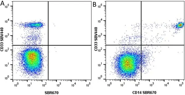
Figure B. StarBright Violet 440 conjugated Mouse anti Human CD14 antibody, clone TüK4 (MCA1568SBV440) and StarBright UltraViolet 605 conjugated Mouse anti Human CD33 antibody, clone WM57 (MCA1271SBUV605). All experiments performed on red cell lysed human blood gated on live single cell mononuclear cells, in the presence of 10% human serum.
Data acquired on the ZE5 Cell analyser.

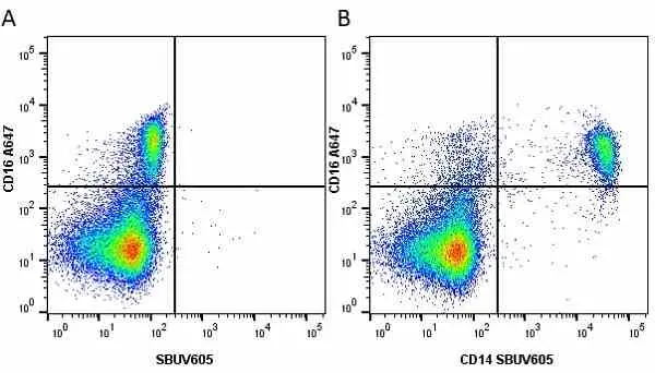
Figure B. Alexa Fluor® 647 conjugated Mouse anti Human CD16 antibody, clone DJ130c (MCA2537A647) and StarBright UltraViolet 605 conjugated Mouse anti Human CD14 antibody, clone TüK4 (MCA1568SBUV605). All experiments performed on red cell lysed human blood gated on live single cell mononuclear cells, in the presence of 10% human serum.
Data acquired on the ZE5 Cell analyser.

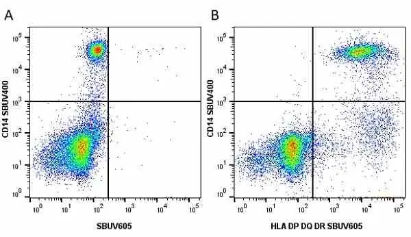
Figure B. StarBright UltraViolet 400 conjugated Mouse anti Human CD14 antibody, clone TüK4 (MCA1568SBUV400) and StarBright UltraViolet 605 conjugated Mouse anti Human HLA DP DQ DR antibody, clone WR18 (MCA477SBUV605). All experiments performed on red cell lysed human blood gated on live single mononuclear cells, in the presence of 10% human serum.
Data acquired on the ZE5 Cell analyser.
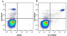
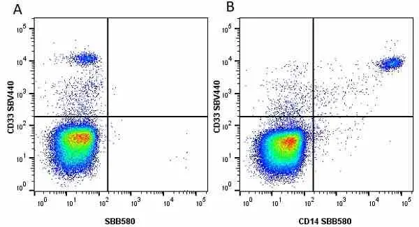
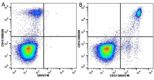
Figure B. StarBright Violet 440 conjugated Mouse anti Human CD33 antibody, clone WM57 (MCA1271SBV440) and StarBright Blue 580 conjugated Mouse anti Human CD14 antibody, clone TüK4 (MCA1568SBB580). All experiments performed on red cell lysed human blood gated on live single mononuclear cells, in the presence of 10% human serum.
Data acquired on the ZE5 Cell analyser.
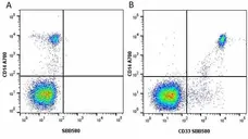
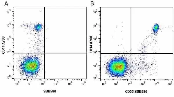
Figure B. Alexa Fluor® 700 conjugated Mouse anti Human CD14 antibody, clone TüK4 (MCA1568A700) and StarBright Blue 580 conjugated Mouse anti Human CD33 antibody, clone WM57 (MCA1271SBB580) All experiments performed on red cell lysed human blood gated on live mononuclear cells, in the presence of 10% human serum.
Data acquired on the ZE5 Cell analyser.
Data acquired on the ZE5 Cell analyser.
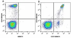
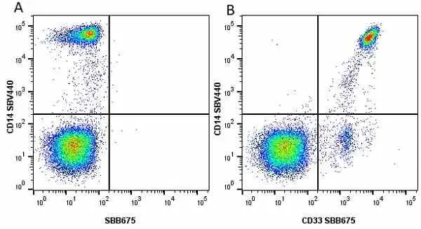
Figure B. StarBright Violet 440 conjugated Mouse anti Human CD14 antibody, clone TüK4 (MCA1568SBV440) and StarBright Blue 675 conjugated Mouse anti Human CD33 antibody, clone WM57 (MCA1271SBB675). All experiments performed on red cell lysed human blood gated on live mononuclear cells, in the presence of 10% human serum.
Data acquired on the ZE5 Cell analyser.

Figure B. StarBright UltraViolet 400 conjugated Mouse anti Human CD33 antibody, clone WM57 (MCA1271SBUV400) and StarBright Blue 675 conjugated Mouse anti Human CD14 antibody, clone TüK4 (MCA1568SBB675). All experiments performed on red cell lysed human blood gated on live single mononuclear cells, in the presence of 10% human serum.
Data acquired on the ZE5 Cell analyser.

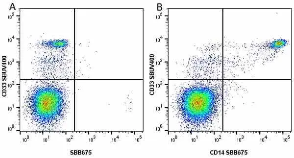
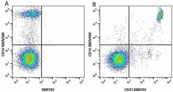
Figure B. StarBright UltraViolet 400 conjugated Mouse anti Human CD14 antibody, clone TüK4 (MCA1568SBUV400) and StarBright Blue 765 conjugated Mouse anti Human CD33 antibody, clone WM57 (MCA1271SBB765). All experiments performed on red cell lysed human blood gated on live mononuclear cells, in the presence of 10% human serum.
Data acquired on the ZE5 Cell analyser.
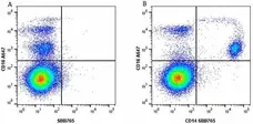
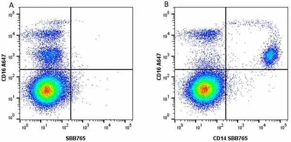
Figure B. Alexa Fluor® 647 conjugated Mouse anti Human CD16 antibody, clone DJ130c (MCA2537A647) and StarBright Blue 765 conjugated Mouse anti Human CD14 antibody, clone TüK4 (MCA1568SBB765). All experiments performed on red cell lysed human blood gated on live single cell lymphocytes, in the presence of 10% human serum.
Data acquired on the ZE5 Cell analyser.
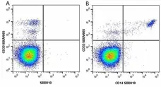
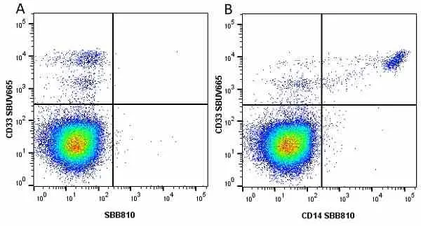
Figure B. StarBright UltraViolet 665 conjugated Mouse anti Human CD33 antibody, clone WM53 (MCA1271SBUV665) and StarBright Blue 810 conjugated Mouse anti Human CD14 antibody, clone TüK4 (MCA1568SBB810). All experiments performed on red cell lysed human blood gated on live single cell lymphocytes, in the presence of 10% human serum.
Data acquired on the ZE5 Cell analyser.
Data acquired on the ZE5 Cell analyser.
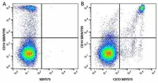
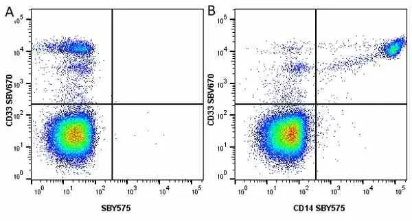
Figure B. StarBright UltraViolet 795 conjugated Mouse anti Human CD14 (MCA1568SBUV795) and StarBright Yellow 575 conjugated Mouse anti Human CD33 antibody, clone WM53 (MCA1271SBY575). All experiments performed on red cell lysed Human blood gated on live single cell lymphocytes, in the presence of 10% Human serum.
Data acquired on the ZE5 Cell analyser.
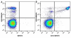
Figure B. StarBright Violet 670 conjugated Mouse anti Human CD33 antibody, clone WM53 (MCA1271SBV670) and StarBright Yellow 575 conjugated Mouse anti Human CD14 antibody, clone TüK4 (MCA1568SBY575). All experiments performed on red cell lysed Human blood gated on live single mononuclear cells, in the presence of 10% Human serum.
Data acquired on the ZE5 Cell analyser.

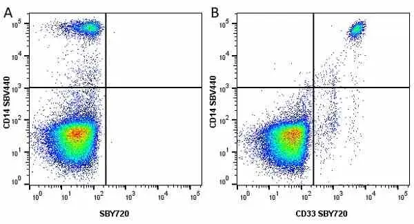
Figure B. StarBright Violet 440 conjugated Mouse anti Human CD14 antibody, clone TüK4 (MCA1568SBV440) and StarBright Yellow 720 conjugated Mouse anti Human CD33 antibody, clone WM53 (MCA1271SBY720). All experiments performed on human blood gated on live single mononuclear cells, in the presence of 10% human serum.
Data acquired on the ZE5 Cell analyser.
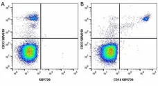
Figure B. StarBright Violet 610 conjugated Mouse anti Human CD33 antibody, clone WM53 (MCA1271SBV610) and StarBright Yellow 720 conjugated Mouse anti Human CD14 antibody, clone TüK4 (MCA1568SBY720). All experiments performed on human blood gated on live single mononuclear cells, in the presence of 10% human serum.
Data acquired on the ZE5 Cell analyser.
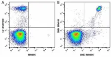
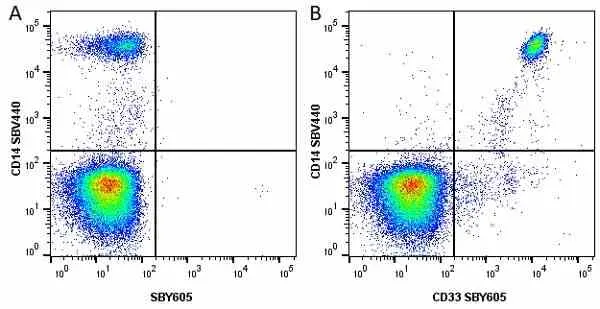
Figure B. StarBright Violet 440 conjugated Mouse anti Human CD14 antibody, clone TüK4 (MCA1568SBV440) and StarBright Yellow 605 conjugated Mouse anti Human CD33 antibody, clone WM53 (MCA1271SBY605). All experiments performed on human blood gated on live single cell lymphocytes, in the presence of 10% human serum.
Data acquired on the ZE5 Cell analyser.
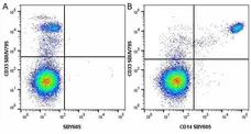
Figure B. StarBright UltraViolet 795 conjugated Mouse anti Human CD33 antibody, clone WM53 (MCA1271SBUV795) and StarBright Yellow 605 conjugated Mouse anti Human CD14 antibody, clone TüK4 (MCA1568SBY605). All experiments performed on human blood gated on live single cell lymphocytes, in the presence of 10% human serum.
Data acquired on the ZE5 Cell analyser.

Figure B. StarBright Violet 570 conjugated Mouse anti Human CD33 antibody, clone WM53 (MCA1271SBV570) and StarBright Yellow 800 conjugated Mouse anti Human CD14 antibody, clone TüK4 (MCA1568SBY800). All experiments performed on red cell lysed human blood gated on live single cell lymphocytes, in the presence of 10% human serum.
Data acquired on the ZE5 Cell analyser.

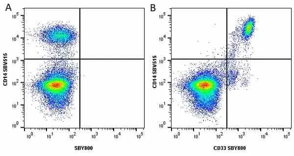
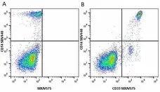
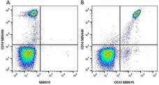
Figure B. StarBright Violet 515 conjugated Mouse anti Human CD14 antibody, clone TüK4 (MCA1568SBV515) and StarBright Yellow 800 conjugated Mouse anti Human CD33 antibody, clone WM53 (MCA1271SBY800). All experiments performed on red cell lysed human blood gated on live single cell lymphocytes, in the presence of 10% human serum.
Data acquired on the ZE5 Cell analyser.
Pacific Blue® conjugated Mouse anti Human CD14 antibody, clone TÜK4 (MCA1568PB) used to label lapine monocytes for analysis by flow cytometry.
Image caption:
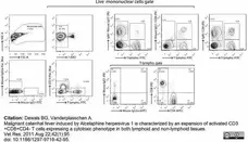
Analysis of rabbit peripheral blood mononuclear cells by multi-colour flow cytometry. PBMC were isolated from K3EDTA-sampled blood of naive rabbits before five-colour flow cytometry analysis. Specific detection of monocytes, B cells and T cell subsets consisted in a 3-step staining procedure. PBMC were first stained with anti-rabbit IgM (or mouse IgG1 isotype control), anti-rabbit CD8 (or mouse IgG1 isotype control) and anti-rabbit CD4 (or mouse IgG2a isotype control). Stainings were revealed with PE-conjugated rat anti-IgG1 or biotinylated rat anti-IgG2a, as secondary staining. Final staining was performed with streptavidin-APC, FITC-conjugated anti-rabbit T lymphocytes (or FITC-conjugated mouse IgG1 isotype control) and anti-human CD14 (or Pacific Blue-conjugated mouse IgG2a isotype control). Live lymphocytes were gated on 7-AAD- cells.
From: Dewals BG, Vanderplasschen A.
Malignant catarrhal fever induced by Alcelaphine herpesvirus 1 is characterized by an expansion of activated CD3+CD8+CD4-
T cells expressing a cytotoxic phenotype in both lymphoid and non-lymphoid tissues.
Vet Res. 2011 Aug 22;42(1):95.
doi: 10.1186/1297-9716-42-95.
This image is from an open access article distributed under terms of a Creative Commons Attribution License.
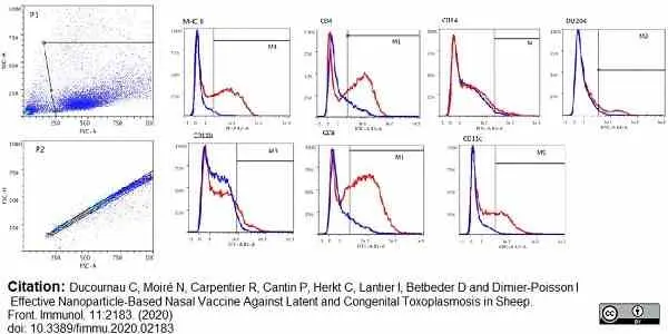
Mouse anti Human CD14 antibody, clone TüK4 (MCA1568) used to label sheep monocytes by flow cytometry.
Image caption:
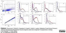
Gating Strategy forFlow Cytometry:
More that 10,000 events were acquired ba a Macsquant flow cytometer for each sample and analysed with Flowlogic software using the following strategy.
The main cell population was selected using side scatter - Area (SS-A) versus FSC-A in P1 gate. Doublet cells were excluded in gate P2. Cells from P1/P2 gate were analsed for the specific staining as shown in histogram overlays showing staining with specific antibodies (in red) in comparison with their respective isotypes (in blue). Markers (M, M1, M2, M3, M4, M5) were set up according to the control isotypes to determine the percentage of positive cells. The same strategy was applied to all samples.
From: Ducournau C, Moiré N, Carpentier R, Cantin P, Herkt C, Lantier I, Betbeder D, Dimier-Poisson I.
Effective Nanoparticle-Based Nasal Vaccine Against Latent and Congenital Toxoplasmosis in Sheep.
Front Immunol. 2020 Sep 9;11:2183.
doi: 10.3389/fimmu.2020.02183.
This image is from an open access article distributed under terms of a Creative Commons Attribution License.
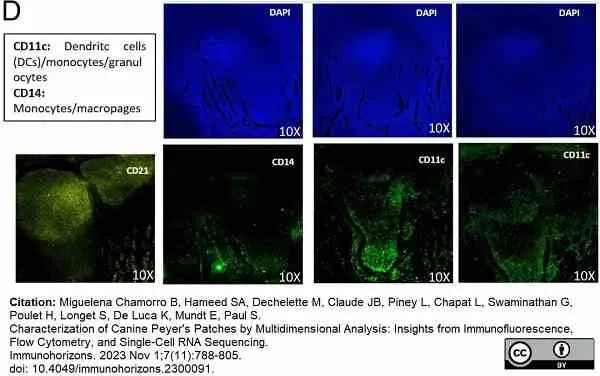
Mouse anti Human CD14 antibody, clone TÜK4 (MCA1568) used to label canine CD14 expressing cells by immunofluorescence.
Image caption:
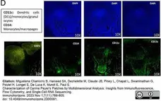
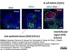
Structural organization of canine Peyer’s patches, according to immunofluorescence data.(D) Myeloid cell staining. Immunostaining of the CD14 and CD11c markers, along with CD21, was used to identify dendritic cells within the SED. Different images from the lower and upper parts of the sample were captured to adequately show the CD11c staining in proximity to both the epithelium and the follicle.
From: Miguelena Chamorro B, Hameed SA, Dechelette M, Claude JB, Piney L, Chapat L, Swaminathan G, Poulet H, Longet S, De Luca K, Mundt E, Paul S.
Characterization of Canine Peyer's Patches by Multidimensional Analysis: Insights from Immunofluorescence, Flow Cytometry, and Single-Cell RNA Sequencing.
Immunohorizons. 2023 Nov 1;7(11):788-805.
doi: 10.4049/immunohorizons.2300091.
This image is from an open access article distributed under terms of a Creative Commons Attribution License.
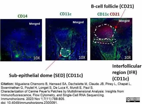
Mouse anti Human CD14 antibody, clone TÜK4 (MCA1568) used to label canine CD14 expressing cells by immunofluorescence.
Image caption:
Structural organization of canine Peyer’s patches, according to immunofluorescence data.(D) Myeloid cell staining. Immunostaining of the CD14 and CD11c markers, along with CD21, was used to identify dendritic cells within the SED. Different images from the lower and upper parts of the sample were captured to adequately show the CD11c staining in proximity to both the epithelium and the follicle.
From: Miguelena Chamorro B, Hameed SA, Dechelette M, Claude JB, Piney L, Chapat L, Swaminathan G, Poulet H, Longet S, De Luca K, Mundt E, Paul S.
Characterization of Canine Peyer's Patches by Multidimensional Analysis: Insights from Immunofluorescence, Flow Cytometry, and Single-Cell RNA Sequencing.
Immunohorizons. 2023 Nov 1;7(11):788-805.
doi: 10.4049/immunohorizons.2300091.
This image is from an open access article distributed under terms of a Creative Commons Attribution License.
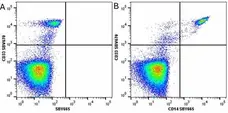
Figure B. StarBright Violet 670 conjugated Mouse anti Human CD33 antibody, clone WM53 (MCA1271SBV670) and StarBright Yellow 665 conjugated Mouse anti Human CD14 antibody, clone TüK4 (MCA1568SBY665). All experiments performed on red cell lysed human blood gated on live single mononuclear cells, in the presence of 10% human serum.
Data acquired on the ZE5 Cell analyser.
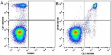
Figure B. StarBright Violet 440 conjugated Mouse anti Human CD14 antibody, clone TüK4 (MCA1568SBV440) and StarBright Yellow 665 conjugated Mouse anti Human CD33 antibody, clone WM53 (MCA1271SBY665). All experiments performed on red cell lysed human blood gated on live single mononuclear cells, in the presence of 10% human serum.
Data acquired on the ZE5 Cell analyser.
StarBright Violet 790 conjugated Mouse anti Human CD14 antibody, clone TüK4 (MCA1568SBV790) used for the gating of milk neutrophils by flow cytometry
Image caption:
Hierarchical gating to identify milk neutrophils bearing the FcμR and expression by migrated neutrophils. Milk cells were isolated by centrifugation from milk samples taken from inflamed glands (> 300 000 cells/mL). (A) The cells were labeled with antibodies to CD14-SBV790, or to granulocytes (MM20A) and F(ab’)2 anti-mouse IgG AF546, or anti-peptide 1 and F(ab’)2 anti-rabbit-PE.
From: Gilbert FB, Rainard P.
Expression of the receptor for IgM (FcμR) by bovine neutrophils.
Dev Comp Immunol. 2024 Jul 30:105235 [Epub ahead of print].
doi: 10.1016/j.dci.2024.105235. ].
This image is from an open access article distributed under terms of a Creative Commons Attribution License.
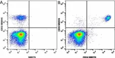
Figure B. StarBright Violet 515 conjugated Mouse anti Human CD33 antibody, clone WM53 (MCA1271SBV515) and StarBright Red 775 conjugated Mouse anti Human CD14 antibody, clone TüK4 (MCA1568SBR775). All experiments performed on red cell lysed human blood gated on live single cell lymphocytes, in the presence of 10% human serum.
Data acquired on the ZE5 Cell analyser.
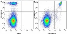
Figure B. StarBright Violet 515 conjugated Mouse anti Human CD14 antibody, clone TüK4 (MCA1568SBV515) and StarBright Red 775 conjugated Mouse anti Human CD33 antibody, clone WM53 (MCA1271SBR775). All experiments performed on red cell lysed human blood gated on live single cell lymphocytes, in the presence of 10% human serum.
Data acquired on the ZE5 Cell analyser.
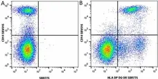
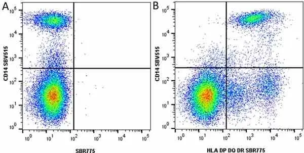
Figure B. StarBright Violet 515 conjugated Mouse anti Human CD14 antibody, clone TüK4 (MCA1568SBV515) and StarBright Red 775 conjugated Mouse anti Human HLA DP DQ DR antibody, clone WR18 (MCA477SBR775). All experiments performed on red cell lysed human blood gated on live single cell lymphocytes, in the presence of 10% human serum.
Data acquired on the ZE5 Cell analyser.

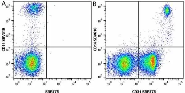
Figure B. StarBright Violet 610 conjugated Mouse anti Human CD14 antibody, clone TüK4 (MCA1568SBV610) and StarBright Red 775 conjugated Mouse anti Human CD31 antibody, clone WM59 (MCA1738SBR775). All experiments performed on red cell lysed human blood gated on live single monocnuclear cells, in the presence of 10% human serum.
Data acquired on the ZE5 Cell analyser.
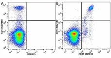
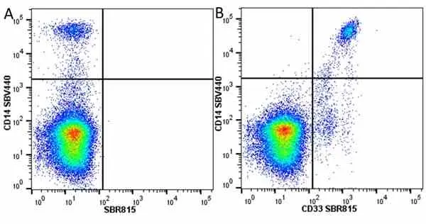
Figure B. StarBright Violet 440 conjugated Mouse anti Human CD14 antibody, clone TüK4 (MCA1568SBV440) and StarBright Red 815 conjugated Mouse anti Human CD33 antibody, clone WM53 (MCA1271SBR815). All experiments performed on red cell lysed human blood gated on live single mononuclear cells, in the presence of 10% human serum.
Data acquired on the ZE5 Cell Analyzer.

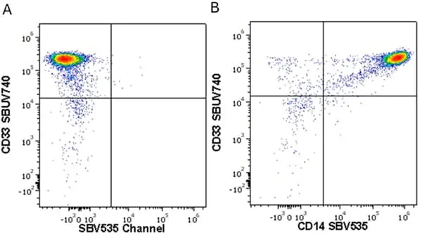
Figure B. StarBright Yellow 605 conjugated Mouse anti Human CD33 antibody, clone WM53 (MCA1271SBY605) and StarBright UltraViolet 740 conjugated Mouse anti Human CD14 antibody, clone TüK4 (MCA1568SBUV740). All experiments performed on red cell lysed human blood gated on live single mononuclear cells, in the presence of 10% human serum.
Data acquired on the ZE5 Cell Analyzer.
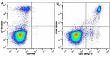
Figure B. StarBright Blue 580 conjugated Mouse anti Human CD14 antibody, clone TüK4 (MCA1568SBB580) and StarBright UltraViolet 740 conjugated Mouse anti Human CD33 antibody, clone WM53 (MCA1271SBUV740). All experiments performed on red cell lysed human blood gated on live single cell lymphocytes, in the presence of 10% human serum.
Data acquired on the ZE5 Cell Analyzer.
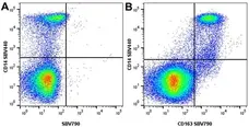
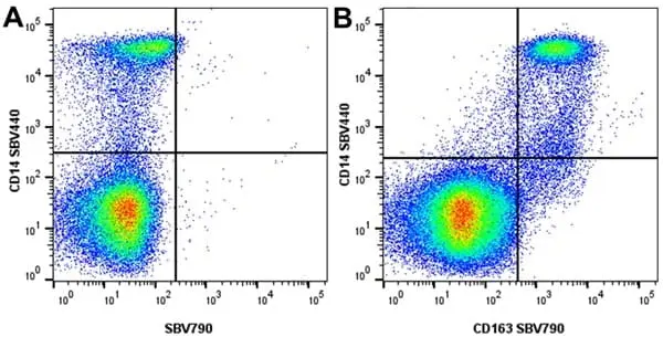
Figure B. StarBright Violet 440 conjugated Mouse anti Human CD14 antibody, clone TüK4 (MCA1568SBV440) and StarBright Violet 790 conjugated Mouse anti Human CD163 antibody, clone EDHu-1 (MCA1853SBV790). All experiments performed on human PBMCs gated on live single mononuclear cells, in the presence of 10% human serum.
Data acquired on the ZE5 Cell Analyzer.

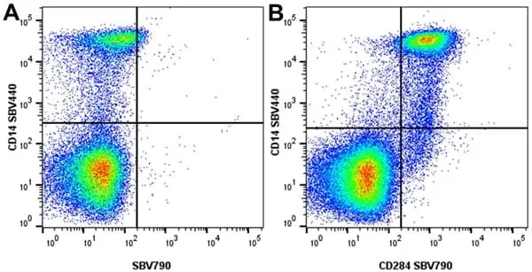
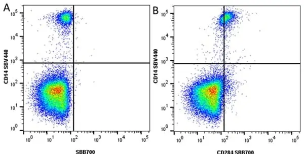
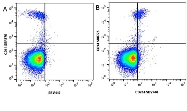
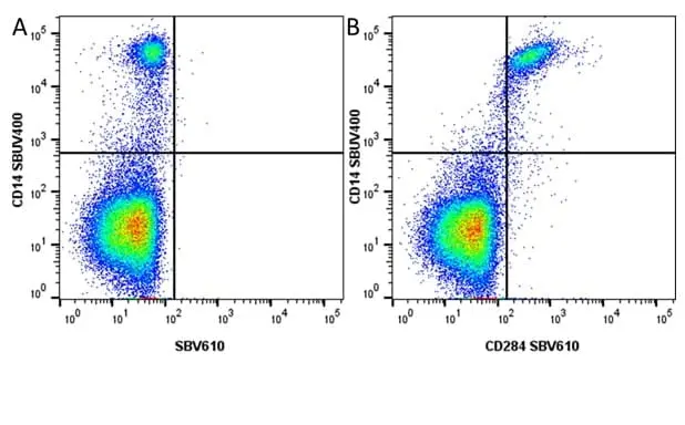
Figure B. StarBright Violet 440 conjugated Mouse anti Human CD14 antibody, clone TüK4 (MCA1568SBV440) and StarBright Violet 790 conjugated Mouse anti Human CD284 antibody, clone HTA125 (MCA2061SBV790). All experiments performed on human PBMCs gated on live single mononuclear cells, in the presence of 10% human serum.
Data acquired on the ZE5 Cell Analyzer.
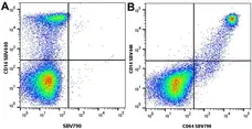
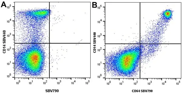
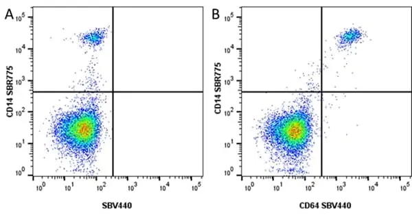
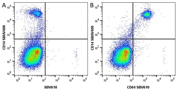
Figure B. StarBright Violet 440 conjugated Mouse anti Human CD14 antibody, clone TüK4 (MCA1568SBV440) and StarBright Violet 790 conjugated Mouse anti Human CD64 antibody, clone 10.1 (MCA756SBV790). All experiments performed on human PBMCs gated on live single mononuclear cells, in the presence of 10% human serum.
Data acquired on the ZE5 Cell Analyzer.

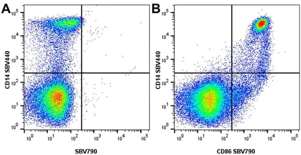
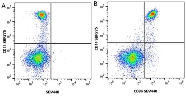
Figure B. StarBright Violet 440 conjugated Mouse anti Human CD14 antibody, clone TüK4 (MCA1568SBV440) and StarBright Violet 790 conjugated Mouse anti Human CD86 antibody, clone Bu63 (MCA1118SBV790). All experiments performed on human PBMCs gated on live single mononuclear cells, in the presence of 10% human serum.
Data acquired on the ZE5 Cell Analyzer.
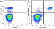
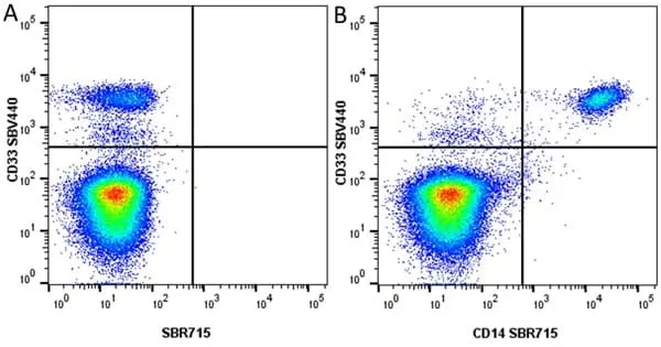
Figure B. StarBright Violet 440 conjugated Mouse anti Human CD33 antibody, WM53 (MCA1271SBV440) and StarBright Red 715 conjugated Mouse anti Human CD14 antibody, clone TüK4 (MCA1568SBR715). All experiments performed on red cell lysed human blood gated on live single mononuclear cells, in the presence of 10% human serum. Gated on live single cells, in the presence of 10% mouse serum.
Data acquired on the ZE5 Cell Analyzer.
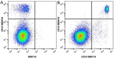
Figure B. StarBright Violet 610 conjugated Mouse anti Human CD14 antibody, clone TüK4 (MCA1568SBV440) and StarBright Red 715 conjugated Mouse anti Human CD33 antibody, clone WM53 (MCA1271SBR715). All experiments performed on human blood gated on live single mononuclear cells, in the presence of 10% human serum.
Data acquired on the ZE5 Cell Analyzer.
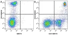
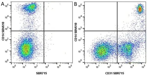
Figure B. StarBright Violet 610 conjugated Mouse anti Human CD14 antibody, clone TüK4 (MCA1568SBV610) and StarBright Red 715 conjugated Mouse anti Human CD31 antibody, clone WM59 (MCA1738SBR715). All experiments performed on human blood gated on live single cell monocytes, in the presence of 10% human serum.
Data acquired on the ZE5 Cell Analyzer.
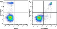
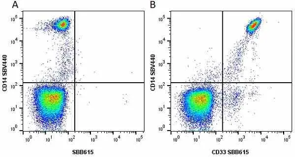
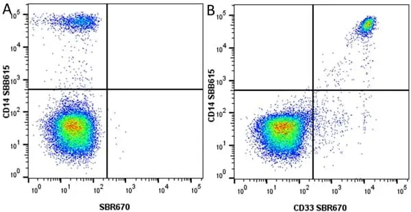
Figure B. StarBright Blue 615 conjugated Mouse anti Human CD14 antibody, clone TüK4 (MCA1568SBB615) and StarBright Red 670 conjugated Mouse anti Human CD33 antibody, clone WM53 (MCA1271SBR670). All experiments performed on red cell lysed human blood gated on live single mononuclear cells, in the presence of 10% human serum.
Data acquired on the ZE5 Cell Analyzer.
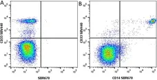
Figure B. StarBright Violet 440 conjugated Mouse anti Human CD33 antibody, clone WM53 (MCA1271SBB615) and StarBright Red 670 conjugated Mouse anti Human CD14 antibody, clone TüK4 (MCA1568SBR670). All experiments performed on red cell lysed human blood gated on live single mononuclear cells, in the presence of 10% human serum.
Data acquired on the ZE5 Cell Analyser.
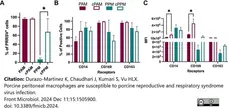
StarBright Violet 610 conjugated Mouse anti Human CD14 antibody, clone TüK4 (MCA1568SBV610) used to evaluate CD14 expression on porcine macrophages by flow cytometry.
Image caption:
Cultured PPMs are more susceptible to PRRSV. Freshly isolated or 24-h cultured PPMs and PAMs were inoculated with the PRRSV isolate RFLP-144 at an MOI of 2. At 24 hpi, cells were analyzed for the expression of viral N protein and the cellular markers CD14, CD163, and CD169 by flow cytometry. (A) Frequency of PRRSV-infected cells. (B) Frequency of cells expressing the indicated cellular markers. (C) MFI of the indicated markers. The experiments were conducted using cells from three different pigs. PAM/PPM, Freshly isolated cells; cPAM/cPPM, Cells cultured for 24 h before infection. *p ≤0.05.
From: Durazo-Martinez K, Chaudhari J, Kumari S, Vu HLX.
Porcine peritoneal macrophages are susceptible to porcine reproductive and respiratory syndrome virus infection.
Front Microbiol. 2024 Dec 11;15:1505900.
doi: 10.3389/fmicb.2024.1505900.
This image is from an open access article distributed under terms of a Creative Commons Attribution License.
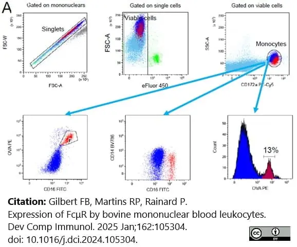
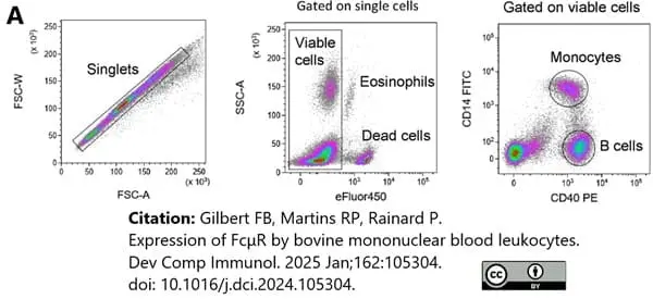
FITC conjugated and StarBright Violet 790 conjugated Mouse anti Human CD14 antibody, clone TüK4 (MCA1568SBV790 & MCA1568F) used for phenotyping of bovine cells by flow cytometry.
Image caption:
Binding of rabbit antibodies to bovine blood leukocytes A) Monocytes (CD172a positive cells) were labeled with anti-OVA (20 µg/mL) and anti-rabbit Ig-PE. Backgating on anti-OVAhigh cells coincided with the CD16high monocytes, showing that the non-specific trapping of rabbit antibodies was essentially by CD16pos monocytes. Representative of 3 cows (with the highest proportion of nonclassical monocytes).
From: Gilbert FB, Martins RP, Rainard P.
Expression of FcμR by bovine mononuclear blood leukocytes.
Dev Comp Immunol. 2025 Jan;162:105304.
doi: 10.1016/j.dci.2024.105304.
This image is from an open access article distributed under terms of a Creative Commons Attribution License.
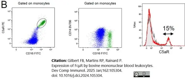
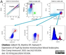
StarBright Violet 790 conjugated Mouse anti Human CD14 antibody, clone Tük4 (MCA1568SBV790) used to label monocytes in bovine blood by flow cytometry.
Image caption:
Binding of rabbit antibodies to bovine blood leukocytes. A) Monocytes (CD172a positive cells) were labeled with anti-OVA (20 μg/mL) and anti-rabbit Ig-PE. Backgating on anti-OVAhigh cells coincided with the CD16high monocytes, showing that the non-specific trapping of rabbit antibodies was essentially by CD16pos monocytes. Representative of 3 cows (with the highest proportion of nonclassical monocytes).
From: Gilbert FB, Martins RP, Rainard P.
Expression of FcμR by bovine mononuclear blood leukocytes.
Dev Comp Immunol. 2025 Jan;162:105304.
doi: 10.1016/j.dci.2024.105304.
This image is from an open access article distributed under terms of a Creative Commons Attribution License.
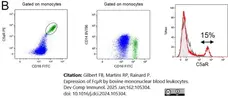
FITC conjugated Mouse anti Human CD14 antibody, clone Tük4 (MCA1568F) used to label monocytes in bovine blood by flow cytometry.
Image caption:
Binding of anti-FcμR antibodies to B lymphocytes and monocytes. A) gating strategy to identify live monocytes and B cells based on expression of CD14 and CD40.
From: Gilbert FB, Martins RP, Rainard P.
Expression of FcμR by bovine mononuclear blood leukocytes.
Dev Comp Immunol. 2025 Jan;162:105304.
doi: 10.1016/j.dci.2024.105304.
This image is from an open access article distributed under terms of a Creative Commons Attribution License.

Figure B. StarBright Violet 440 conjugated Mouse anti Human CD33 antibody, clone WM53 (MCA1271SBV440) and StarBright Red 815 conjugated Mouse anti Human CD14 antibody, clone TüK4 (MCA1568SBR815). All experiments performed on red cell lysed human blood gated on live single mononuclear cells, in the presence of 10% human serum.
Data acquired on the ZE5 Cell Analyzer.

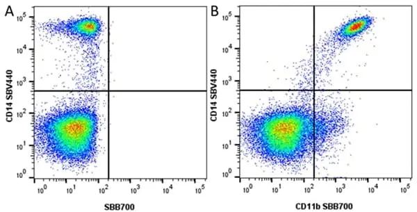
Figure B. StarBright Violet 440 conjugated Mouse anti Human CD14 antibody, clone TüK4 (MCA1568SBV440)and StarBright Blue 700 conjugated Mouse anti Human CD11b antibody, clone ICRF44 (MCA551SBB700).
All experiments performed on red cell lysed human blood gated on live single mononuclear cells, in the presence of 10% human serum. Data acquired on the ZE5 Cell Analyzer.
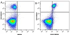
Figure B. StarBright Violet 515 conjugated Mouse anti Human CD14 antibody, clone TüK4 (MCA1568SBV515) and StarBright Blue 700 conjugated Mouse anti Human CD163 antibody, clone EDHu-1 (MCA1853SBB700).
All experiments performed on red cell lysed human blood gated on live single mononuclear cells, in the presence of 10% human serum. Data acquired on the ZE5 Cell Analyzer.
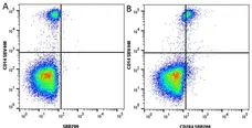
Figure B. StarBright Violet 440 conjugated Mouse anti Human CD14 (MCA1568SBV440) and StarBright Blue 700 conjugated Mouse anti Human CD284 antibody, clone HTA125 (MCA2061SBB700).
All experiments performed on red cell lysed human blood gated on live single mononuclear cells, in the presence of 10% human serum. Data acquired on the ZE5 Cell Analyzer.
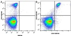
Figure B. StarBright Violet 440 conjugated Mouse anti Human CD14 antibody, clone TüK4 (MCA1568SBV440) and StarBright Blue 700 conjugated Mouse anti Human CD64 antibody, clone 10.1 (MCA756SBB700).
All experiments performed on red cell lysed human blood gated on live single mononuclear cells, in the presence of 10% human serum. Data acquired on the ZE5 Cell Analyzer.

Figure B. StarBright Red 670 conjugated Mouse anti Human CD14 antibody, clone TüK4 (MCA1568SBR670) and StarBright Violet 570 conjugated Mouse anti Human CD11b antibody, clone ICRF44 (MCA551SBV570). All experiments performed on red cell lysed human blood gated on live single mononuclear cells, in the presence of 10% human serum.
Data acquired on the ZE5 Cell Analyzer.

Data acquired on the ZE5 Cell Analyzer.

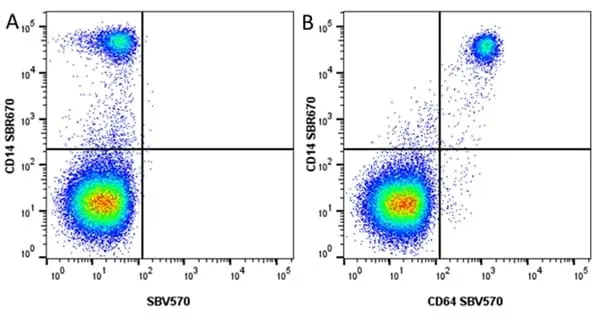
Figure B. StarBright Red 670 conjugated Mouse anti Human CD14 antibody, clone TüK4 (MCA1568SBR670) and StarBright Violet 570 conjugated Mouse anti Human CD64 antibody, clone 10.1 (MCA756SBV570). All experiments performed on human blood gated on live single mononuclear cells, in the presence of 10% human serum.
Data acquired on the ZE5 Cell Analyzer.
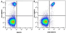
Figure B. StarBright Red 670 conjugated Mouse anti Human CD14 antibody, clone TüK4 (MCA1568SBR670) and StarBright Violet 570 conjugated Mouse anti Human CD86 antibody, clone BU63 (MCA1118SBV570). All experiments performed on red cell lysed human blood gated on live mononuclear cells, in the presence of 10% human serum.
Data acquired on the ZE5 Cell Analyzer.

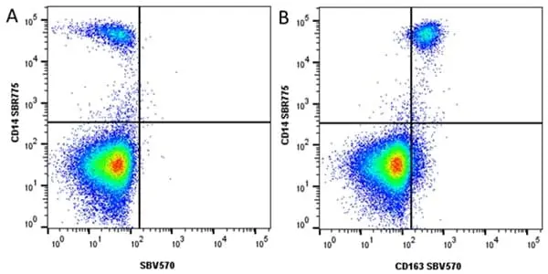
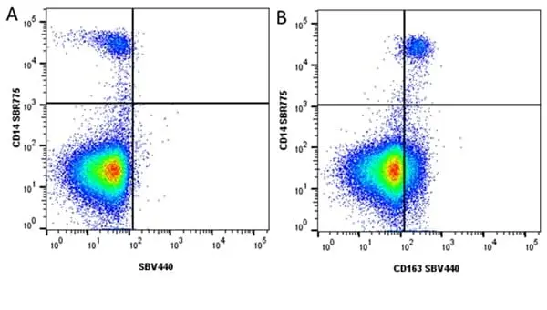
Figure B. StarBright Red 775 conjugated Mouse anti Human CD14 (MCA1568SBR775) and StarBright Violet 440 conjugated Mouse anti Human CD163 (MCA1853SBV440). All experiments performed on red blood lysed human blood gated on live single cell lymphocytes, in the presence of 10% human serum.
Data acquired on the ZE5 Cell Analyzer.
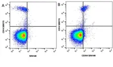
Figure B. StarBright Red 775 conjugated Mouse anti Human CD14 antibody, clone TüK4 (MCA1568SBR775) and StarBright Violet 440 conjugated Mouse anti Human CD284 antibody, clone HTA125 (MCA2061SBV440). All experiments performed on red blood lysed human blood gated on live single cell granulocytes, in the presence of 10% human serum.
Data acquired on the ZE5 Cell Analyzer.
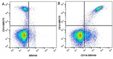
Figure B. StarBright Red 775 conjugated Mouse anti Human CD14 antibody, clone TüK4 (MCA1568SBR775) and StarBright Violet 440 conjugated Mouse anti Human CD11b antibody, clone ICRF44 (MCA551SBV440). All experiments performed on red cell lysed human blood gated on live myeloid cells, in the presence of 10% human serum.
Data acquired on the ZE5 Cell Analyzer.

Figure B. StarBright Red 775 conjugated Mouse anti Human CD14 antibody, clone Tü4 (MCA1568SBR775) and StarBright Violet 440 conjugated Mouse anti Human CD64 antibody, clone 10.1 (MCA756SBV440). All experiments performed on red cell lysed human blood gated on live single mononuclear cells, in the presence of 10% human serum.
Data acquired on the ZE5 Cell Analyzer.
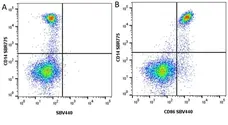
Figure B. StarBright Red 775 conjugated Mouse anti Human CD14 antibody, clone TüK4 (MCA1568SBR775) and StarBright Violet 440 conjugated Mouse anti Human CD86 antibody, clone Bu63 (MCA1118SBV440). All experiments performed on red cell lysed human blood gated on live single mononuclear cells, in the presence of 10% human serum.
Data acquired on the ZE5 Cell Analyzer.
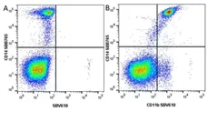
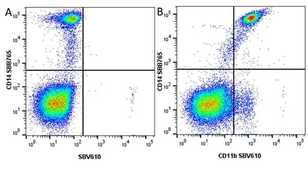
Figure B. StarBright Blue 765 conjugated Mouse anti Human CD14 antibody, clone TüK4 (MCA1568SBB765) and StarBright Violet 610 conjugated Mouse anti Human CD11b antibody, clone ICRF44 (MCA551SBV610). All experiments performed on red blood lysed human blood gated on live single mononuclear cells, in the presence of 10% human serum.
Data acquired on the ZE5 Cell Analyzer.

Figure B. StarBright UltraViolet 400 conjugated Mouse anti Human CD14 antibody, clone TüK4 (MCA1568SBUV400) and StarBright Violet 610 conjugated Mouse anti Human CD64 antibody, clone 10.1 (MCA756SBV610). All experiments performed on red blood lysed human blood gated on live single mononuclear cells, in the presence of 10% human serum.
Data acquired on the ZE5 Cell Analyzer.
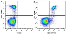
Figure B. StarBright Yellow 800 conjugated Mouse anti Human CD14 antibody, clone TüK4 (MCA1568SBY800) and StarBright Violet 610 conjugated Mouse anti Human CD86 antibody, clone Bu63 (MCA1118SBV610). All experiments performed on red blood lysed human blood gated on live single mononuclear cells, in the presence of 10% human serum.
Data acquired on the ZE5 Cell Analyzer.

Figure B. StarBright UltraViolet 400 conjugated Mouse anti Human CD14 antibody, clone TüK4 (MCA1568SBUV400) and StarBright Violet 610 conjugated Mouse anti Human CD284 antibody, clone HTA125 (MCA2061SBV610). All experiments performed on red blood lysed human blood gated on live single mononuclear cells, in the presence of 10% human serum.
Data acquired on the ZE5 Cell Analyzer.
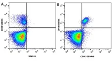
Figure B. StarBright Blue 765 conjugated Mouse anti Human CD14 antibody, clone TüK4 (MCA1568SBB765) and StarBright Violet 610 conjugated Mouse anti Human CD163 antibody, clone EDHu-1 (MCA1853SBV610). All experiments performed on red blood lysed human blood gated on live single cell lymphocytes, in the presence of 10% human serum.
Data acquired on the ZE5 Cell Analyzer.
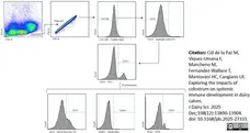
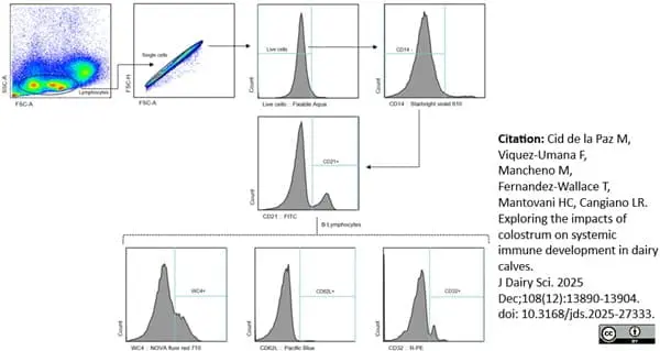
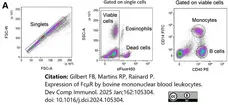
StarBright Violet 610 conjugated Mouse anti Human CD14 antibody, clone TüK4 (MCA1568SBV610) as part of the gating strategy for bovine B lymphocytes by flow cytometry.
Image caption:
Gating strategy for B lymphocytes. Lymphocytes were identified by a scatter plot, followed by single cells, live cells, and then CD14- Lymphocytes, CD21+ Lymphocytes, WC4, CD62L, and CD32. SSC = side scatter; FSC = forward scatter; A = signal area; H = signal height.
From: Cid de la Paz M, Viquez-Umana F, Mancheno M, Fernandez-Wallace T, Mantovani HC, Cangiano LR.
Exploring the impacts of colostrum on systemic immune development in dairy calves.
J Dairy Sci. 2025 Dec;108(12):13890-13904.
doi: 10.3168/jds.2025-27333.
This image is from an open access article distributed under terms of a Creative Commons Attribution License.
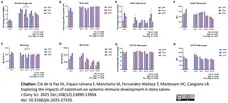
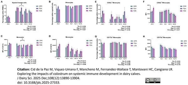
StarBright Violet 610 conjugated Mouse anti Human CD14 antibody, clone TüK4 (MCA1568SBV610) used for the enumeration of monocytes in bovine cell samples by flow cytometry.
Image caption:
Gating strategy for B lymphocytes. Lymphocytes were identified by a scatter plot, followed by single cells, live cells, and then CD14- Lymphocytes, CD21+ Lymphocytes, WC4, CD62L, and CD32. SSC = side scatter; FSC = forward scatter; A = signal area; H = signal height.
From: Cid de la Paz M, Viquez-Umana F, Mancheno M, Fernandez-Wallace T, Mantovani HC, Cangiano LR.
Exploring the impacts of colostrum on systemic immune development in dairy calves.
J Dairy Sci. 2025 Dec;108(12):13890-13904.
doi: 10.3168/jds.2025-27333.
This image is from an open access article distributed under terms of a Creative Commons Attribution License.
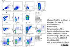
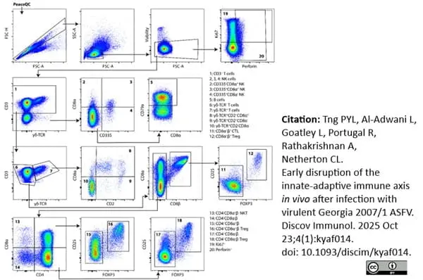
StarBright Ultraviolet 400 conjugated Mouse anti Human CD14 antibody, clone TüK4 (MCA1568SBUV400) used to label mononuclear cells in porcine blood samples by flow cytometry
Image caption:
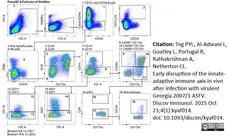
Gating strategy for volumetric whole blood staining. Representative gating of whole blood sample from AZ33 at 2 dpi. (1, 3 - 6) Granulocytes, (2) monocytes and lymphocytes, (7) CD3+ T cells, (9) γδ-TCR+T cells, (10) γδ-TCR- T cells, (11) CD8α+ γδ-TCR+ T cells, (13) CTLs and CD4-CD8α+NKT, (14) unconventional CD4-CD8α- T cells, (15) CD4+CD8α+ activated T-helper and Treg cells, (16) naïve CD4+CD8α- T-helper and Treg cells, (17) CD4+CD8α+CD25+ Treg containing population, (18) CD4+CD8α-CD25+ Treg containing population, (19) NK cells, (21) SLAII+CD172a-/lo, (23) CD21+CD25- B cells, (24) SLAII+CD172a-/loCD21-CD25-, (25) CD21+CD25+ B cells, (26) SLAII+CD172a-/loCD21-CD25+, (27) CD172a, (28) CD172a+CD14-, (29) CD172a++CD14-, and (30) CD172a++CD14+ monocytes.
From: Tng PYL, Al-Adwani L, Goatley L, Portugal R, Rathakrishnan A, Netherton CL.
Early disruption of the innate-adaptive immune axis in vivo after infection with virulent Georgia 2007/1 ASFV.
Discov Immunol. 2025 Oct 23;4(1):kyaf014.
doi: 10.1093/discim/kyaf014.
This image is from an open access article distributed under terms of a Creative Commons Attribution License.
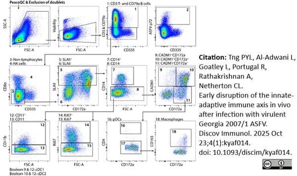
StarBright Ultraviolet 400 conjugated Mouse anti Human CD14 antibody, clone TüK4 (MCA1568SBUV400) used to label mononuclear cells in porcine blood samples by flow cytometry.
Image caption:
Gating strategy for (3) non-lymphocyte tSNE and conventional gating analysis. Representative gating of spleen sample from AZ48 on day 4 post-inoculation with Georgia 2007/1. (1) CD3+ T cells and CD79a+ B cells, (3) non-lymphocytes after removal of CD3-CD8α+, CD3-CD335+ NK cells, (5) SLAII+ non-lymphocytes, (7) monocytes, (8) CD14-SLAII+ non-lymphocytes, (Boolean of 9 and 12) CD11b+ cDC1 cells, (Boolean of 10 and 12) CD11b+ cDC2 cells, (Boolean of 9 and 13) CD11b- DC1 cells, (Boolean of 10 and 13) CD11b- DC2 cells, (16) pDCs, (17) CD14- macrophage containing population, (18) CD14-CD163+ macrophages, subsequent Boolean gating with 14 for frequencies of proliferating cells and Boolean gating with (2) for ASFV p72+ cells.
From: Tng PYL, Al-Adwani L, Goatley L, Portugal R, Rathakrishnan A, Netherton CL.
Early disruption of the innate-adaptive immune axis in vivo after infection with virulent Georgia 2007/1 ASFV.
Discov Immunol. 2025 Oct 23;4(1):kyaf014.
doi: 10.1093/discim/kyaf014.
This image is from an open access article distributed under terms of a Creative Commons Attribution License.
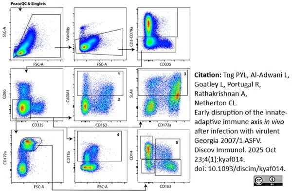
Filter by Application:
F E IF C Reset| Mouse anti Human CD14 antibody, clone TÜK4 recognizes the human CD14 cell surface antigen. CD14 is a ~55 kDa glycoprotein that contains multiple leucine-rich repeats. It is anchored to the cell membrane via a glycosylphosphatidylinositol (GPI) linkage (Simmons et al. 1989), a soluble form of CD14 also exists (Bazil et al. 1986). CD14 is strongly expressed on the surface of monocytes and macrophages but has also been shown to be expressed on the surface of non-myeloid cells (Jersmann 2005). CD14 functions as a pattern recognition receptor (Pugin et al. 1994, Dziarski et al. 1998) in innate immunity for a variety of ligands, in particular for the LPS (endotoxin) of Gram-negative bacteria. Mouse anti human CD14 antibody, clone TÜK4 has been shown to block SDF-induced chemotaxis of U937 cells in a dose –dependent manner (Yang et al. 2003). Use of the anti-human CD14 antibody, Low Endotoxin format is recommended for this purpose. |
- Target Species
- Human
- Species Cross-Reactivity
-
Target Species Cross Reactivity Dog Goat Cat Rabbit Mink Bovine Pig Sheep Cynomolgus monkey Llama - N.B. Antibody reactivity and working conditions may vary between species.
- Product Form
- Purified IgG conjugated to Alexa Fluor® 647 - liquid
- Preparation
- Purified IgG prepared by affinity chromatography on Protein A from tissue culture supernatant
- Buffer Solution
- Phosphate buffered saline
- Preservative Stabilisers
- 0.09% sodium azide (NaN3)
1% bovine serum albumin - Approx. Protein Concentrations
- IgG concentration 0.05 mg/ml
- Max Ex/Em
-
Fluorophore Excitation Max (nm) Emission Max (nm) Alexa Fluor®647 650 665 - Regulatory
- For research purposes only
- Guarantee
- 12 months from date of despatch
- Acknowledgements
- This product is provided under an intellectual property licence from Life Technologies Corporation. The transfer of this product is contingent on the buyer using the purchase product solely in research, excluding contract research or any fee for service research, and the buyer must not sell or otherwise transfer this product or its components for (a) diagnostic, therapeutic or prophylactic purposes; (b) testing, analysis or screening services, or information in return for compensation on a per-test basis; (c) manufacturing or quality assurance or quality control, or (d) resale, whether or not resold for use in research. For information on purchasing a license to this product for purposes other than as described above, contact Life Technologies Corporation, 5791 Van Allen Way, Carlsbad CA 92008 USA or outlicensing@thermofisher.com
This product is shipped at ambient temperature. It is recommended to aliquot and store at -20°C on receipt. When thawed, aliquot the sample as needed. Keep aliquots at 2-8°C for short term use (up to 4 weeks) and store the remaining aliquots at -20°C.
Avoid repeated freezing and thawing as this may denature the antibody. Storage in frost-free freezers is not recommended. This product is photosensitive and should be protected from light.
Avoid repeated freezing and thawing as this may denature the antibody. Storage in frost-free freezers is not recommended. This product is photosensitive and should be protected from light.
This product has been reported to work in the following applications. This information is derived from testing within our laboratories, peer-reviewed publications or personal communications from the originators. Please refer to references indicated for further information. For general protocol recommendations, please visit the antibody protocols page.
| Application Name | Verified | Min Dilution | Max Dilution |
|---|---|---|---|
| Flow Cytometry |  |
Neat | 1/10 |
Where this product has not been tested for use in a particular technique this does not necessarily exclude its use in such procedures. Suggested working dilutions are given as a guide only. It is recommended that the user titrates the product for use in their own system using appropriate negative/positive controls.
- Flow Cytometry
- Use 5μl of the suggested working dilution to label 106 cells or 100μl whole blood
How to Use the Spectraviewer
Watch the Tool Tutorial Video ▸- Start by selecting the application you are interested in, with the option to select an instrument from the drop down menu or create a customized instrument
- Select the fluorophores or fluorescent proteins you want to include in your panel to check compatibility
- Select the lasers and filters you wish to include
- Select combined or multi-laser view to visualize the spectra
| Description | Product Code | Applications | Pack Size | List Price | Your Price | Quantity | |
|---|---|---|---|---|---|---|---|
| Mouse IgG2a Negative Control:Alexa Fluor® 647 | MCA929A647 | F | 100 Tests/1ml |

|
Log in | ||
| List Price | Your Price | ||||||
|
|
Log in | ||||||
| Description | Mouse IgG2a Negative Control:Alexa Fluor® 647 | ||||||
| Description | Product Code | Applications | Pack Size | List Price | Your Price | Quantity | |
|---|---|---|---|---|---|---|---|
| Human Seroblock | BUF070A | F | 50 Test | Log in | |||
| List Price | Your Price | ||||||
| Log in | |||||||
| Description | Human Seroblock | ||||||
| Human Seroblock | BUF070B | F | 200 Test | Log in | |||
| List Price | Your Price | ||||||
| Log in | |||||||
| Description | Human Seroblock | ||||||
References for CD14 antibody
-
Jacobsen, C.N. et al. (1993) Reactivities of 20 anti-human monoclonal antibodies with leucocytes from ten different animal species.
Vet Immunol Immunopathol. 39 (4): 461-6. -
Gupta, V.K. et al. (1996) Identification of the sheep homologue of the monocyte cell surface molecule--CD14.
Vet Immunol Immunopathol. 51 (1-2): 89-99. -
Sopp, P. & Howard, C.J. (1997) Cross-reactivity of monoclonal antibodies to defined human leucocyte differentiation antigens with bovine cells.
Vet Immunol Immunopathol. 56 (1-2): 11-25. -
Werling, D. et al. (1998) Analysis of the phenotype and phagocytic activity of monocytes/macrophages from cattle infected with the bovine leukaemia virus.
Vet Immunol Immunopathol. 62 (3): 185-95. -
Weiss, D.J. (2001) Evaluation of proliferative disorders in canine bone marrow by use of flow cytometric scatter plots and monoclonal antibodies.
Vet Pathol. 38: 512-8. -
Bryan, S.A. et al. (2002) Responses of leukocytes to chemokines in whole blood and their antagonism by novel CC-chemokine receptor 3 antagonists.
Am J Respir Crit Care Med. 165: 1602-9. -
Yang, H. et al. (2003) Antibody to CD14 like CXCR4-specific antibody 12G5 could inhibit CXCR4-dependent chemotaxis and HIV Env-mediated cell fusion.
Immunol Lett. 88 (1): 27-30. -
Schenk, M. et al. (2005) Macrophages expressing triggering receptor expressed on myeloid cells-1 are underrepresented in the human intestine.
J Immunol. 174 (1): 517-24.
View The Latest Product References
-
Fulton, B.E. Jr. et al. (2006) Dissemination of bovine leukemia virus-infected cells from a newly infected sheep lymph node.
J Virol. 80: 7873-84. -
Willett, B.J. et al. (2007) Probing the interaction between feline immunodeficiency virus and CD134 by using the novel monoclonal antibody 7D6 and the CD134 (Ox40) ligand.
J Virol. 81: 9665-79. -
Dewals, B.G. & Vanderplasschen, A. (2011) Malignant catarrhal fever induced by Alcelaphine herpesvirus 1 is characterized by an expansion of activated CD3+CD8+CD4- T cells expressing a cytotoxic phenotype in both lymphoid and non-lymphoid tissues.
Vet Res. 42 (1): 95. -
Dalli J et al. (2008) Annexin 1 mediates the rapid anti-inflammatory effects of neutrophil-derived microparticles.
Blood. 112 (6): 2512-9. -
Martel, C.J. & Aasted, B. (2009) Characterization of antibodies against ferret immunoglobulins, cytokines and CD markers.
Vet Immunol Immunopathol. 132:109-15. -
Lybeck, K.R. et al. (2009) Neutralization of interleukin-10 from CD14(+) monocytes enhances gamma interferon production in peripheral blood mononuclear cells from Mycobacterium avium subsp. paratuberculosis-infected goats.
Clin Vaccine Immunol. 16 (7): 1003-11. -
Ferret-Bernard, S. et al. (2010) Cellular and molecular mechanisms underlying the strong neonatal IL-12 response of lamb mesenteric lymph node cells to R-848.
PLoS One. 5: e13705. -
Xiong, W. et al. (2010) Human Flt3L generates dendritic cells from canine peripheral blood precursors: implications for a dog glioma clinical trial.
PLoS One. 5: e11074. -
Kallapur, S.G. et al. (2011) Pulmonary and systemic inflammatory responses to intra-amniotic IL-1α in fetal sheep.
Am J Physiol Lung Cell Mol Physiol. 301 (3): L285-95. -
Gelain, M.E. et al. (2014) CD44 in canine leukemia: analysis of mRNA and protein expression in peripheral blood.
Vet Immunol Immunopathol. 159 (1-2): 91-6. -
Schaut, R.G. et al. (2015) Bovine viral diarrhea virus type 2 in vivo infection modulates TLR4 responsiveness in differentiated myeloid cells which is associated with decreased MyD88 expression.
Virus Res. 208: 44-55. -
Novacco, M. et al. (2016) Prognostic factors in canine acute leukaemias: a retrospective study.
Vet Comp Oncol. 14 (4): 409-16. -
Gibson, A.J. et al. (2016) Differential macrophage function in Brown Swiss and Holstein Friesian cattle.
Vet Immunol Immunopathol. 181: 15-23. -
Krueger, L.A. et al. (2016) Gamma delta T cells are early responders to Mycobacterium avium ssp. paratuberculosis in colostrum-replete Holstein calves.
J Dairy Sci. 99 (11): 9040-50. -
Lund, H. et al. (2016) Transient Migration of Large Numbers of CD14(++) CD16(+) Monocytes to the Draining Lymph Node after Onset of Inflammation.
Front Immunol. 7: 322. -
Westover, A.J. et al. (2016) An Immunomodulatory Device Improves Insulin Resistance in Obese Porcine Model of Metabolic Syndrome.
J Diabetes Res. 2016: 3486727. -
Pomeroy, B. et al. (2017) Counts of bovine monocyte subsets prior to calving are predictive for postpartum occurrence of mastitis and metritis.
Vet Res. 48 (1): 13. -
Martini, V. et al. (2018) Flow cytometry for feline lymphoma: a retrospective study regarding pre-analytical factors possibly affecting the quality of samples.
J Feline Med Surg. 20 (6): 494-501. -
Feng, P.H. et al. (2018) S100A9+ MDSC and TAM-mediated EGFR-TKI resistance in lung adenocarcinoma: the role of RELB.
Oncotarget. 9 (7): 7631-43. -
Higgins, J.L. et al. (2018) Cell mediated immune response in goats after experimental challenge with the virulent Brucella melitensis strain 16M and the reduced virulence strain Rev. 1.
Vet Immunol Immunopathol. 202: 74-84. -
Lessard, M. et al. (2018) Piglet weight gain during the first two weeks of lactation influences the immune system development.
Vet Immunol Immunopathol. 206: 25-34. -
Moncada-Saucedo, N.K. et al. (2019) A Bioactive Cartilage Graft of IGF1-Transduced Adipose Mesenchymal Stem Cells Embedded in an Alginate/Bovine Cartilage Matrix Tridimensional Scaffold.
Stem Cells Int. 2019: 9792369. -
Kolar, Q.K. et al. (2020) Anatomical distribution of respiratory tract leukocyte cell subsets in neonatal calves.
Vet Immunol Immunopathol. 227: 110090. -
Risalde, M.A. et al. (2020) BVDV permissiveness and lack of expression of co-stimulatory molecules on PBMCs from calves pre-infected with BVDV.
Comp Immunol Microbiol Infect Dis. 68: 101388. -
Muñoz-Silvestre, A. et al. (2020) Pathogenesis of Intradermal Staphylococcal Infections: Rabbit Experimental Approach to Natural Staphylococcus aureus Skin Infections.
Am J Pathol. 190 (6): 1188-210. -
Sipka, A.S. et al. (2020) The effect of ex vivo. lipopolysaccharide stimulation and nutrient availability on transition cow innate immune cell AKT/mTOR pathway responsiveness.
J Dairy Sci. 103 (2): 1956-1968. -
Mas, A. et al. (2020) A further investigation of the leishmaniosis outbreak in Madrid (Spain): low-infectivity phenotype of the Leishmania infantum BOS1FL1 isolate to establish infection in canine cells.
Vet Immunol Immunopathol. 230: 110148. -
Schwarz, E.R. et al. (2020) Experimental Infection of Mid-Gestation Pregnant Female and Intact Male Sheep with Zika Virus.
Viruses. 12 (3): 291. -
Penadés, M. et al. (2020) Early deviations in performance, metabolic and immunological indicators affect stayability in rabbit females.
Animal. 14 (4): 780-9. -
Tuohy, J.L. et al. (2020) Immune dysregulation and osteosarcoma: Staphylococcus aureus. downregulates TGF-β and heightens the inflammatory signature in human and canine macrophages suppressed by osteosarcoma.
Vet Comp Oncol. 18 (1): 64-75. -
Park, D.S. et al. (2021) Dynamic changes in blood immune cell composition and function in Holstein and Jersey steers in response to heat stress.
Cell Stress Chaperones. 26 (4): 705-20. -
Grudzien, M. et al. (2021) A newly established canine NK-type cell line and its cytotoxic properties.
Vet Comp Oncol. 19 (3): 567-77. -
Jaensch, S.M. et al. (2022) Clinicopathologic and immunophenotypic features in dogs with presumptive large granular lymphocyte leukaemia.
Aust Vet J. 100 (11): 527-32. -
Riccardo, F. et al. (2022) Antigen mimicry as an effective strategy to induce CSPG4-targeted immunity in dogs with oral melanoma: a veterinary trial.
J Immunother Cancer. 10(5):e004007. -
Shiue, S.J. et al. (2022) Arthrospira Enhances Seroclearance in Patients with Chronic Hepatitis B Receiving Nucleos(t)ide Analogue through Modulation of TNF-α/IFN-γ Profile.
Nutrients. 14 (14): 2790. -
Wee, J.H. et al. (2022) Stem cell laden nano and micro collagen/PLGA bimodal fibrous patches for myocardial regeneration.
Biomater Res. 26 (1): 79. -
Arnaud-Franco, Á. et al. (2022) Effect of Adipose-Derived Mesenchymal Stem Cells (ADMSCs) Application in Achilles-Tendon Injury in an Animal Model.
Curr Issues Mol Biol. 44 (12): 5827-38. -
Ashwood, P. (2022) Preliminary Evidence of Differentially Induced Immune Responses by Microparticle-adsorbed LPS in Patients with Crohn's Disease.
J Cell Immunol. 4 (6): 211-218. -
Rotolo, A. et al. (2023) Unedited allogeneic iNKT cells show extended persistence in MHC-mismatched canine recipients.
Cell Rep Med. 4 (10): 101241. -
Rütgen, B.C. et al. (2022) Composition of lymphocyte subpopulations in normal and mildly reactive peripheral lymph nodes in cats.
J Feline Med Surg. 24 (2): 77-90. -
Ducournau, C. et al. (2020) Effective Nanoparticle-Based Nasal Vaccine Against Latent and Congenital Toxoplasmosis in Sheep
Front Immunol. 11:2183. -
Sheng, R. et al. (2023) Prognostic significance of CD25 expression in dogs with a noninvasive diagnosis of B-cell lymphoma treated with CHOP chemotherapy.
Vet Comp Oncol. 21 (1): 28-35. -
Miguelena Chamorro, B. et al. (2023) Characterization of Canine Peyer's Patches by Multidimensional Analysis: Insights from Immunofluorescence, Flow Cytometry, and Single-Cell RNA Sequencing.
Immunohorizons. 7 (11): 788-805. -
Mason, N.J. et al. (2021) Development of a fully canine anti-canine CTLA4 monoclonal antibody for comparative translational research in dogs with spontaneous tumors.
MAbs. 13 (1): 2004638. -
Gilbert, F.B. & Rainard, P. (2024) Expression of the receptor for IgM (FcμR) by bovine neutrophils.
Dev Comp Immunol. : 105235. 30 Jul [Epub ahead of print]. -
delaO-Escamilla, A. et al. (2024) Comparison of microneedling and CO(2) laser with adipose-derived stem cells for facial rejuvenation: a randomized split-face study.
Int J Dermatol. Oct 31 [Epub ahead of print]. -
Durazo-Martinez, K. et al. (2024) Porcine peritoneal macrophages are susceptible to porcine reproductive and respiratory syndrome virus infection.
Front Microbiol. 15: 1505900. -
Gilbert, F.B. et al. (2025) Expression of FcμR by bovine mononuclear blood leukocytes.
Dev Comp Immunol. 162: 105304. -
Rogato, F. et al. (2024) Leukemia cutis as a prominent clinical sign in a dog with acute myeloid leukemia.
Vet Clin Pathol. 53 (4): 448-57. -
Moreno-Grua, E. et al. (2023) Effect of selection for growth rate on the rabbit (Oryctolagus cuniculus) immune system and its response after experimental Staphylococcus aureus infection.
Vet Res Commun. 47 (3): 1547-1560. -
Cid de la Paz M, et al. (2025) Exploring the impacts of colostrum on systemic immune development in dairy calves.
J Dairy Sci. 108 (12): 13890-13904. -
Tng, P.Y.L. et al. (2025) Early disruption of the innate-adaptive immune axis in vivo after infection with virulent Georgia 2007/1 ASFV.
Discov Immunol. 4 (1): kyaf014. -
Peixoto-Gonçalves, C. et al. (2025) Immunological studies on new rabbit paternal lines with different potentials for growth rate and resilience: pathways towards healthier animals.
Vet Res. 56 (1): 226. -
Blockeel, A. et al. (2025) CD94 as a novel marker for immunophenotyping of leukemia and lymphoma in dogs.
Front Vet Sci. 12: 1716800.
Further Reading
-
Bazil, V. et al. (1986) Biochemical characterization of a soluble form of the 53-kDa monocyte surface antigen.
Eur J Immunol. 16:1583-9. -
Simmons, D. L. et al. (1989) Monocyte antigen CD14 is a phospholipid anchored membrane protein.
Blood. 73:284-9. -
Pugin, J. et al. (1994) CD14 is a pattern recognition receptor.
Immunity.1:509-16. -
Dziarski, R. et al. (1998) Binding of bacterial peptidoglycan to CD14.
J Biol Chem. 273:8680-90. -
Jersmann, H.P. (2005) Time to abandon dogma: CD14 is expressed by non-myeloid lineage cells.
Immunol Cell Biol. 83:462-7. -
Piriou-Guzylack, L. (2008) Membrane markers of the immune cells in swine: an update.
Vet Res. 39: 54.
- RRID
- AB_566517
- UniProt
- P08571
- Entrez Gene
- CD14
- GO Terms
- GO:0005886 plasma membrane
- GO:0001530 lipopolysaccharide binding
- GO:0001847 opsonin receptor activity
- GO:0006915 apoptosis
- GO:0006909 phagocytosis
- GO:0006954 inflammatory response
- GO:0008063 Toll signaling pathway
- GO:0031225 anchored to membrane
- GO:0016019 peptidoglycan receptor activity
- View More GO Terms
- GO:0032760 positive regulation of tumor necrosis factor production
- GO:0045087 innate immune response
- GO:0070891 lipoteichoic acid binding
- GO:0071222 cellular response to lipopolysaccharide
- GO:0071223 cellular response to lipoteichoic acid
View more products with CD14 specificity
Please Note: All Products are "FOR RESEARCH PURPOSES ONLY"
View all Anti-Human ProductsAlways be the first to know.
When we launch new products and resources to help you achieve more in the lab.
Yes, sign me up