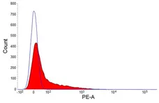
IgM antibody | NRBM
Mouse anti Rabbit IgM (B Cell Marker):RPE
- Product Type
- Monoclonal Antibody
- Clone
- NRBM
- Isotype
- IgG1
- Specificity
- IgM
- Region
- (B-CELL MARKER)


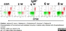
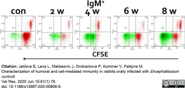
Mouse anti Rabbit IgM antibody, clone NRBM (MCA812GA) used to assess lapine splenocyte IgM expression following infection with Encephalitozoon cuniculi.
Image caption:
Representative flow cytometry analysis of CFSE labelled splenocytes in control uninfected rabbit (con) and rabbits in 2, 4, 6 and 8 weeks after E. cuniculioral infection. Splenocytes were stimulated with E. cuniculi spores and after 6 days of incubation stained with specific monoclonal antibodies against cell surface markers.
From: Jeklova E, Leva L, Matiasovic J, Ondrackova P, Kummer V, Faldyna M.
Characterization of humoral and cell-mediated immunity in rabbits orally infected with Encephalitozoon cuniculi..
Vet Res. 2020 Jun 15;51(1):79.
doi: 10.1186/s13567-020-00806-9.
This image is from an open access article distributed under terms of a Creative Commons Attribution License.

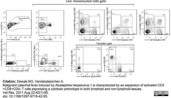
Mouse anti Rabbit IgM antibody, clone NRBM (MCA812GA) used to label lapine B lymphocytes for analysis by flow cytometry.
Image caption:
Analysis of rabbit peripheral blood mononuclear cells by multi-colour flow cytometry. PBMC were isolated from K3EDTA-sampled blood of naive rabbits before five-colour flow cytometry analysis. Specific detection of monocytes, B cells and T cell subsets consisted in a 3-step staining procedure. PBMC were first stained with anti-rabbit IgM (or mouse IgG1 isotype control), anti-rabbit CD8 (or mouse IgG1 isotype control) and anti-rabbit CD4 (or mouse IgG2a isotype control). Stainings were revealed with PE-conjugated rat anti-IgG1 or biotinylated rat anti-IgG2a, as secondary staining. Final staining was performed with streptavidin-APC, FITC-conjugated anti-rabbit T lymphocytes (or FITC-conjugated mouse IgG1 isotype control) and anti-human CD14 (or Pacific Blue-conjugated mouse IgG2a isotype control). Live lymphocytes were gated on 7-AAD- cells.
From: Dewals BG, Vanderplasschen A.
Malignant catarrhal fever induced by Alcelaphine herpesvirus 1 is characterized by an expansion of activated CD3+CD8+CD4-
T cells expressing a cytotoxic phenotype in both lymphoid and non-lymphoid tissues.
Vet Res. 2011 Aug 22;42(1):95.
doi: 10.1186/1297-9716-42-95.
This image is from an open access article distributed under terms of a Creative Commons Attribution License.
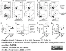
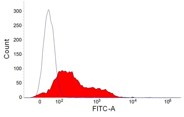
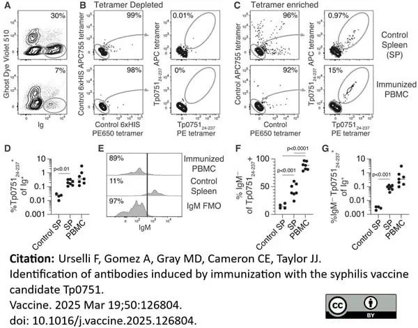
FITC conjugated Mouse anti Rabbit IgM antibody, clone NRBM (MCA812F) used to identify rabbit B lymphocytes by flow cytometry.
Image caption:
Representative flow cytometric gating of (A) live Ig+ cells that bind Tp075124–237 PE and APC tetramers but not control 6xHIS PE650 and APC755 tetramers in fractions from PBMC from immunized rabbits or spleen cells from control rabbits (B) depleted or (C) enriched for tetramer-binding B cells using microbeads specific for PE and APC prior to analysis. D. Combined data from three experiments showing the frequency of Tp075124–237 tetramer-binding cells amongst the total Ig+ population for individual rabbits in PBMC seven days after the final immunization, and in the spleen of immunized and control rabbits forty days after T. pallidum challenge. E. Representative flow cytometric gating of IgM− Tp075124–237 tetramer-binding B cells using an FMO control. Of note, cells were stained with antibodies specific for IgM, CD4, and CD8 in the same fluorescent channel but only IgM was left out of the FMO to exclude non-B cells expressing CD8 or CD4 from the population of interest. F. Combined data from three experiments showing the frequency of Tp075124–237 tetramer-binding cells that were IgM− in individual rabbits. G. Combined data from three experiments showing the frequency of IgM− Tp075124–237 tetramer-binding cells amongst the total Ig+ population in individual rabbits. In D, F, and G the lines represent the mean and p values were generated using a two-tailed Student’s t-test.
From: Urselli F, Gomez A, Gray MD, Cameron CE, Taylor JJ.
Identification of antibodies induced by immunization with the syphilis vaccine candidate Tp0751.
Vaccine. 2025 Mar 19;50:126804.
doi: 10.1016/j.vaccine.2025.126804,
This image is from an open access article distributed under terms of a Creative Commons Attribution License.
Filter by Application:
F Reset| Mouse anti Rabbit IgM (B Cell Marker) antibody, clone NRBM recognizes rabbit IgM. Mammalian IgM is produced and secreted by plasma cells located in bone marrow, lymph nodes and spleen. IgM is present in both a secreted polymeric form and as cell surface monomeric form on B cells. Mouse anti Rabbit IgM antibody, clone NRBM labels IgM+ve B cells (Dewals et al. 2011, Waclavicek et al. 2009) and as such can be considered a reliable marker of lagomorph B cells for flow cytometry. |
- Target Species
- Rabbit
- Product Form
- Purified IgG conjugated to R. Phycoerythrin (RPE) - lyophilized
- Reconstitution
- Reconstitute with 1.0 ml distilled water
- Preparation
- Purified IgG prepared by affinity chromatography on Protein A from tissue culture supernatant
- Buffer Solution
- Phosphate buffered saline
- Preservative Stabilisers
- 0.09% Sodium Azide (NaN3)
1% Bovine Serum Albumin
5% Sucrose - Fusion Partners
- Spleen cells from immunized mice were fused with cells of the Mouse P3X63Ag8.653 myeloma cell line.
- Max Ex/Em
-
Fluorophore Excitation Max (nm) Emission Max (nm) RPE 488nm laser 496 578 - Regulatory
- For research purposes only
- Guarantee
- 12 months from date of despatch
This product is shipped at ambient temperature.
Prior to reconstitution store at +4oC.
After reconstitution store at +4oC.
DO NOT FREEZE. This product should be stored undiluted. This product is photosensitive and should be protected from light.
Prior to reconstitution store at +4oC.
After reconstitution store at +4oC.
DO NOT FREEZE. This product should be stored undiluted. This product is photosensitive and should be protected from light.
This product has been reported to work in the following applications. This information is derived from testing within our laboratories, peer-reviewed publications or personal communications from the originators. Please refer to references indicated for further information. For general protocol recommendations, please visit the antibody protocols page.
| Application Name | Verified | Min Dilution | Max Dilution |
|---|---|---|---|
| Flow Cytometry |  |
Neat |
Where this product has not been tested for use in a particular technique this does not necessarily exclude its use in such procedures. Suggested working dilutions are given as a guide only. It is recommended that the user titrates the product for use in their own system using appropriate negative/positive controls.
- Flow Cytometry
- Use 10ul of the suggested working dilution to label 1x10⁶ cells in 100ul.
How to Use the Spectraviewer
Watch the Tool Tutorial Video ▸- Start by selecting the application you are interested in, with the option to select an instrument from the drop down menu or create a customized instrument
- Select the fluorophores or fluorescent proteins you want to include in your panel to check compatibility
- Select the lasers and filters you wish to include
- Select combined or multi-laser view to visualize the spectra
| Description | Product Code | Applications | Pack Size | List Price | Your Price | Quantity | |
|---|---|---|---|---|---|---|---|
| Mouse anti Rabbit T Lymphocytes:FITC | MCA800F | F | 100 Tests |

|
Log in | ||
| List Price | Your Price | ||||||
|
|
Log in | ||||||
| Description | Mouse anti Rabbit T Lymphocytes:FITC | ||||||
References for IgM antibody
-
Idogawa, H. et al. (1997) Progression of articular destruction and the production of tumour necrosis factor-alpha in antigen-induced arthritis in rabbits.
Scand J Immunol. 46 (6): 572-80. -
Dewals, B. et al. (2008) Malignant catarrhal fever induced by alcelaphine herpesvirus 1 is associated with proliferation of CD8+ T cells supporting a latent infection.
PLoS One 3: e1627. -
Gillet, L. et al. (2009) Anchoring tick salivary anti-complement proteins IRAC I and IRAC II to membrane increases their immunogenicity.
Vet Res. 40: 51. -
Stich, N. et al. (2010) Staphylococcal superantigen (TSST-1) mutant analysis reveals that t cell activation is required for biological effects in the rabbit including the cytokine storm.
Toxins (Basel). 2 (9): 2272-88. -
Waclavicek, M. et al. (2009) Analysis of the early response to TSST-1 reveals Vbeta-unrestricted extravasation, compartmentalization of the response, and unresponsiveness but not anergy to TSST-1.
J Leukoc Biol. 85 (1): 44-54. -
Anderson, I.E. et al. (2008) Production and utilization of interleukin-15 in malignant catarrhal fever.
J Comp Pathol. 138: 131-44. -
Dewals, B.G. and Vanderplasschen, A. (2011) Malignant catarrhal fever induced by Alcelaphine herpesvirus 1 is characterized by an expansion of activated CD3+CD8+CD4- T cells expressing a cytotoxic phenotype in both lymphoid and non-lymphoid tissues.
Vet Res. 42: 95. -
Dewals, B. et al. (2011) Ex vivo bioluminescence detection of alcelaphine herpesvirus 1 infection during malignant catarrhal fever.
J Virol. 85: 6941-54.
View The Latest Product References
-
Milanovic, V. et al. (2017) Histological and immunological changes in uterus during the different reproductive stages at Californian rabbit (Oryctolagus cuniculus).
Kafkas Univ Vet Fak Derg, 23, 137-44. -
Ondruska, L. et al. (2016) Decrease in C-reactive protein levels in rabbits after vaccination with a live attenuated myxoma virus vaccine.
Veterinární Medicína. 61 (No. 10): 571-6. -
Myster, F. et al. (2015) Viral semaphorin inhibits dendritic cell phagocytosis and migration but is not essential for gammaherpesvirus-induced lymphoproliferation in malignant catarrhal fever.
J Virol. 89 (7): 3630-47. -
Sorel, O. et al. (2017) Macavirus latency-associated protein evades immune detection through regulation of protein synthesis in cis depending upon its glycin/glutamate-rich domain.
PLoS Pathog. 13 (10): e1006691. -
Jeklova, E. et al. (2020) Characterization of humoral and cell-mediated immunity in rabbits orally infected with Encephalitozoon cuniculi..
Vet Res. 51 (1): 79. -
Niedżwiedzka-Rystwej, P. et al. (2020) B and T lymphocytes in rabbits change according to the sex and throughout the year.
Pol J Vet Sci. 23 (1): 37-42. -
Muñoz-Silvestre, A. et al. (2020) Pathogenesis of Intradermal Staphylococcal Infections: Rabbit Experimental Approach to Natural Staphylococcus aureus Skin Infections.
Am J Pathol. 190 (6): 1188-210. -
Niedżwiedzka-Rystwej, P. et al. (2022) Reactivity of selected markers of innate and adaptive immunity in rabbits experimentally infected with antigenic variants of RHD (Lagovirus europaeus/GI.1a).
Vet Res Commun. 46 (1): 233-42. -
Urselli, F. et al. (2025) Identification of antibodies induced by immunization with the syphilis vaccine candidate Tp0751.
Vaccine. 50: 126804.
- UniProt
- P04221
- P03988
- GO Terms
- GO:0003823 antigen binding
- GO:0005886 plasma membrane
- GO:0016021 integral to membrane
MCA812PE
If you cannot find the batch/lot you are looking for please contact our technical support team for assistance.
View more products with IgM specificity
Please Note: All Products are "FOR RESEARCH PURPOSES ONLY"
View all Anti-Rabbit ProductsAlways be the first to know.
When we launch new products and resources to help you achieve more in the lab.
Yes, sign me up