T Lymphocytes antibody | KEN-5
Mouse anti Rabbit T Lymphocytes:FITC
- Product Type
- Monoclonal Antibody
- Clone
- KEN-5
- Isotype
- IgG1
- Specificity
- T Lymphocytes

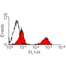

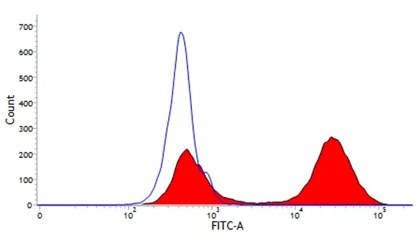
Mouse anti Rabbit T lymphocytes antibody, clone KEN-5 (MCA800G) used to assess CD5 expression levels on rabbit mononuclear cells by flow cytometry.
Image caption:
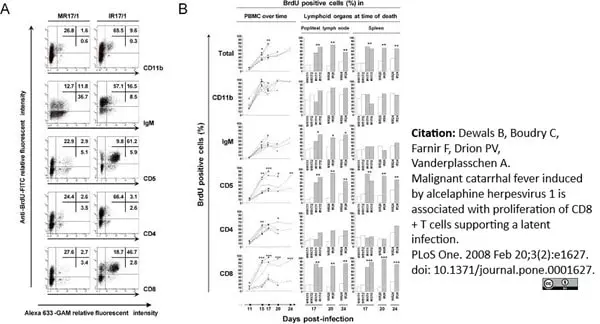
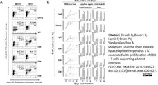
Analysis of in vivo BrdU incorporation.
Rabbits were treated as described in Fig. 1. PBMCs were collected at days 11, 15, 17, 20 and 24 post-inoculation, while mononuclear cells were isolated from popliteal lymph node and spleen at the time of death. Cells were labelled with anti-CD11b, IgM, CD5, CD4 and CD8 mAbs as the primary antibodies. Alexa 633-GAM was used as the secondary antibody. In vivo BrdU incorporation was revealed by immunofluorescent staining as described in Methods. After staining, cells were analysed by flow cytometry. A. Representative flow cytometry dot plots are shown for each double staining, they illustrate the data obtained at day 17 post-infection for the PBMC of rabbits MR17/1 and IR17/1. The data represent the percentages of BrdU positive cells (y-axis) calculated based on the acquisition of 10,000 cells expressing the indicated cell marker (x-axis). B. The percentage of BrdU positive cells amongst the indicated cellular subset was determined and compared between AlHV-1 infected (left column: bold lines; middle and right columns: hatched bars) and mock infected (left column: dotted lines; middle and right columns: open bars) groups (* P<0.05; **P<0.005, *** P<0.0001). In the left column the following symbols were used: ▴, MR17/1 and IR17/1; ▭, MR17/2 and IR17/2; ●, MR20 and IR20; ○, MR24 and IR24.
From: Dewals B, Boudry C, Farnir F, Drion P-V, Vanderplasschen A (2008)
Malignant Catarrhal Fever Induced by Alcelaphine herpesvirus 1 Is Associated with Proliferation of CD8+ T Cells Supporting a Latent Infection.
PLoS ONE 3(2): e1627.
doi: 10.1371/journal.pone.0001627.
This image is from an open access article distributed under terms of a Creative Commons Attribution License.

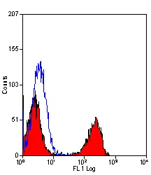
FITC conjugated Mouse anti Rabbit T lymphocytes, clone KEN-5 (MCA800F) used to label lapine T lymphocytes for analysis by flow cytometry.
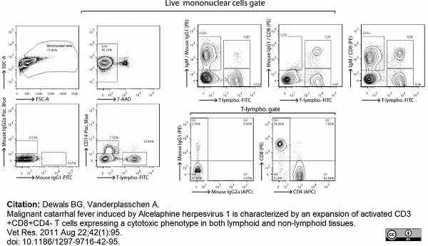
Image caption:
Analysis of rabbit peripheral blood mononuclear cells by multi-colour flow cytometry. PBMC were isolated from K3EDTA-sampled blood of naive rabbits before five-colour flow cytometry analysis. Specific detection of monocytes, B cells and T cell subsets consisted in a 3-step staining procedure. PBMC were first stained with anti-rabbit IgM (or mouse IgG1 isotype control), anti-rabbit CD8 (or mouse IgG1 isotype control) and anti-rabbit CD4 (or mouse IgG2a isotype control). Stainings were revealed with PE-conjugated rat anti-IgG1 or biotinylated rat anti-IgG2a, as secondary staining. Final staining was performed with streptavidin-APC, FITC-conjugated anti-rabbit T lymphocytes (or FITC-conjugated mouse IgG1 isotype control) and anti-human CD14 (or Pacific Blue-conjugated mouse IgG2a isotype control). Live lymphocytes were gated on 7-AAD- cells.
From: Dewals BG, Vanderplasschen A.
Malignant catarrhal fever induced by Alcelaphine herpesvirus 1 is characterized by an expansion of activated CD3+CD8+CD4-
T cells expressing a cytotoxic phenotype in both lymphoid and non-lymphoid tissues.
Vet Res. 2011 Aug 22;42(1):95.
doi: 10.1186/1297-9716-42-95.
This image is from an open access article distributed under terms of a Creative Commons Attribution License.
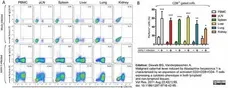
Mouse anti Rabbit T-lymphocytes antibody, clone KEN-5 (MCA800GA) used to label lapine T-lymphocytes for analysis by flow cytometry.
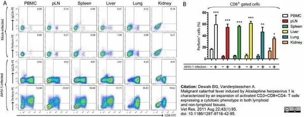
Image caption:
CD8+CD4- T cells are the prominent cellular phenotype in lymphoid and non-lymphoid organs during WD-MCF in rabbits. At time of euthanasia, analysis by multi-colour flow cytometry was conducted on PBMC and mononuclear leukocytes isolated from pLN, spleen, liver, lung and kidney of mock-infected or AlHV-1-infected rabbits developing WD-MCF. (A) Flow cytometry dot plots obtained by multi-colour staining of PBMC and mononuclear leukocytes isolated from pLN, spleen, liver, lung and kidney of one representative rabbit of each group at time of euthanasia.
From: Dewals BG, Vanderplasschen A.
Malignant catarrhal fever induced by Alcelaphine herpesvirus 1 is characterized by an expansion of activated CD3+CD8+CD4-
T cells expressing a cytotoxic phenotype in both lymphoid and non-lymphoid tissues.
Vet Res. 2011 Aug 22;42(1):95.
doi: 10.1186/1297-9716-42-95.
This image is from an open access article distributed under terms of a Creative Commons Attribution License.
Filter by Application:
F Reset| Mouse anti Rabbit T lymphocytes, clone KEN-5, recognizes rabbit (lapine) T-cells. Clone KEN-5 recognizes >90% thymocytes and approximately 40-45% mesenteric lymph node cells and immunoprecipitates a ~67 kDa protein from rabbit thymocytes. In contrast to humans and mice where only a very small population of B-cells express the CD5 antigen, in rabbits it is widely expressed by peripheral blood B cells. However, the KEN-5 antibody, while originally reported as recognizing rabbit CD5 (Kotani et al.1993), does not bind to rabbit CD5 transfectants. Furthermore, clone KEN-5, unlike other known antibodies recognizing rabbit CD5 and anti human cross reactive CD5 antibodies, shows very limited binding to rabbit B-cells, which in adult rabbits express CD5 (Raman & Knight 1992) Clone KEN-5 may recognize a epitope that is dependant on glycosylation to maintain its structural configuration which may explain why this antibody does not recognize recombinant rabbit CD5 produced in insect cells which contain different glycans from those found in mammalian cells. KEN-5 may distinguish between different isoforms of CD5 in lapine T and B cells (Pospisil et al. 2009). |
- Target Species
- Rabbit
- Species Cross-Reactivity
-
Target Species Cross Reactivity Mouse - N.B. Antibody reactivity and working conditions may vary between species.
- Product Form
- Purified IgG conjugated to Fluorescein Isothiocyanate Isomer 1 (FITC) - liquid
- Preparation
- Purified IgG prepared by affinity chromatography on Protein A from tissue culture supernatant
- Buffer Solution
- Phosphate buffered saline
- Preservative Stabilisers
0.09% Sodium Azide 1% Bovine Serum Albumin - Immunogen
- Rabbit Thymocytes
- Approx. Protein Concentrations
- IgG concentration 0.1 mg/ml
- Fusion Partners
- Spleen cells from immunised BALB/c mice were fused with cells of the PAI mouse myeloma cell line
- Max Ex/Em
-
Fluorophore Excitation Max (nm) Emission Max (nm) FITC 490 525 - Regulatory
- For research purposes only
- Guarantee
- 12 months from date of despatch
This product is shipped at ambient temperature. It is recommended to aliquot and store at -20°C on receipt. When thawed, aliquot the sample as needed. Keep aliquots at 2-8°C for short term use (up to 4 weeks) and store the remaining aliquots at -20°C.
Avoid repeated freezing and thawing as this may denature the antibody. Storage in frost-free freezers is not recommended. This product is photosensitive and should be protected from light.
Avoid repeated freezing and thawing as this may denature the antibody. Storage in frost-free freezers is not recommended. This product is photosensitive and should be protected from light.
This product has been reported to work in the following applications. This information is derived from testing within our laboratories, peer-reviewed publications or personal communications from the originators. Please refer to references indicated for further information. For general protocol recommendations, please visit the antibody protocols page.
| Application Name | Verified | Min Dilution | Max Dilution |
|---|---|---|---|
| Flow Cytometry |  |
Neat |
Where this antibody has not been tested for use in a particular technique this does not necessarily exclude its use in such procedures. Suggested working dilutions are given as a guide only. It is recommended that the user titrates the antibody for use in their own system using appropriate negative/positive controls.
- Flow Cytometry
- Use 10ul of the suggested working dilution to label 106 cells or 100ul whole blood
How to Use the Spectraviewer
Watch the Tool Tutorial Video ▸- Start by selecting the application you are interested in, with the option to select an instrument from the drop down menu or create a customized instrument
- Select the fluorophores or fluorescent proteins you want to include in your panel to check compatibility
- Select the lasers and filters you wish to include
- Select combined or multi-laser view to visualize the spectra
| Description | Product Code | Applications | Pack Size | List Price | Your Price | Quantity | |
|---|---|---|---|---|---|---|---|
| Mouse IgG1 Negative Control:FITC | MCA928F | F | 100 Tests |

|
Log in | ||
| List Price | Your Price | ||||||
|
|
Log in | ||||||
| Description | Mouse IgG1 Negative Control:FITC | ||||||
Source Reference
-
Kotani, M. et al. (1993) Generation and characterization of monoclonal antibodies against rabbit CD4, CD5 and CD11a antigens.
J Immunol Methods. 157 (1-2): 241-52.
References for T Lymphocytes antibody
-
Matsumura, T. et al. (1999) Suppression of atherosclerotic development in Watanabe heritable hyperlipidemic rabbits treated with an oral antiallergic drug, tranilast.
Circulation. 99 (7): 919-24. -
Gu, W. et al. (2004) Immune response in rabbit ovaries following infection of a recombinant myxoma virus expressing rabbit zona pellucida protein B.
Virology. 318 (2): 516-23. -
Hoefer, I.E. et al. (2005) Leukocyte subpopulations and arteriogenesis: specific role of monocytes, lymphocytes and granulocytes.
Atherosclerosis. 181 (2): 285-93. -
Dewals, B. et al. (2008) Malignant catarrhal fever induced by alcelaphine herpesvirus 1 is associated with proliferation of CD8+ T cells supporting a latent infection.
PLoS One. 3 (2): e1627. -
Gillet, L. et al. (2009) Anchoring tick salivary anti-complement proteins IRAC I and IRAC II to membrane increases their immunogenicity.
Vet Res. 40: 51. -
Guerrero, I. et al. (2010) Evolution of the peripheral blood lymphocyte populations in multiparous rabbit does with two reproductive management rhythms.
Vet Immunol Immunopathol. 140: 75-81. -
Milanovic, V. et al. (2017) Histological and immunological changes in uterus during the different reproductive stages at Californian rabbit (Oryctolagus cuniculus).
Kafkas Univ Vet Fak Derg, 23, 137-44. -
Penadés, M. et al. (2018) Long-term implications of feed energy source in different genetic types of reproductive rabbit females. II. Immunologic status.
Animal. 12 (9): 1877-85.
View The Latest Product References
-
Gates, K.V. & Griffiths, L.G. (2018) Chronic graft-specific cell-mediated immune response toward candidate xenogeneic biomaterial.
Immunol Res. 66 (2): 288-98. -
Penadés, M.et al. (2019) Early deviations in performance, metabolic and immunological indicators affect stayability in rabbit females.
Animal. : 1-10. -
Niedźwiedzka-Rystwej, P. et al. (2020) B and T lymphocytes in rabbits change according to the sex and throughout the year.
Pol J Vet Sci. 23 (1): 37-42. -
Muñoz-Silvestre, A. et al. (2020) Pathogenesis of Intradermal Staphylococcal Infections: Rabbit Experimental Approach to Natural Staphylococcus aureus Skin Infections.
Am J Pathol. 190 (6): 1188-210. -
Lin, W. et al. (2020) Rapid identification of anti-idiotypic mAbs with high affinity and diverse epitopes by rabbit single B-cell sorting-culture and cloning technology.
PLoS One. 15 (12): e0244158. -
Niedźwiedzka-Rystwej, P. et al. (2021) Reactivity of selected markers of innate and adaptive immunity in rabbits experimentally infected with antigenic variants of RHD (Lagovirus europaeus/GI.1a).
Vet Res Commun. Oct 29 [Epub ahead of print]. -
Noreng, S. et al. (2022) Structure of the core human NADPH oxidase NOX2.
Nat Commun. 13 (1): 6079. -
Dewals, B.G. & Vanderplasschen, A. (2011) Malignant catarrhal fever induced by Alcelaphine herpesvirus 1 is characterized by an expansion of activated CD3+CD8+CD4- T cells expressing a cytotoxic phenotype in both lymphoid and non-lymphoid tissues.
Vet Res. 42 (1): 95. -
Myster, F. et al. (2015) Viral semaphorin inhibits dendritic cell phagocytosis and migration but is not essential for gammaherpesvirus-induced lymphoproliferation in malignant catarrhal fever.
J Virol. 89 (7): 3630-47. -
Moreno-Grua, E. et al. (2023) Effect of selection for growth rate on the rabbit (Oryctolagus cuniculus) immune system and its response after experimental Staphylococcus aureus infection.
Vet Res Commun. 47 (3): 1547-1560. -
Peixoto-Gonçalves, C. et al. (2025) Immunological studies on new rabbit paternal lines with different potentials for growth rate and resilience: pathways towards healthier animals.
Vet Res. 56 (1): 226.
Further Reading
-
Raman, C. & Knight, K.L. (1992) CD5+ B cells predominate in peripheral tissues of rabbit.
J Immunol. 149 (12): 3858-64.
- Synonyms
- CD5-LIKE
- RRID
- AB_321388
MCA800F
If you cannot find the batch/lot you are looking for please contact our technical support team for assistance.
View more products with T LYMPHOCYTES specificity
Please Note: All Products are "FOR RESEARCH PURPOSES ONLY"
View all Anti-Rabbit ProductsAlways be the first to know.
When we launch new products and resources to help you achieve more in the lab.
Yes, sign me up