CD44 antibody | W4/86
Mouse anti Rabbit CD44
- Product Type
- Monoclonal Antibody
- Clone
- W4/86
- Isotype
- IgG1
- Specificity
- CD44

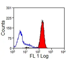
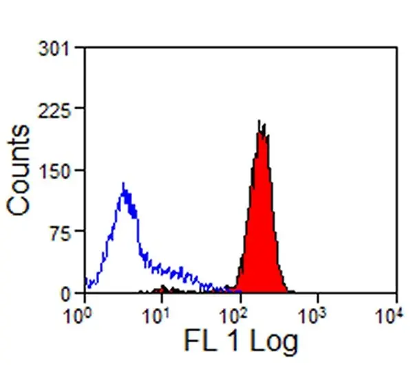
Mouse anti Rabbit CD44 antibody, clone W4/86 (MCA806GA) used for the evaluation of CD44 expression on CD8 expressing cells harveted from different lymphoid and non-lymphoid organs by flow cytometry.
Image caption:
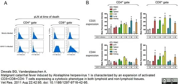
CD25 and CD44 expression in CD8+ T cells isolated from lymphoid and non-lymphoid organs during WD-MCF in rabbits. At time of euthanasia, analysis by multi-colour flow cytometry was conducted on PBMC and mononuclear leukocytes isolated from pLN, spleen, liver, lung and kidney of mock-infected or AlHV-1-infected rabbits developing WD-MCF. (A) Half-offset histograms of CD25 or CD44 expression by CD4+ (left panel) or CD8+ cells (right panel) in mononuclear leukocytes isolated from pLN of one representative rabbit of each group. Data were obtained by triple-staining of CD4 or CD8 with isotype control (grey histograms) or CD25 and CD44 (blue histograms) as described in Methods. (B) Mean fluorescence intensities of CD25 and CD44 in CD4+ (left panel) or CD8+ cells (right panel) obtained by multi-colour flow cytometry analysis. The percentages of CD4+ or CD8+ cells in the kidney of mock-infected animals were below 3% and were therefore not plotted. Data are representative of two independent experiments (n = 3). Bars show means ± SEM. *p < 0.05, **p < 0.01.
From: Dewals BG, Vanderplasschen A.
Malignant catarrhal fever induced by Alcelaphineherpesvirus 1 is characterized by an expansion of activated CD3+CD8+CD4- T cells expressing a cytotoxic phenotype in both lymphoid and non-lymphoid tissues.
Vet Res. 2011 Aug 22;42:95.
doi: 10.1186/1297-9716-42-95
This image is from an open access article distributed under terms of a Creative Commons Attribution License.
Filter by Application:
F Reset| Mouse anti Rabbit CD44 antibody, clone W4/86 recognizes the rabbit CD44 cell surface antigen, a ~95 kDa glycoprotein expressed by all leucocytes. In immunohistochemical staining the antibody labels the medullary area strongly and the cortical area weakly. Mouse anti Rabbit CD44 antibody, clone W4/86 has been reported as being suitable for use in western blotting (Blackford 1996). |
- Target Species
- Rabbit
- Product Form
- Purified IgG - liquid
- Preparation
- Purified IgG prepared by affinity chromatography on Protein A from tissue culture supernatant
- Buffer Solution
- Phosphate buffered saline
- Preservative Stabilisers
0.09% Sodium Azide - Carrier Free
- Yes
- Immunogen
- RL-5 T cell line glycoproteins.
- Approx. Protein Concentrations
- IgG concentration 1.0mg/ml
- Fusion Partners
- Spleen cells from an immunized mouse were fused with cells of the mouse P3.X63 Ag8 myeloma cell line.
- Regulatory
- For research purposes only
- Guarantee
- 12 months from date of despatch
This product is shipped at ambient temperature. It is recommended to aliquot and store at -20°C on receipt. When thawed, aliquot the sample as needed. Keep aliquots at 2-8°C for short term use (up to 4 weeks) and store the remaining aliquots at -20°C.
Avoid repeated freezing and thawing as this may denature the antibody. Storage in frost-free freezers is not recommended.
Avoid repeated freezing and thawing as this may denature the antibody. Storage in frost-free freezers is not recommended.
This product has been reported to work in the following applications. This information is derived from testing within our laboratories, peer-reviewed publications or personal communications from the originators. Please refer to references indicated for further information. For general protocol recommendations, please visit the antibody protocols page.
| Application Name | Verified | Min Dilution | Max Dilution |
|---|---|---|---|
| Flow Cytometry |  |
1/25 | 1/200 |
| Immunohistology - Frozen | |
||
| Immunoprecipitation | |
Where this antibody has not been tested for use in a particular technique this does not necessarily exclude its use in such procedures. Suggested working dilutions are given as a guide only. It is recommended that the user titrates the antibody for use in their own system using appropriate negative/positive controls.
- Flow Cytometry
- Use 10ul of the suggested working dilution to label 106 cells in 100ul.
| Description | Product Code | Applications | Pack Size | List Price | Your Price | Quantity | |
|---|---|---|---|---|---|---|---|
| Rabbit F(ab')2 anti Mouse IgG:RPE | STAR12A | F | 1 ml |

|
Log in | ||
| List Price | Your Price | ||||||
|
|
Log in | ||||||
| Description | Rabbit F(ab')2 anti Mouse IgG:RPE | ||||||
| Rabbit F(ab')2 anti Mouse IgG:HRP (Human Adsorbed) | STAR13B | C E P RE WB | 1 mg |
|
Log in | ||
| List Price | Your Price | ||||||
|
|
Log in | ||||||
| Description | Rabbit F(ab')2 anti Mouse IgG:HRP (Human Adsorbed) | ||||||
| Rabbit F(ab')2 anti Mouse IgG:FITC | STAR9B | F | 1 mg |
|
Log in | ||
| List Price | Your Price | ||||||
|
|
Log in | ||||||
| Description | Rabbit F(ab')2 anti Mouse IgG:FITC | ||||||
| Description | Product Code | Applications | Pack Size | List Price | Your Price | Quantity | |
|---|---|---|---|---|---|---|---|
| Mouse IgG1 Negative Control | MCA928 | F | 100 Tests |
|
Log in | ||
| List Price | Your Price | ||||||
|
|
Log in | ||||||
| Description | Mouse IgG1 Negative Control | ||||||
Source Reference
-
Wilkinson, J.M. et al. (1984) Cell surface glycoproteins of rabbit lymphocytes: characterization with monoclonal antibodies.
Mol Immunol. 21 (1): 95-103.
References for CD44 antibody
-
Jackson, S. et al. (1983) Differentiation antigens identify subpopulations of rabbit T and B lymphocytes. Definition by flow cytometry.
J Exp Med. 157 (1): 34-46. -
Galea-Lauri, J. et al. (1993) Characterization of monoclonal antibodies against rabbit CD44: evidence of a role for CD44 in modulating synoviocyte metabolism.
Mol Immunol. 30 (15): 1383-92. -
Dewals, B.G. and Vanderplasschen, A. (2011) Malignant catarrhal fever induced by Alcelaphine herpesvirus 1 is characterized by an expansion of activated CD3+CD8+CD4- T cells expressing a cytotoxic phenotype in both lymphoid and non-lymphoid tissues.
Vet Res. 42: 95. -
Yagi, M. et al. (2010) Hyaluronan modulates proliferation and migration of rabbit fibroblasts derived from flexor tendon epitenon and endotenon.
J Hand Surg Am. 35: 791-6. -
Zhang, J. et al. (2016) Bone mesenchymal stem cells differentiate into myofibroblasts in the tumor microenvironment.
Oncol Lett. 12 (1): 644-50. -
Kováč, M. et al. (2016) Cryopreservation of Amniotic Fluid Stem Cells Derived From Zobor Rabbits.
Slovak J Anim Sci., 49,(2): 62–67. -
Sugaya, H. et al. (2016) Fate of bone marrow mesenchymal stromal cells following autologous transplantation in a rabbit model of osteonecrosis.
Cytotherapy. 18 (2): 198-204. -
Kováč, M. et al. (2017) Phenotype and ultrastructure of stem cells derived from amniotic fluid of Nitra rabbit
J Cent Euro Agric. 18 (1): 226-34.
View The Latest Product References
-
Honda, H. et al. (2017) Hyaluronic Acid Accelerates Tendon-to-Bone Healing After Rotator Cuff Repair.
Am J Sports Med. 45 (14): 3322-30. -
Kim, H.J. et al. (2019) Intra-articular delivery of synovium-resident mesenchymal stem cells via BMP-7-loaded fibrous PLGA scaffolds for cartilage repair.
J Control Release. 302: 169-80. -
Desando, G. et al. (2018) Short-Term Homing of Hyaluronan-Primed Cells: Therapeutic Implications for Osteoarthritis Treatment.
Tissue Eng Part C Methods. 24 (2): 121-33. -
Kulikova, B. et al. (2019) Survivability of rabbit amniotic fluid-derived mesenchymal stem cells post slow-freezing or vitrification.
Acta Histochem. 121 (4): 491-9. -
Kim, D.H. et al. (2019) Rabbit palatum-derived mesenchymal progenitor cells tri-lineage differentiation on 2D substrates and 3D printed constructs.
J Appl Biomater Funct Mater. 17 (3): 2280800019834520.
- Synonyms
- H-CAM
- PGP-1
MCA806GA
If you cannot find the batch/lot you are looking for please contact our technical support team for assistance.
View more products with CD44 specificity
Please Note: All Products are "FOR RESEARCH PURPOSES ONLY"
View all Anti-Rabbit ProductsAlways be the first to know.
When we launch new products and resources to help you achieve more in the lab.
Yes, sign me up