CD9 antibody | MM2/57


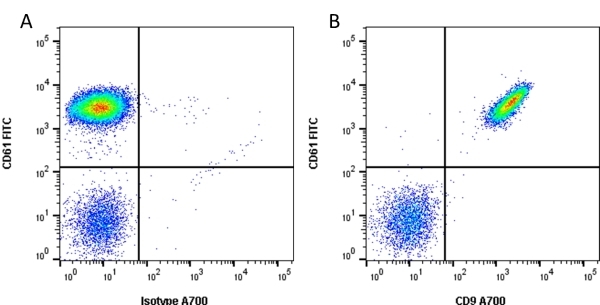
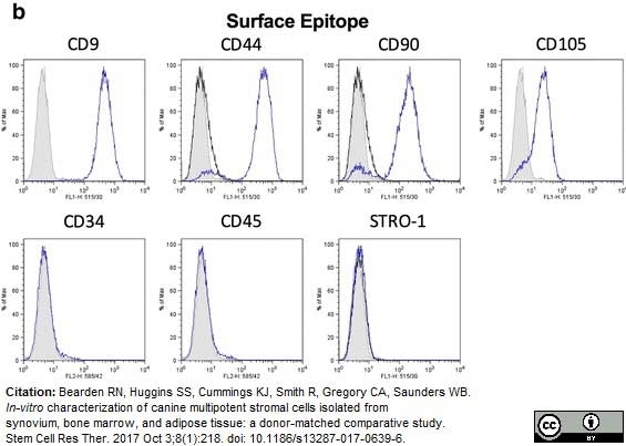
Mouse anti Human CD9 antibody, clone MM2/57 (MCA469GA) used as part of a panel of antibodies to demonstrate the phenotype of canine mesenchymal stem cells by flow cytometry together with mesenchymal markers CD44, CD90 and CD105. Negative results for lineage cell markers CD34, CD45 and STRO-1 are shown.
Image caption:
b. Representative histograms demonstrating positive and negative staining of marrow cMSCs from a single donor
From: Bearden RN, Huggins SS, Cummings KJ, Smith R, Gregory CA, Saunders WB.
In-vitro characterization of canine multipotent stromal cells isolated from synovium, bone marrow, and adipose tissue: a donor-matched comparative study.
Stem Cell Res Ther. 2017 Oct 3;8(1):218.
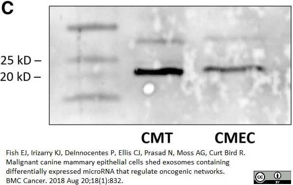
Mouse anti Human CD9 antibody, clone MM2/57 (MCA649G) used for the evaluation of CD9 expression in canine cell line lysates by western blotting.
Image caption:
Insert image caption
From: Fish EJ, Irizarry KJ, DeInnocentes P, Ellis CJ, Prasad N, Moss AG, Curt Bird R.
Malignant canine mammary epithelial cells shed exosomes containing differentially expressed microRNA that regulate oncogenic networks.
BMC Cancer. 2018 Aug 20;18(1):832.
This image is from an open access article distributed under the terms of the Creative Commons Attribution License.
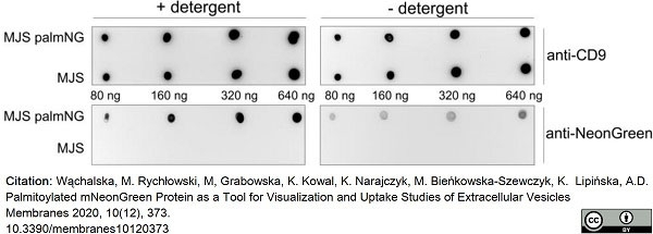
Mouse anti Human CD9 antibody, clone MM2/57 (MCA469G) used for the evaluation of CD9 expression in lysates from human melanoma derived extracellular vesicles by immunoblotting.
Image caption:
Localization of palmNG in EVs. Various amounts of EVs from MJS or MJSpalmNG cells were dot-blotted in a dose range on a nitrocellulose membrane and analyzed by immunoblotting with the use of anti-mNeonGreen or anti-CD9 antibody in the presence or absence of a detergent.
From: Wąchalska, M. et al.
Palmitoylated mNeonGreen Protein as a Tool for Visualization and Uptake Studies of Extracellular Vesicles.
Membranes 2020, 1(12),373.
10.3390/membranes10120373..
This image is from an open access article distributed under terms of a Creative Commons Attribution License.
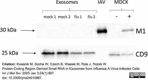
Mouse anti Human CD9 antibody, clone MM2/57 (MCA469G) used to evaluate exosome levels in virally infected MDCK cell lysates by western blotting.
Image caption:
Identification and characterization of exosomes.(c) Immunoblotting of exosomal marker (CD9) and IAV matrix protein (M1). Exosomes secreted by mock–infected (mock 1, 2) and IAV infected (flu 1, 2) cells; IAV–influenza virions; MDCK–mock–infected (−) and IAV infected cells (+).
From: Kwasnik M, Socha W, Czech B, Wasiak M, Rola J, Rozek W.
Protein-Coding Region Derived Small RNA in Exosomes from Influenza A Virus-Infected Cells.
Int J Mol Sci. 2023 Jan 3;24(1):867.
doi: 10.3390/ijms24010867.
This image is from an open access article distributed under terms of a Creative Commons Attribution License.
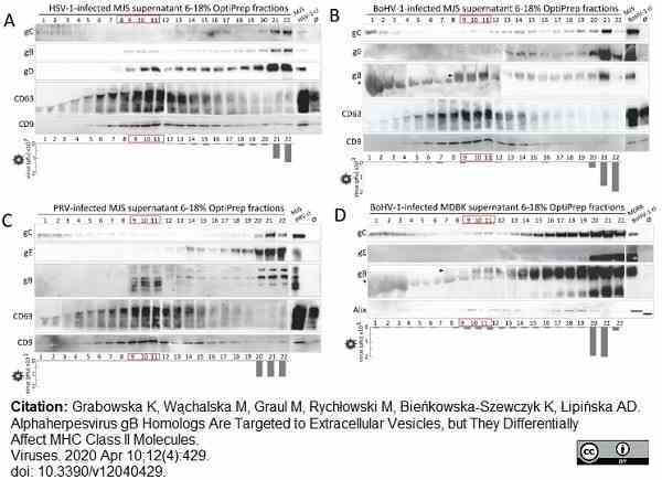
Mouse anti Human CD9 antibody, clone MM2/57 (MCA469G) used to label extracellular vesicles (EVs) from MDBK and MJS cells by western blotting.
Image caption:
Alphaherpesvirus gB localizes to EVs-enriched fractions isolated from virus-infected MJS cells and BoHV-1-infected MDBK. OptiPrep gradient-purification of EVs-enriched and virion-enriched fractions from HSV-1-infected MJS (A), BoHV-1-infected MJS (B), PRV-infected MJS (C), and BoHV-1-infected MDBK (D) supernatants. MJS cells were infected with HSV-1 at a multiplicity of infection (moi) of 0.1, with BoHV-1/PRV at an moi of 0.5; MDBK cells were infected at an moi of 0.1. After 50 h post-infection, 0.5 mL fractions were collected from 6%–18% iodixanol (Optiprep) gradient by ultracentrifugation. The 40 μL samples were analyzed in denaturing (for viral glycoprotein C-gC, glycoprotein B-gB, glycoprotein D-gD, for HSV-1, glycoprotein E-gE, for BoHV-1 and PRV or EVs marker Alix) or non-denaturing (for EVs markers CD63 and CD9) SDS-PAGE, and immunoblotted with specific mAbs or polyclonal anti-PRV gB serum. Uninfected (ø) or virus-infected cell lysates (cl) were analyzed as controls. Bottom panels represent virus titres in each fraction measured by plaque assay.
From: Grabowska K, Wąchalska M, Graul M, Rychłowski M, Bieńkowska-Szewczyk K, Lipińska AD.
Alphaherpesvirus gB Homologs Are Targeted to Extracellular Vesicles, but They Differentially Affect MHC Class II Molecules.
Viruses. 2020 Apr 10;12(4):429.
doi: 10.3390/v12040429.
This image is from an open access article distributed under terms of a Creative Commons Attribution License.
Mouse anti Human CD9:FITC
- Product Type
- Monoclonal Antibody
- Clone
- MM2/57
- Isotype
- IgG2b
- Specificity
- CD9
| Mouse anti Human CD9 antibody, clone MM2/57 recognizes human leukocyte antigen MIC3 also known as MRP-1 or CD9. CD9 is a 228 amino acid multi pass membrane glycoprotein belonging to the tetraspanin family with a molecular weight of ~24 kDa expressed by platelets, monocytes, some lymphocytes and endothelial cells. Mouse anti Human CD9 antibody, clone MM2/57 recognizes a conserved epitope on CD9 present on a wide range of mammalian species. |
- Target Species
- Human
- Species Cross-Reactivity
-
Target Species Cross Reactivity Cat Rhesus Monkey Bovine Dog Rabbit Horse Pig Mustelid Expected from Sequence Mink Llama Ferret - N.B. Antibody reactivity and working conditions may vary between species.
- Product Form
- Purified IgG conjugated to Fluorescein Isothiocyanate Isomer 1 (FITC) - liquid
- Preparation
- Purified IgG prepared by affinity chromatography on Protein A from tissue culture supernatant.
- Buffer Solution
- Phosphate buffered saline
- Preservative Stabilisers
0.09% Sodium Azide 1% Bovine Serum Albumin - Immunogen
- Human platelet membranes
- Approx. Protein Concentrations
- IgG concentration 0.1 mg/ml
- Fusion Partners
- Spleen cells from immunised BALB/c mice were fused with cells from the SP2/0 mouse myeloma line
- Max Ex/Em
-
Fluorophore Excitation Max (nm) Emission Max (nm) FITC 490 525 - Regulatory
- For research purposes only
- Guarantee
- 12 months from date of despatch
This product is shipped at ambient temperature. It is recommended to aliquot and store at -20°C on receipt. When thawed, aliquot the sample as needed. Keep aliquots at 2-8°C for short term use (up to 4 weeks) and store the remaining aliquots at -20°C.
Avoid repeated freezing and thawing as this may denature the antibody. Storage in frost-free freezers is not recommended. This product is photosensitive and should be protected from light.
Avoid repeated freezing and thawing as this may denature the antibody. Storage in frost-free freezers is not recommended. This product is photosensitive and should be protected from light.
This product has been reported to work in the following applications. This information is derived from testing within our laboratories, peer-reviewed publications or personal communications from the originators. Please refer to references indicated for further information. For general protocol recommendations, please visit the antibody protocols page.
| Application Name | Verified | Min Dilution | Max Dilution |
|---|---|---|---|
| Flow Cytometry | Neat |
Where this antibody has not been tested for use in a particular technique this does not necessarily exclude its use in such procedures. Suggested working dilutions are given as a guide only. It is recommended that the user titrates the antibody for use in their own system using appropriate negative/positive controls.
- Flow Cytometry
- Use 10ul of the suggested working dilution to label 106 cells in 100ul.
| Description | Product Code | Applications | Pack Size | List Price | Your Price | Quantity | |
|---|---|---|---|---|---|---|---|
| Mouse IgG2b Negative Control:FITC | MCA691F | F | 100 Tests |

|
Log in | ||
| List Price | Your Price | ||||||
|
|
Log in | ||||||
| Description | Mouse IgG2b Negative Control:FITC | ||||||
| Description | Product Code | Applications | Pack Size | List Price | Your Price | Quantity | |
|---|---|---|---|---|---|---|---|
| Human Seroblock | BUF070A | F | 50 Test | Log in | |||
| List Price | Your Price | ||||||
| Log in | |||||||
| Description | Human Seroblock | ||||||
| Human Seroblock | BUF070B | F | 200 Test | Log in | |||
| List Price | Your Price | ||||||
| Log in | |||||||
| Description | Human Seroblock | ||||||
References for CD9 antibody
-
Ed Knapp W. et al. (1989) Leucocyte Typing IV
Oxford University Press. -
Jennings, L. K. et al. (1995) CD9 cluster workshop report: cell surface binding and functional analysis.
In S.F. Sclossman. et al. Editors. 1995. Leucocyte Typing V. White Cell Differentiation Antigens. Oxford University Press, New York, NY. 1249-1251. -
Löffler, S. et al. (1997) CD9, a tetraspan transmembrane protein, renders cells susceptible to canine distemper virus.
J Virol. 71: 42-9. -
Brodersen, R. et al. (1998) Analysis of the immunological cross reactivities of 213 well characterized monoclonal antibodies with specificities against various leucocyte surface antigens of human and 11 animal species.
Vet Immunol Immunopathol. 64 (1): 1-13. -
Ferrer, M. et al. (1998) Pattern of expression of tetraspanin antigen genes in Burkitt lymphoma cell lines.
Clin Exp Immunol. 113: 346-52. -
Kao, Y.R. et al. (2003) Tumor-associated antigen L6 and the invasion of human lung cancer cells.
Clin Cancer Res. 9: 2807-16. -
Aasted, B. et al. (2007) Reactivity of monoclonal antibodies to human CD antigens with cells from mink.
Vet Immunol Immunopathol. 119: 27-37. -
Davis, W.C. et al. (2007) Use of flow cytometry to identify monoclonal antibodies that recognize conserved epitopes on orthologous leukocyte differentiation antigens in goats, llamas, and rabbits.
Vet Immunol Immunopathol. 119: 123-30.
View The Latest Product References
-
Meister, R.K. et al. (2007) Progress in the discovery and definition of monoclonal antibodies for use in feline research.
Vet Immunol Immunopathol. 119: 38-46. -
Martel, C.J. & Aasted, B. (2009) Characterization of antibodies against ferret immunoglobulins, cytokines and CD markers.
Vet Immunol Immunopathol. 132:109-15. -
Müller, T. et al. (2009) A novel embryonic stem cell line derived from the common marmoset monkey (Callithrix jacchus) exhibiting germ cell-like characteristics.
Hum Reprod. 24: 1359-72. -
Kubota, H. et al. (2011) Glial cell line-derived neurotrophic factor and endothelial cells promote self-renewal of rabbit germ cells with spermatogonial stem cell properties.
FASEB J. 25 (8): 2604-14. -
Hogue, I.B. et al. (2011) Gag induces the coalescence of clustered lipid rafts and tetraspanin-enriched microdomains at HIV-1 assembly sites on the plasma membrane.
J Virol. 85 (19): 9749-66. -
Viswanathan, K. et al. (2017) Quantitative membrane proteomics reveals a role for tetraspanin enriched microdomains during entry of human cytomegalovirus.
PLoS One. 12 (11): e0187899. -
Bearden, R.N. et al. (2017) In-vitro characterization of canine multipotent stromal cells isolated from synovium, bone marrow, and adipose tissue: a donor-matched comparative study.
Stem Cell Res Ther. 8 (1): 218. -
Jackson, C.E. et al. (2017) Effects of Inhibiting VPS4 Support a General Role for ESCRTs in Extracellular Vesicle Biogenesis.
Biophys J. 113 (6): 1342-1352. -
Fish, E.J. et al. (2018) Malignant canine mammary epithelial cells shed exosomes containing differentially expressed microRNA that regulate oncogenic networks.
BMC Cancer. 18 (1): 832. -
Wąchalska, M. et al. (2020) Palmitoylated mNeonGreen Protein as a Tool for Visualization and Uptake Studies of Extracellular Vesicles.
Membranes (Basel). 10 (12): 373. -
Fu, T.S. et al. (2023) Biomimetic vascularized adipose-derived mesenchymal stem cells bone-periosteum graft enhances angiogenesis and osteogenesis in a male rabbit spine fusion model.
Bone Joint Res. 12 (12): 722-33. -
Kwasnik, M. et al. (2023) Protein-Coding Region Derived Small RNA in Exosomes from Influenza A Virus-Infected Cells.
Int J Mol Sci. 24 (1): 867. -
Grabowska, K. et al. (2020) Alphaherpesvirus gB Homologs Are Targeted to Extracellular Vesicles, but They Differentially Affect MHC Class II Molecules.
Viruses. 12 (4): 429.
- Synonyms
- MRP-1
- RRID
- AB_321492
- UniProt
- P21926
- Entrez Gene
- CD9
- GO Terms
- GO:0005515 protein binding
- GO:0002576 platelet degranulation
- GO:0007155 cell adhesion
- GO:0005887 integral to plasma membrane
- GO:0006928 cellular component movement
- GO:0007342 fusion of sperm to egg plasma membrane
- GO:0030168 platelet activation
- GO:0030913 paranodal junction assembly
- GO:0031092 platelet alpha granule membrane
- View More GO Terms
MCA469FT
MCA469F
If you cannot find the batch/lot you are looking for please contact our technical support team for assistance.
View more products with CD9 specificity
Please Note: All Products are "FOR RESEARCH PURPOSES ONLY"
View all Anti-Human ProductsAlways be the first to know.
When we launch new products and resources to help you achieve more in the lab.
Yes, sign me up