MHC Class II DR antibody | CC108
Mouse anti Bovine MHC Class II DR:RPE
- Product Type
- Monoclonal Antibody
- Clone
- CC108
- Isotype
- IgG1
- Specificity
- MHC Class II DR


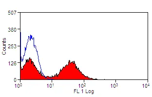
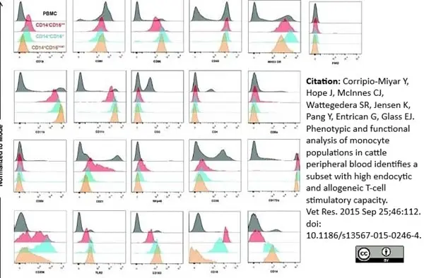
Mouse anti Bovine MHC Class II DR antibody, clone CC108 (MCA5656) used for the evaluation of MHC class II expression on bovine myeloid cell sub-populations by flow cytometry.
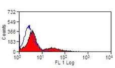
Image caption:
Phenotypic profiles of bovine myeloid cell sub-populations. Live gated PBMC were assessed for expression of CD16 and CD14 and a panel of molecules associated with antigen presentation, co-stimulation or specific cell lineages (see Table 1) by 3 colour flow cytometry. PBMC were stained with primary mAb to the specific molecules indicated and then with an isotype-specific PE conjugated secondary, followed by CD16 and CD14 conjugated to FITC or Alexa Fluor 647 respectively. Live, single PBMC were gated based on the expression of CD16 and CD14 as detailed in Figures 1 and and 2C.2C. Histograms show the levels of expression of selected markers in the cell populations studied, CD14−CD16++ (red), CD14+CD16+ (blue) and CD14+CD16low/- (orange) compared to PBMC (black). Data shown are for one representative animal of four animals.
From: Corripio-Miyar Y, Hope J, McInnes CJ, Wattegedera SR, Jensen K, Pang Y,Entrican G, Glass EJ.
Phenotypic and functional analysis of monocyte populations in cattle peripheral blood identifies a subset with high endocytic and allogeneicT-cell stimulatory capacity.
Vet Res. 2015 Sep 25;46:112.
This image is from an open access article distributed under terms of a Creative Commons Attribution License.
Mouse anti Bovine MHC class II DR antibody, clone CC108 (MCA5656) used for the evaluation of MHC class II expression on peripheral blood cells by flow cytometry.
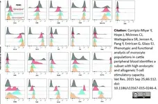
Image caption:
Monocyte/macrophages and MoDCs express IL-10 and TGF-β, and induce proliferation of IL-10+ γδ T cells. Peripheral blood monocyte/macrophages expressing CD14 (A) and expression of MHC class II/CD1b (B) in CD14+ cells. After a 3-d culture in the presence of GM-CSF and IL-4, the cells increased expression of MHC class II and CD1b (C). IL-10 and TGF-β expression in ex vivo CD14+ cells (D) and cultured MoDCs (E). Both monocyte/macrophages and MoDCs induced the expansion of autologous IL-10–expressing γδ T cells (F). Bar graph shows means (n = 10) and error bars indicate SEMs. Representative plots of cells obtained from 10 different animals analyzed in duplicate. Quadrants were placed based on isotype and fluorochrome controls.
From: Guzman E, Hope J, Taylor G, Smith AL, Cubillos-Zapata C, Charleston B.
Bovine γδ T cells are a major regulatory T cell subset.
J Immunol. 2014 Jul 1;193(1):208-22.
This image is from an open access article distributed under terms of a Creative Commons Attribution License.
Mouse anti Bovine MHC class II DR antibody, clone CC108 (MCA5656) used for the evaluation of MHC class II expression on peripheral blood cells by flow cytometry.
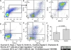
Image caption:
CD8α− SIRPα− ALDCs express cytokines that induce γδ T with a regulatory phenotype. Phenotypic analysis of bovine ALDC subsets includes FSChigh MHC class II (A), SIRPα and CD11c (B), and SIRPα and CD8α (C). Cells were fixed, permeabilized, and stained for intracellular TGF-β and IL-10. DCs were gated on SIRPα+ CD8α− (D), SIRPα+ CD8α+ (E), and SIRPα− CD8α− double negative (F). Dot plots are representative of cells from five different animals. (G–I) SIRPα− CD8α− ALDCs are capable of inducing γδ T cells with suppressive phenotype. (G) Subpopulations of ALDCs were FACS sorted and cocultured with autologous MACS-sorted γδ TCR+ T cells for 5 d. Proliferation was measured by CFSE dilution and intracellular staining of IL-10 by flow cytometry. (H) FMDV-specific proliferation of CD4+ T cells in the absence or presence of in vitro–expanded autologous γδ T cells by SIRPα− CD8α− ALDCs and in Transwell plates with or without blocking anti–IL-10. (I) FMDV-specific IFN-γ responses in CD4+ T cells in the presence or absence of in vitro–expanded autologous γδ T cells SIRPα− CD8α− ALDCs and in Transwell plates with or without blocking anti–IL-10. Bars indicate means of cells from four different animals analyzed in triplicate, and error bars indicate SEMs.
From: Guzman E, Hope J, Taylor G, Smith AL, Cubillos-Zapata C, Charleston B.
Bovine γδ T cells are a major regulatory T cell subset.
J Immunol. 2014 Jul 1;193(1):208-22.
This image is from an open access article distributed under terms of a Creative Commons Attribution License.

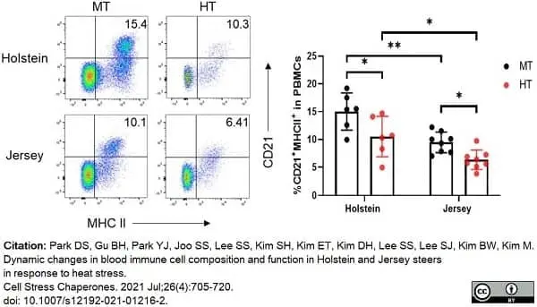
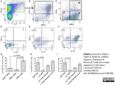
FITC conjugated Mouse anti Bovine MHC class II DR antibody, clone CC108 (MCA5656F) used to discriminate lymphocyte populations in bovine blood samples by flow cytometry.
Image caption:
Changes in T and B lymphocytes among PBMCs in Holstein and Jersey steers subjected to heat stress. Flow cytometry analysis to identify lymphocytes subset population. Heat stress caused reduction in lymphocyte populations in the PBMCs of both Holstein and Jersey steers. The lymphocytes comprised CD21+MHCII+ B cells. MT indicates moderate THI condition and HT indicates high THI condition. * = P <0.05, ** = P <0.01
From: Park DS, Gu BH, Park YJ, Joo SS, Lee SS, Kim SH, Kim ET, Kim DH, Lee SS, Lee SJ, Kim BW, Kim M.
Dynamic changes in blood immune cell composition and function in Holstein and Jersey steers in response to heat stress.
Cell Stress Chaperones. 2021 Jul;26(4):705-20.
doi: 10.1007/s12192-021-01216-2.
This image is from an open access article distributed under terms of a Creative Commons Attribution License.
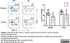
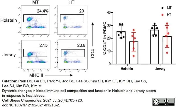
FITC conjugated Mouse anti Bovine MHC class II DR antibody, clone CC108 (MCA5656F) used to identify T cell populations in bovine blood samples by flow cytometry.
Image caption:
Changes in T and B lymphocytes among PBMCs in Holstein and Jersey steers subjected to heat stress. Flow cytometry analysis to identify lymphocytes subset population. Heat stress caused reduction in lymphocyte populations in the PBMCs of both Holstein and Jersey steers. The lymphocytes comprised CD4+ T cells. MT indicates moderate THI condition and HT indicates high THI condition. * = P <0.05, ** = P <0.01
From: Park DS, Gu BH, Park YJ, Joo SS, Lee SS, Kim SH, Kim ET, Kim DH, Lee SS, Lee SJ, Kim BW, Kim M.
Dynamic changes in blood immune cell composition and function in Holstein and Jersey steers in response to heat stress.
Cell Stress Chaperones. 2021 Jul;26(4):705-20.
doi: 10.1007/s12192-021-01216-2.
This image is from an open access article distributed under terms of a Creative Commons Attribution License.
Filter by Application:
F Reset| Mouse anti Bovine MHC class II DR antibody, clone CC108 recognizes Bovine MHC Class II DR. MHC Class II molecules are constitutively expressed on antigen presenting cells such as dendritic cells, B lymphocytes, monocytes, macrophages, activated T lymphocytes and may be induced on a range of other cell types by interferon gamma. The major histocompatibility complex (MHC) is a cluster of genes some of which are important in the immune response to infections. In cattle, this complex is referred to as the bovine leukocyte antigen (BoLA) region. There are 2 major types of MHC class IIa molecules encoded by the BoLA which are DR and DQ each composed of an alpha and beta chain. |
- Target Species
- Bovine
- Product Form
- Purified IgG conjugated to R. Phycoerythrin (RPE) - lyophilized
- Reconstitution
- Reconstitute with 1.0ml distilled water
- Preparation
- Purified IgG prepared by affinity chromatography on Protein G from tissue culture supernatant
- Buffer Solution
- Phosphate buffered saline
- Preservative Stabilisers
- 0.09% Sodium Azide (NaN3)
1% Bovine Serum Albumin
5% Sucrose - Fusion Partners
- Spleen cells from immunized BALB/c mice were fused with cells of the Mouse NS1 myeloma cell line.
- Max Ex/Em
-
Fluorophore Excitation Max (nm) Emission Max (nm) RPE 488nm laser 496 578 - Regulatory
- For research purposes only
- Guarantee
- 12 months from date of despatch
This product is shipped at ambient temperature.
Prior to reconstitution store at +4oC.
After reconstitution store at +4oC.
DO NOT FREEZE. This product should be stored undiluted. This product is photosensitive and should be protected from light. Should this product contain a precipitate we recommend microcentrifugation before use.
Prior to reconstitution store at +4oC.
After reconstitution store at +4oC.
DO NOT FREEZE. This product should be stored undiluted. This product is photosensitive and should be protected from light. Should this product contain a precipitate we recommend microcentrifugation before use.
This product has been reported to work in the following applications. This information is derived from testing within our laboratories, peer-reviewed publications or personal communications from the originators. Please refer to references indicated for further information. For general protocol recommendations, please visit the antibody protocols page.
| Application Name | Verified | Min Dilution | Max Dilution |
|---|---|---|---|
| Flow Cytometry |  |
Neat |
Where this product has not been tested for use in a particular technique this does not necessarily exclude its use in such procedures. Suggested working dilutions are given as a guide only. It is recommended that the user titrates the product for use in their own system using appropriate negative/positive controls.
- Flow Cytometry
- Use 10ul of the suggested working dilution to label 1x10⁶ cells in 100ul.
How to Use the Spectraviewer
Watch the Tool Tutorial Video ▸- Start by selecting the application you are interested in, with the option to select an instrument from the drop down menu or create a customized instrument
- Select the fluorophores or fluorescent proteins you want to include in your panel to check compatibility
- Select the lasers and filters you wish to include
- Select combined or multi-laser view to visualize the spectra
| Description | Product Code | Applications | Pack Size | List Price | Your Price | Quantity | |
|---|---|---|---|---|---|---|---|
| Mouse IgG1 Negative Control:RPE | MCA928PE | F | 100 Tests | Log in | |||
| List Price | Your Price | ||||||
| Log in | |||||||
| Description | Mouse IgG1 Negative Control:RPE | ||||||
References for MHC Class II DR antibody
-
Stephens, S.A. & Howard, C.J. (2002) Infection and transformation of dendritic cells from bovine afferent lymph by Theileria annulata.
Parasitology. 124 (Pt 5): 485-93. -
Yamakawa, Y. et al. (2008) Identification and functional characterization of a bovine orthologue to DC-SIGN.
J Leukoc Biol. 83 (6): 1396-403. -
Corripio-Miyar, Y. et al. (2015) Phenotypic and functional analysis of monocyte populations in cattle peripheral blood identifies a subset with high endocytic and allogeneic T-cell stimulatory capacity.
Vet Res. 46: 112. -
Guzman, E. et al. (2014) Bovine γδ T cells are a major regulatory T cell subset.
J Immunol. 193 (1): 208-22. -
Childerstone, A.J. et al. (1999) Demonstration of bovine CD8+ T-cell responses to foot-and-mouth disease virus.
J Gen Virol. 80 ( Pt 3): 663-9. -
Sopp, P. et al. (1994) Detection of bovine viral diarrhoea virus p80 protein in subpopulations of bovine leukocytes.
J Gen Virol. 75 ( Pt 5): 1189-94. -
Bembridge, G.P. et al. (1995) CD45RO expression on bovine T cells: relation to biological function.
Immunology. 86 (4): 537-44. -
Gibson, A.J. et al. (2016) Differential macrophage function in Brown Swiss and Holstein Friesian cattle.
Vet Immunol Immunopathol. 181: 15-23.
View The Latest Product References
-
Corripio-Miyar, Y. et al. (2017) 1,25-Dihydroxyvitamin D3 modulates the phenotype and function of Monocyte derived dendritic cells in cattle.
BMC Vet Res. 13 (1): 390. -
Risalde, M.A. et al. (2020) BVDV permissiveness and lack of expression of co-stimulatory molecules on PBMCs from calves pre-infected with BVDV.
Comp Immunol Microbiol Infect Dis. 68: 101388. -
Park, D.S. et al. (2021) Dynamic changes in blood immune cell composition and function in Holstein and Jersey steers in response to heat stress.
Cell Stress Chaperones. 26 (4): 705-20.
- RRID
- AB_11152779
- UniProt
- Q30309
- P79464
MCA5656PE
If you cannot find the batch/lot you are looking for please contact our technical support team for assistance.
View more products with MHC CLASS II specificity
Please Note: All Products are "FOR RESEARCH PURPOSES ONLY"
View all Anti-Bovine ProductsAlways be the first to know.
When we launch new products and resources to help you achieve more in the lab.
Yes, sign me up