AIF1 antibody
Goat anti AIF1 (C-Terminal)
- Product Type
- Polyclonal Antibody
- Isotype
- Polyclonal IgG
- Specificity
- AIF1
- Region
- (C-TERMINAL)

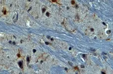

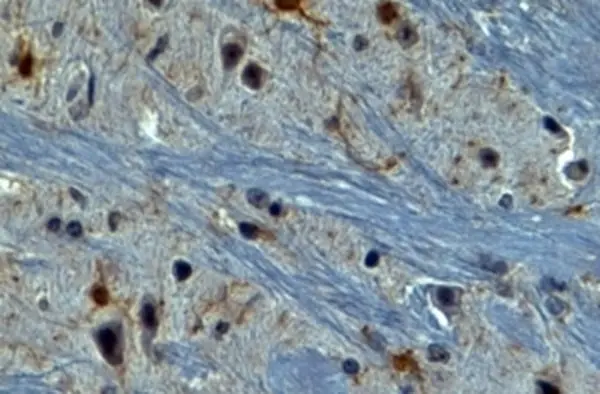
Goat anti AIF1 antibody (AHP2024) used to label microglial cells in rat hippocampus by immunofluorescence.
Image caption:
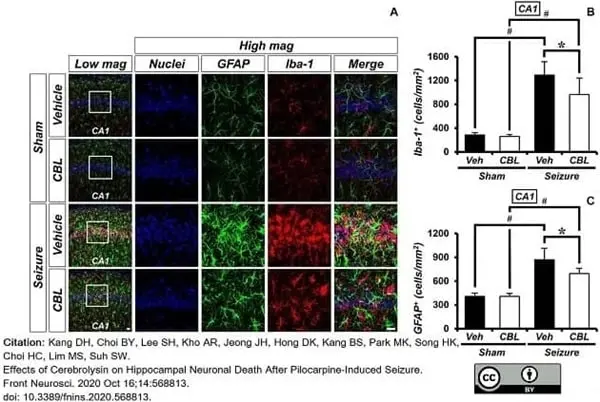
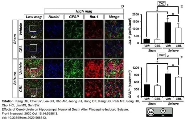
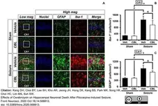
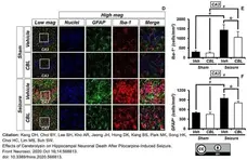
Cerebrolysin decreases the density of glial cells after pilocarpine-induced seizure. The administration of cerebrolysin decreased the density of glial cells after pilocarpine-induced seizure. (A,D) Iba-1 (red), GFAP (green), and DAPI (blue) in the hippocampal CA1 (A) and CA3 (D) regions. The administration of cerebrolysin after a seizure decreased the density of glial cells in the hippocampal CA1 and CA3 regions compared to the seizure-vehicle groups. Scale bar = 20 μm. (B,C,E,F) A graph of the density of glial cells according to the standard. The data are the mean ± SEM, n = 5, from each sham group; n = 5–7 for each seizure group. *p <0.05 vs. vehicle-treated group; #p <0.05 vs. sham-operated group [Kruskal–Wallis test with post hoc test: (B) Chi square = 17.244, df = 3, p = 0.001, (C) Chi square = 17.153, df = 3, p <0.001, (E) Chi square = 17.456, df = 3, p = 0.001, (F) Chi square = 18.292, df = 3, p < 0.001].
From: Kang DH, Choi BY, Lee SH, Kho AR, Jeong JH, Hong DK, Kang BS, Park MK, Song HK, Choi HC, Lim MS, Suh SW.
Effects of Cerebrolysin on Hippocampal Neuronal Death After Pilocarpine-Induced Seizure.
Front Neurosci. 2020 Oct 16;14:568813.
doi: 10.3389/fnins.2020.568813.
This image is from an open access article distributed under terms of a Creative Commons Attribution License.
Goat anti AIF1 antibody (AHP2024) used to label microglial cells in rat hippocampus by immunofluorescence.
Image caption:
Cerebrolysin decreases the density of glial cells after pilocarpine-induced seizure. The administration of cerebrolysin decreased the density of glial cells after pilocarpine-induced seizure. (A,D) Iba-1 (red), GFAP (green), and DAPI (blue) in the hippocampal CA1 (A) and CA3 (D) regions. The administration of cerebrolysin after a seizure decreased the density of glial cells in the hippocampal CA1 and CA3 regions compared to the seizure-vehicle groups. Scale bar = 20 μm. (B,C,E,F) A graph of the density of glial cells according to the standard. The data are the mean ± SEM, n = 5, from each sham group; n = 5–7 for each seizure group. *p <0.05 vs. vehicle-treated group; #p <0.05 vs. sham-operated group [Kruskal–Wallis test with post hoc test: (B) Chi square = 17.244, df = 3, p = 0.001, (C) Chi square = 17.153, df = 3, p <0.001, (E) Chi square = 17.456, df = 3, p = 0.001, (F) Chi square = 18.292, df = 3, p < 0.001].
From: Kang DH, Choi BY, Lee SH, Kho AR, Jeong JH, Hong DK, Kang BS, Park MK, Song HK, Choi HC, Lim MS, Suh SW.
Effects of Cerebrolysin on Hippocampal Neuronal Death After Pilocarpine-Induced Seizure.
Front Neurosci. 2020 Oct 16;14:568813.
doi: 10.3389/fnins.2020.568813.
This image is from an open access article distributed under terms of a Creative Commons Attribution License.
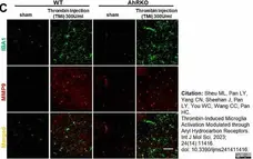
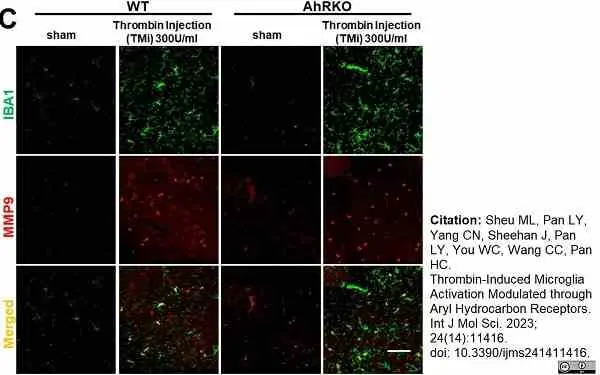
Goat anti AIF1 antibody (AHP2024) used to label murine microglia by immunofluorescence
Image caption:
Aryl hydrocarbon receptor deficiency (AhRKO) after thrombin injection in vivo increased MMP9 activity, but not MMP2. (C) Representative images of brain sections at 72 h after thrombin injection, showing double staining with both microglia cell marker Iba1 (green) and MMP 9 (red). Scale bars: 50 μm.
From: Sheu ML, Pan LY, Yang CN, Sheehan J, Pan LY, You WC, Wang CC, Pan HC.
Thrombin-Induced Microglia Activation Modulated through Aryl Hydrocarbon Receptors.
Int J Mol Sci. 2023 Jul 13;24(14):11416.
doi: 10.3390/ijms241411416.
This image is from an open access article distributed under terms of a Creative Commons Attribution License.
Filter by Application:
P WB IF Reset| Goat anti AIF1 antibody recognizes human allograft inflammatory factor 1 (AIF1), otherwise known as Ionized calcium-binding adapter molecule 1 (IBA1). AIF1 is a 147 amino acid ~17kDa actin-binding protein involved in macrophage activation, vascular smooth muscle cell proliferation and activation and T-lymphocyte proliferation (UniProt P55008). Goat anti AIF1 antibody recognizes epitopes at the C-terminal region of AIF1 and is expected to recognize AIF1 isoform 1 and isoform 3 formed by alternative splicing. AIF1 was first identified in rat cardiac allografts with chronic rejection (Utans et al. 1995), is expressed by activated monocytes and macrophages (Orsmark et al. 2007) and plays a significant role in vascular inflammation (Chen et al. 2004) Goat anti AIF1 has been used successfully for the identification of AIF1 expressing cells in an inflammatory model in formalin fixed mouse brain following heat mediated antigen retrieval with sodium citrate buffer by immunohistochemistry (Wang et al. 2014). |
- Target Species
- Human
- Species Cross-Reactivity
-
Target Species Cross Reactivity Mouse Pig Expected from Sequence Rat - N.B. Antibody reactivity and working conditions may vary between species.
- Product Form
- Purified IgG - liquid
- Antiserum Preparation
- Antiserum to human AIF1 (CT) was raised by repeated immunisation of goats with highly purified antigen. Purified IgG was prepared by affinity chromatography.
- Buffer Solution
- TRIS buffered saline
- Preservative Stabilisers
- 0.02% Sodium Azide (NaN3)
0.5% Bovine Serum Albumin - Immunogen
- Synthetic peptide sequence C-TGPPAKKAISELP from the C-terminal region of AIF1 (NP_116573.1; NP_001614.3).
- Approx. Protein Concentrations
- IgG concentration 0.5mg/ml
- Regulatory
- For research purposes only
- Guarantee
- 12 months from date of despatch
This product is shipped at ambient temperature. It is recommended to aliquot and store at -20°C on receipt. When thawed, aliquot the sample as needed. Keep aliquots at 2-8°C for short term use (up to 4 weeks) and store the remaining aliquots at -20°C.
Avoid repeated freezing and thawing as this may denature the antibody. Storage in frost-free freezers is not recommended.
Avoid repeated freezing and thawing as this may denature the antibody. Storage in frost-free freezers is not recommended.
This product has been reported to work in the following applications. This information is derived from testing within our laboratories, peer-reviewed publications or personal communications from the originators. Please refer to references indicated for further information. For general protocol recommendations, please visit the antibody protocols page.
| Application Name | Verified | Min Dilution | Max Dilution |
|---|---|---|---|
| ELISA |  |
1/32000 | |
| Immunohistology - Paraffin 1 | |
2.0ug/ml | |
| Western Blotting | |
1.0 | 3.0ug/ml |
- 1This antibody requires heat-mediated antigen retrieval prior to staining paraffin sections. Tris/EDTA buffer pH9.0 is recommended for this purpose.
Where this product has not been tested for use in a particular technique this does not necessarily exclude its use in such procedures. Suggested working dilutions are given as a guide only. It is recommended that the user titrates the product for use in their own system using appropriate negative/positive controls.
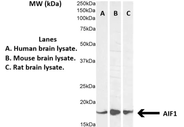
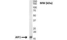
- Western Blotting
- AHP2024 detects a band of approximately 16kDa in rat brain cell lysates.
| Description | Product Code | Applications | Pack Size | List Price | Your Price | Quantity | |
|---|---|---|---|---|---|---|---|
| Rabbit anti Goat IgG (Fc):FITC | STAR122F | F | 1 mg |

|
Log in | ||
| List Price | Your Price | ||||||
|
|
Log in | ||||||
| Description | Rabbit anti Goat IgG (Fc):FITC | ||||||
| Rabbit anti Goat IgG (Fc):HRP | STAR122P | C E WB | 1 mg |
|
Log in | ||
| List Price | Your Price | ||||||
|
|
Log in | ||||||
| Description | Rabbit anti Goat IgG (Fc):HRP | ||||||
| Description | Product Code | Applications | Pack Size | List Price | Your Price | Quantity | |
|---|---|---|---|---|---|---|---|
| Antigen Retrieval Buffer, pH8.0 | BUF025A | P | 500 ml | Log in | |||
| List Price | Your Price | ||||||
| Log in | |||||||
| Description | Antigen Retrieval Buffer, pH8.0 | ||||||
References for AIF1 antibody
-
Kang, D.H. et al. (2020) Effects of Cerebrolysin on Hippocampal Neuronal Death After Pilocarpine-Induced Seizure.
Front Neurosci. 14: 568813. -
Wu, C. et al. (2022) E3 ubiquitin ligase Triad1 promotes neuronal apoptosis by regulating the p53-caspase3 pathway after spinal cord injury.
Somatosens Mot Res. 39 (1): 21-8. -
Zhou, C. et al. (2022) Mild Hypothermia Protects Brain Injury After Intracerebral Hemorrhage in Mice Via Enhancing the Nrdp1/MyD88 Signaling Pathway.
Neurotox Res. 40 (6): 1664-72. -
Sheu, M.L. et al. (2023) Thrombin-Induced Microglia Activation Modulated through Aryl Hydrocarbon Receptors.
Int J Mol Sci.24 (14):11416. -
Lu, X. et al. (2018) HAX1 is associated with neuronal apoptosis and astrocyte proliferation after spinal cord injury.
Tissue Cell. 54: 1-9. -
Yu, M. et al. (2023) PU.1 interaction with p50 promotes microglial-mediated inflammation in secondary spinal cord injury in SCI rats.
Int J Neurosci. 133 (4): 389-402. -
Lassen, L.B. et al. (2022) Mutation of Tyrosine Sites in the Human Alpha-Synuclein Gene Induces Neurotoxicity in Transgenic Mice with Soluble Alpha-Synuclein Oligomer Formation.
Cells. 11 (22): 3673. -
Kardash, E. et al. (2025) Pro-cognitive efficacy of Prospekta in a rat model of age-associated cognitive impairment.
BMC Neurosci. 26 (1): 35.
- Synonyms
- IBA1
- RRID
- AB_2224406
- UniProt
- P55008
- P55009
- Entrez Gene
- AIF1
- Aif1
- GO Terms
- GO:0005509 calcium ion binding
- GO:0051015 actin filament binding
- GO:0051017 actin filament bundle assembly
- GO:0005634 nucleus
- GO:0005737 cytoplasm
- GO:0005856 cytoskeleton
- GO:0006954 inflammatory response
- GO:0007050 cell cycle arrest
- GO:0008285 negative regulation of cell proliferation
- View More GO Terms
- GO:0032587 ruffle membrane
AHP2024
If you cannot find the batch/lot you are looking for please contact our technical support team for assistance.
Request a different product with this specificity
Please Note: All Products are "FOR RESEARCH PURPOSES ONLY"
View all Anti-Human ProductsAlways be the first to know.
When we launch new products and resources to help you achieve more in the lab.
Yes, sign me up