RPE Conjugation Kit
LYNX Rapid RPE Antibody Conjugation Kit
- Product Type
- Conjugation Kit
- Specificity
- RPE Conjugation Kit
| Product Code | Applications | Pack Size | List Price | Your Price | Qty | ||||||||||
|---|---|---|---|---|---|---|---|---|---|---|---|---|---|---|---|
|
|||||||||||||||
|
|||||||||||||||
|
|||||||||||||||
|
|||||||||||||||


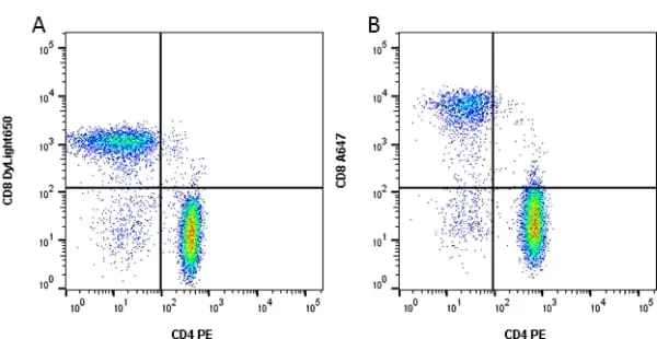
A. Purified CD4 (MCA1267) and CD8 (MCA1226) were labeled with LYNX Rapid RPE Antibody Conjugation Kit (LNK021RPE) and LYNX Rapid Plus DyLight 650 Antibody Conjugation Kit (LNK241D650) and used to stain human peripheral blood. B. Alternatively directly labeled antibodies (MCA1267PE and MCA1226A647) were used. The staining shown are lymphocytes gated on the CD3 positive population. CD4 and CD8 positive T cells can be identified in both plots.
Data acquired on the ZE5 Cell Analyzer.
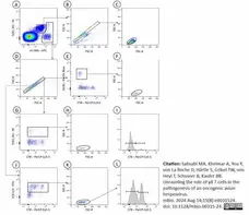
LYNX Rapid RPE Antibody Conjugation Kit (LNK02) used to conjugate fluorochrome to Mouse anti Chicken TCRαβantibody prior to analysis of chicken blood cells.
Image caption:
Flow cytometry gating
EDTA-blood was stained with mix of anti-TCRαβ/Vβ1-FITC), anti-TCRαβ/Vβ2-FITC (clone TCR3), anti-TCRγδ-PE (clone TCR1), anti-Bu1-Pacific Blue (clone AV20), anti-CD8-PerCP-Cy5.5 (clone CT8, anti-CD45-APC (clone UM16-6,) and thrombocyte marker in a no-lyse no-wash onetube procedure and subsequently analyzed by flow cytometry.
We first separated thrombocytes and leukocytes (A) followed by a single cell gate (B and D). Leukocytes were subdivided in B cells (E), yd-T cells (G) and aß-T cells (J). T cell subpopulations were further separated in CD8pos and CD8neg cells (I and L). CD8neg aß-T cells were addressed as CD4pos T cells. To exclude potentially contaminating erythrocytes, for all cell populations an additional FSC/SSC gating was performed (C, F, H and K).
From: Sabsabi MA, Kheimar A, You Y, von La Roche D, Härtle S, Göbel TW, von Heyl T, Schusser B, Kaufer BB.
Unraveling the role of γδ T cells in the pathogenesis of an oncogenic avian herpesvirus.
mBio. 2024 Aug 14;15(8):e0031524.
doi: 10.1128/mbio.00315-24.
This image is from an open access article distributed under terms of a Creative Commons Attribution License.
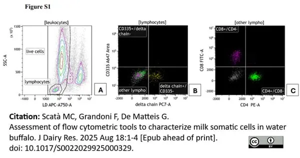
Mouse anti Bovine CD4 antibody, clone IL-A11A conjugated to phycoerythrin using the Lynx Rapid PE conjugation kit (LNK024RPE) prior to use in flow cytometric analysis of buffalo lymphocyte populations.
Image caption:
Gating strategy for the identification of T lymphocytes. A morphological gate was drawn to highlight lymphocytes (A); the lymphocytes were gated in γδ TCR vs CD335 plot, to identify γδ and NK cells (B); the double negative population (other lymphocytes) were plotted on CD4 vs CD8 to identify T helper (CD4+) and T cytotoxic (CD8+) lymphocytes (C).
From: Scatà MC, Grandoni F, De Matteis G.
Assessment of flow cytometric tools to characterize milk somatic cells in water buffalo.
J Dairy Res. 2025 92 (1): 69-72.
doi: 10.1017/S0022029925000329. Epub ahead of print. PMID: 40820690.
This image is from an open access article distributed under terms of a Creative Commons Attribution License.
Filter by Application:
CJ Reset| LYNX Rapid RPE Anitbody Conjugation Kit® enables the rapid conjugation of a pre-prepared lyophilized mixture containing R-Phycoerythrin (RPE) label to an antibody or protein. Activation of proprietary reagents within the antibody-label solution results in directional covalent bonding of RPE to the antibody. The LYNX Rapid Conjugation kit® can be used to label small quantities of antibody/protein at near neutral pH, allowing a high conjugation efficiency with 100% antibody recovery. |
- Reagents In The Kit
-
LNK024RPE: 3 Vials of 10ug LYNX lyophilized RPE mix
1 Vial LYNX Modifier reagent
1 Vial LYNX Quencher reagent
-
LNK023RPE: 1 Vial of 1mg LYNX lyophilized RPE mix
1 Vial LYNX Modifier reagent
1 Vial LYNX Quencher reagent
-
LNK022RPE: 3 Vials of 100ug LYNX lyophilized RPE mix
1 Vial LYNX Modifier reagent
1 Vial LYNX Quencher reagent
-
LNK021RPE: 1 Vial of 100ug LYNX lyophilized RPE mix
1 Vial LYNX Modifier reagent
1 Vial LYNX Quencher reagent
- Preparing The Antibody
-
LNK024RPE
The following buffer solutions are recommended for preparing the antibody:
10-50mM amine-free buffer (e.g HEPES, MES, MOPS and phosphate) pH range 6.5-8.5, although moderate concentrations of Tris buffer (<20mM) may be tolerated.
If possible, avoid buffers containing nucleophilic components such as primary amines and thiols (e.g. thiomersal/thimerosal) since they may react with LYNX chemicals. Azide (0.02-0.1%), EDTA, up to 50% Glycerol and common non-buffering salts and sugars have little or no effect on conjugation efficiency.
Due to the large size of RPE (240kDa), the quantity of RPE is in slight molar excess. Approximately 10ug of IgG will give a 1:1 molar ratio of antibody:RPE. For optimal results the antibody should be at a concentration of 1mg/ml, with a maximum volume of 10ul and a maximum antibody amount of 10ug. Antibody at a concentration of greater than 1mg/ml requires dilution. Antibody below 1mg/ml can still be used as long as the maximum volume is not exceeded. Using less than the recommended amount of antibody may result in unbound label, but this will be removed during subsequent application wash steps. Antibody below 0.5mg/ml should be concentrated before use with the kit. -
LNK023RPE
The following buffer solutions are recommended for preparing the antibody:
10-50mM amine-free buffer (e.g HEPES, MES, MOPS and phosphate) pH range 6.5-8.5, although moderate concentrations of Tris buffer (<20mM) may be tolerated.
If possible, avoid buffers containing nucleophilic components such as primary amines and thiols (e.g. thiomersal/thimerosal) since they may react with LYNX chemicals. Azide (0.02-0.1%), EDTA, up to 50% Glycerol and common non-buffering salts and sugars have little or no effect on conjugation efficiency.
Due to the large size of RPE (240kDa), it is recommended that 50-60ug of antibody be used for every 100ug RPE, to ensure a slight RPE molar excess. For optimal results the antibody should be at a concentration of 1mg/ml, with a maximum volume of 600ul and a maximum antibody amount of 600ug. Antibody at a concentration of greater than 1mg/ml requires dilution. Antibody below 1mg/ml can still be used as long as the maximum volume is not exceeded. Using less than the recommended amount of antibody may result in unbound label, but this will be removed during subsequent application wash steps. Antibody below 0.5mg/ml should be concentrated before use with the kit. -
LNK022RPE, LNK021RPE
The following buffer solutions are recommended for preparing the antibody:
10-50mM amine-free buffer (e.g HEPES, MES, MOPS and phosphate) pH range 6.5-8.5, although moderate concentrations of Tris buffer (<20mM) may be tolerated.
If possible, avoid buffers containing nucleophilic components such as primary amines and thiols (e.g. thiomersal/thimerosal) since they may react with LYNX chemicals. Azide (0.02-0.1%), EDTA, up to 50% Glycerol and common non-buffering salts and sugars have little or no effect on conjugation efficiency.
Due to the large size of RPE (240kDa), it is recommended that 50-60ug of antibody be used for every 100ug RPE, to ensure a slight RPE molar excess. For optimal results the antibody should be at a concentration of 1mg/ml, with a maximum volume of 60ul and a maximum antibody amount of 60ug. Antibody at a concentration of greater than 1mg/ml requires dilution. Antibody below 1mg/ml can still be used as long as the maximum volume is not exceeded. Using less than the recommended amount of antibody may result in unbound label, but this will be removed during subsequent application wash steps. Antibody below 0.5mg/ml should be concentrated before use with the kit. - Regulatory
- For research purposes only
- Guarantee
- 12 months from date of despatch
- Licensed Use
- These products and the methodology of conjugation are patent protected under United Kingdom patent number 2446088 and associated international patent applications. The purchase of this product conveys to the buyer the limited, non exclusive non-transferable right (without the right to resell repackage or further sublicense) under these patents to use the product to make conjugates for research and development purposes only. The purchaser cannot sell or otherwise transfer this product, or its components, or materials or data made using this product, or its components to a third party. Further information on purchasing licenses for diagnostic and other uses may be obtained by contacting Bio-Rad, at. Endeavour House, Langford Business Park, Langford Lane, Kidlington, Oxon. OX5 1GE UNITED KINGDOM. Tel: +44 1865 852 700. E-mail: antibodies@bio-rad.com
This kit contains lyophilized hygroscopic components that are moisture-sensitive. This kit is shipped under ambient conditions with silica packets to avoid exposure to moisture. On receipt, Bio-Rad recommend that the kit is stored at -20oC and protected from moisture. Storage in frost-free freezers is not recommended.This product should be stored undiluted. Avoid repeated freezing and thawing. Before opening, allow the components to reach room temperature to minimize condensation.
This product has been reported to work in the following applications. This information is derived from testing within our laboratories, peer-reviewed publications or personal communications from the originators. Please refer to references indicated for further information. For general protocol recommendations, please visit the antibody protocols page.
We recommend that for each conjugation the user determines the best antibody:conjugate ratio.
| Application Name | Verified | Min Dilution | Max Dilution |
|---|---|---|---|
| Conjugation |  |
We recommend that for each conjugation the user determines the best antibody:conjugate ratio.
- Instructions For Use
-
LNK024RPE, LNK023RPE, LNK021RPE
1. To the antibody sample add 1ul of the Modifier reagent for every 10ul of antibody and mix gently.
2. Pipette the mixed antibody-modifier sample directly onto the LYNX lyophilized mix and gently pipette up and down twice to resuspend.
3. Replace cap onto vial and incubate in the dark at room temperature (20-25oC) for 3 hours, or overnight if preferred.
4. After incubation, add 1ul of Quencher reagent for every 10ul of antibody used. Leave to stand for 30 minutes before use.
-
LNK022RPE
1. To the antibody sample add 1ul of the Modifier reagent for every 10ul of antibody and mix gently.
2. Pipette the mixed antibody-modifier sample directly onto the LYNX lyophilized mix and gently pipette up and down twice to resuspend.
3. Replace cap onto vial and incubate in the dark at room temperature (20-25oC) for 3 hours, or overnight if preferred.
4. After incubation, add 1ul of Quencher reagent for every 10ul of antibody used. Leave to stand for 30 minutes before use.
References for RPE Conjugation Kit
-
Li, X. et al. (2010) Design of a potent CD1d-binding NKT cell ligand as a vaccine adjuvant.
Proc Natl Acad Sci U S A. 107: 13010-5. -
Campbell, J.E. et al. (2010) Cellular regulation of blood coagulation: a model for venous stasis.
Blood. 116: 6082-91. -
Tighe, R.M. et al. (2011) Ozone Inhalation Promotes CX3CR1-Dependent Maturation of Resident Lung Macrophages That Limit Oxidative Stress and Inflammation.
J Immunol. 187: 4800-8. -
Dutertre, C.A. et al. (2008) A novel subset of NK cells expressing high levels of inhibitory FcgammaRIIB modulating antibody-dependent function.
J Leukoc Biol. 84: 1511-20. -
Wielgosz, M.M. et al. (2015) Generation of a lentiviral vector producer cell clone for human Wiskott-Aldrich syndrome gene therapy.
Mol Ther Methods Clin Dev. 2: 14063. -
Hofer, C.C. et al. (2015) Infection of Mice with Influenza A/WSN/33 (H1N1) Virus Alters Alveolar Type II Cell Phenotype.
Am J Physiol Lung Cell Mol Physiol. ajplung.00373.2014. -
Welinder, C. et al. (2015) Cytokeratin 20 improves the detection of circulating tumor cells in patients with colorectal cancer.
Cancer Lett. 358:43-6. -
Shive, C.L. et al. (2014) Inflammatory cytokines drive CD4+ T-cell cycling and impaired responsiveness to interleukin 7: implications for immune failure in HIV disease.
J Infect Dis. 210: 619-29.
View The Latest Product References
-
Hofer, S. et al. (2016) RAGE-mediated inflammation in patients with septic shock.
J Surg Res. 202 (2): 315-27. -
Attatippaholkun, N. et al. (2017) Dengue Virus and Its Relation to Human Glycoprotein IIb/IIIa Revealed by Fluorescence Microscopy and Flow Cytometry.
Viral Immunol. 30 (9): 654-61. -
Botha, J. et al. (2022) Lipid-based strategies used to identify extracellular vesicles in flow cytometry can be confounded by lipoproteins: Evaluations of annexin V, lactadherin, and detergent lysis.
J Extracell Vesicles. 11 (4): e12200. -
Jax, E. et al. (2023) Evaluating Effects of AIV Infection Status on Ducks Using a Flow Cytometry-Based Differential Blood Count.
Microbiol Spectr. 11 (4): e0435122. -
Haach, V. et al. (2023) A polyvalent virosomal influenza vaccine induces broad cellular and humoral immunity in pigs.
Virol J. 20 (1): 181. -
Sabsabi, M.A. et al. (2024) Unraveling the role of γδ T cells in the pathogenesis of an oncogenic avian herpesvirus.
mBio. 15 (8): e0031524. -
Scatà, M.C. et al. (2025) Assessment of flow cytometric tools to characterize milk somatic cells in water buffalo.
J Dairy Res. : 1-4. Aug 18 [Epub ahead of print].
LNK024RPE
LNK023RPE
LNK022RPE
LNK021RPE
If you cannot find the batch/lot you are looking for please contact our technical support team for assistance.
View more products with CONJUGATION KIT specificity
Please Note: All Products are "FOR RESEARCH PURPOSES ONLY"
Always be the first to know.
When we launch new products and resources to help you achieve more in the lab.
Yes, sign me up