RIP3 antibody
Rabbit anti RIP3
- Product Type
- Polyclonal Antibody
- Isotype
- Polyclonal IgG
- Specificity
- RIP3


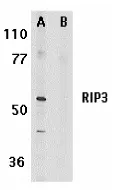
Rabbit anti RIP3 antibody (AHP1797) used to evaluate RIP3 expression in mouse intestinal organoid lysates by western blotting. mouse
Image caption:
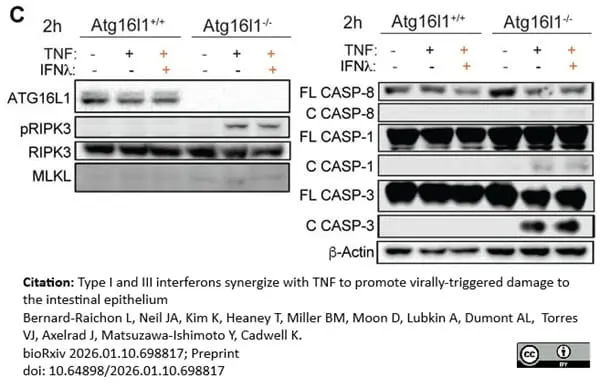
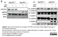
TNF and IFNλ synergize to promote RIPK1-dependent cell death in small intestinal organoids.
(C) Representative western blot of indicated proteins in lysates from Atg16l1+/+ and Atg16l1-/- organoids stimulated for 2h with 20 ng/ml TNF alone or combine d with 1ng/ml IFNl2. C and FL designate cleaved and full length, respectively.
From: Bernard-Raichon, L. et al.
Type I and III interferons synergize with 1 TNF to promote virally-triggered damage to the intestinal epithelium.
bioRχiv. 2026 12 Jan [Preprint].
doi: 10.64898/2026.01.10.698817
This image is from an open access article distributed under terms of a Creative Commons Attribution License.
Filter by Application:
WB P Reset| Rabbit anti RIP3 antibody recognizes mouse serine-threonine kinase receptor-interacting protein 3 (RIP3). RIP3 is related to RIP and RIP2 but does not posses a death domain or CARD motif at its C-terminus. RIP3 activates NF-Kappa B and potently induces apoptosis. RIP3 is expressed in a range of tissues including the spleen, liver, testis, heart, brain and lung. |
- Target Species
- Mouse
- Species Cross-Reactivity
-
Target Species Cross Reactivity Rat - N.B. Antibody reactivity and working conditions may vary between species.
- Product Form
- Purified IgG - liquid
- Antiserum Preparation
- Antisera to mouse RIP3 were raised by repeated immunisation of rabbits with highly purified antigen. Purified IgG prepared from whole serum by affinity chromatography.
- Buffer Solution
- Phosphate buffered saline
- Preservative Stabilisers
- 0.02% Sodium Azide (NaN3)
- Immunogen
- Synthetic peptide sequence corresponding to amino acids 473-486 of mouse RIP3.
- Approx. Protein Concentrations
- IgG concentration 1.0mg/ml
- Regulatory
- For research purposes only
- Guarantee
- 12 months from date of despatch
This product is shipped at ambient temperature. It is recommended to aliquot and store at -20°C on receipt. When thawed, aliquot the sample as needed. Keep aliquots at 2-8°C for short term use (up to 4 weeks) and store the remaining aliquots at -20°C.
Avoid repeated freezing and thawing as this may denature the antibody. Storage in frost-free freezers is not recommended.
Avoid repeated freezing and thawing as this may denature the antibody. Storage in frost-free freezers is not recommended.
This product has been reported to work in the following applications. This information is derived from testing within our laboratories, peer-reviewed publications or personal communications from the originators. Please refer to references indicated for further information. For general protocol recommendations, please visit the antibody protocols page.
| Application Name | Verified | Min Dilution | Max Dilution |
|---|---|---|---|
| Immunohistology - Paraffin 1 |  |
5ug/ml | |
| Western Blotting | |
0.5 | 1.0ug/ml |
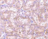
- 1This product requires antigen retrieval using heat treatment prior to staining of paraffin sections.Sodium citrate buffer pH 6.0 is recommended for this purpose.
Where this product has not been tested for use in a particular technique this does not necessarily exclude its use in such procedures. Suggested working dilutions are given as a guide only. It is recommended that the user titrates the product for use in their own system using appropriate negative/positive controls.
- Histology Positive Control Tissue
- Rat kidney
- Western Blotting
- AHP1797 detects a band of approximately 56 kDa in Mouse 3T3 whole cell lysate
| Description | Product Code | Applications | Pack Size | List Price | Your Price | Quantity | |
|---|---|---|---|---|---|---|---|
| Antigen Retrieval Buffer, pH8.0 | BUF025A | P | 500 ml | Log in | |||
| List Price | Your Price | ||||||
| Log in | |||||||
| Description | Antigen Retrieval Buffer, pH8.0 | ||||||
| TidyBlot Western Blot Detection Reagent:HRP | STAR209P | WB * | 0.5 ml | Log in | |||
| List Price | Your Price | ||||||
| Log in | |||||||
| Description | TidyBlot Western Blot Detection Reagent:HRP | ||||||
References for RIP3 antibody
-
Feng, S. et al. (2007) Cleavage of RIP3 inactivates its caspase-independent apoptosis pathway by removal of kinase domain.
Cell Signal. 19 (10): 2056-67. -
Günther, C. et al. (2011) Caspase-8 regulates TNF-α-induced epithelial necroptosis and terminal ileitis.
Nature. 477: 335-9. -
Mizumura, K. et al. (2014) Mitophagy-dependent necroptosis contributes to the pathogenesis of COPD.
J Clin Invest. 124 (9): 3987-4003. -
Huang, S.C. et al. (2014) Tumor necrosis factor suppresses NR5A2 activity and intestinal glucocorticoid synthesis to sustain chronic colitis.
Sci Signal. 7 (314): ra20. -
Meng, L. et al. (2015) RIP3-mediated necrotic cell death accelerates systematic inflammation and mortality.
Proc Natl Acad Sci U S A. 112 (35): 11007-12. -
Mizumura, K. et al. (2018) Sphingolipid regulation of lung epithelial cell mitophagy and necroptosis during cigarette smoke exposure.
FASEB J. 32 (4): 1880-90. -
Najafov, A. et al. (2019) TAM Kinases Promote Necroptosis by Regulating Oligomerization of MLKL.
Mol Cell. 75 (3): 457-468.e4. -
Matsuzawa-Ishimoto, Y. et al. (2022) The γδ IEL effector API5 masks genetic susceptibility to Paneth cell death.
Nature. 610 (7932): 547-54.
View The Latest Product References
-
Bernard-Raichon, L. et al. (2026) Type I and III interferons synergize with TNF to promote virally-triggered damage to the intestinal epithelium
bioRχiv. 12 Jan [Preprint].
Further Reading
-
Yu, P.W. et al. (1999) Identification of RIP3, a RIP-like kinase that activates apoptosis and NFkappaB.
Curr Biol. 9 (10): 539-42. -
Sun, X., et al (1999) RIP3, a novel apoptosis-inducing kinase.
J Biol Chem. 274:16871-5. -
Pazdernik, N.J., et al (1999) Mouse receptor interacting protein 3 does not contain a caspase-recruiting or a death domain but induces apoptosis and activates NF-κB.
Mol Cell Bio.19:6500-8
- RRID
- AB_2178676
- UniProt
- Q9QZL0
- Entrez Gene
- Ripk3
- GO Terms
- GO:0005515 protein binding
- GO:0006915 apoptosis
- GO:0004704 NF-kappaB-inducing kinase activity
- GO:0005524 ATP binding
- GO:0005737 cytoplasm
- GO:0006468 protein phosphorylation
- GO:0006917 induction of apoptosis
- GO:0007249 I-kappaB kinase/NF-kappaB cascade
View more products with RIP3 specificity
Please Note: All Products are "FOR RESEARCH PURPOSES ONLY"
View all Anti-Mouse ProductsAlways be the first to know.
When we launch new products and resources to help you achieve more in the lab.
Yes, sign me up