MHC Class II RT1Bu/L antibody | OX-3
Mouse anti Rat MHC Class II RT1Bu/L:RPE
- Product Type
- Monoclonal Antibody
- Clone
- OX-3
- Isotype
- IgG1
- Specificity
- MHC Class II RT1Bu/L


Mouse anti Rat MHC class II antibody, clone OX-3 (MCA45R) used to label MHC class II expressing cells in formalin fixed mouse brain cryosections by immunohistochemistry.
Image caption:
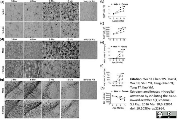
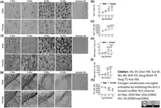
(a) Representative immunostained micrographs show Iba1+ cells in the SN of 3-, 6-, 9-, and 12-month-old female and male mice. Scale bar: 10 μm. Quantitative results of Iba1+ cell areas (b) and the number of Iba1+ cells (c) in the SN of female and male mice at different ages (n = 5). *(p <0.05), **(p <0.01), ***(p <0.001): versus the opposite sex. (d) Representative immunostained micrographs show MHC class II+ cells in the SN of 3-, 6-, 9-, and 12-month-old female and male mice. Scale bar: 10 μm. Quantitative results of MHC class II+ cell areas (e) and the number of MHC class II+ cells (f) in the SN of female and male mice at different ages (n = 5). ***(p <0.001): versus the opposite sex. (g) Representative immunostained micrographs show TH+ cells in the SN of 3-, 6-, 9-, and 12-month-old female and male mice. Scale bar: 200 μm. (h) Quantitative results of TH+ cells in the SN of female and male mice at different ages (n = 5). *(p <0.05), **(p <0.01): versus the opposite sex.
From: Wu SY, Chen YW, Tsai SF, Wu SN, Shih YH, Jiang-Shieh YF, Yang TT, Kuo YM.
Estrogen ameliorates microglial activation by inhibiting the Kir2.1 inward-rectifier K(+) channel.
Sci Rep. 2016; 6: 22864.
doi: 10.1038/srep22864.
This image is from an open access article distributed under terms of a Creative Commons Attribution License.
Mouse anti Rat MHC Class II RT1Bu/L antibody, clone OX-3 (MCA45R) used to demonstrate MHC Class II staining on rat brain by immunohistochemistry on cryosections.
Image caption:
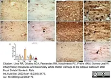
Increased immunoreactivity for MHC-II in the CC after focal striatal ischemia. Photomicrographs represent control animal injected with sterile saline (A,B) or ischemic animals injected with ET-1 at 3 (C,D); 7 (E,F); 14 (G,H); and 30 (I,J) PLDs. Maximum MHC-II immunoreactivity was present at 7 days (E,F), which has been confirmed by quantitative analysis (K). The results of quantitative analysis (K) are expressed as mean ± standard error of the mean (SEM). One-way ANOVA with Tukey’s post-hoc test (p <0.05). Similar overwritten letters did not show significant statistical differences. Left-sided photomicrographs in lower magnification with a scale bar of 20 μm, and right-sided ones, with higher magnification with a scale bar of 100 μm.
From:Lima RR, Oliveira ACA, Fernandes RM, Nascimento PC, Freire MAM, Gomes-Leal W.
Inflammatory Response and Secondary White Matter Damage to the Corpus Callosum after Focal Striatal Stroke in Rats.
Int J Mol Sci. 2022 Mar 16;23(6):3179.
doi: 10.3390/ijms23063179.
This image is from an open access article distributed under terms of a Creative Commons Attribution License.
Filter by Application:
P C Reset| Mouse anti Rat MHC Class II RT1Bu/L antibody, clone OX-3 recognizes a polymorphic determinant of the rat RT1B MHC class II antigen, reacting with haplotypes u and l. The literature reports reactivity with Lewis, Wistar and AO strain rats but not BN, DA or PVG/c strains. This antibody is useful for distinguishing RT1B positive cells from different rat strains, e.g. for recognising cells of donor origin in bone marrow reconstituted radiation chimaeras. The major histocompatibility complex (MHC) is a cluster of genes that are important in the immune response to infections. In rats, this complex is referred to as the RT1 region. In mice, this complex is referred to as the H-2 region. Mouse anti Rat MHC Class II RT1Bu/L antibody, clone OX-3 also cross reacts with mouse strains of the H-2 haplotypes b and s. Analysis of recombinant mouse strains has mapped the OX-3 determinant to the H-2I-A region. This product is routinely tested in flow cytometry on Lewis rat splenocytes. |
- Target Species
- Rat
- Species Cross-Reactivity
-
Target Species Cross Reactivity Mouse - N.B. Antibody reactivity and working conditions may vary between species.
- Product Form
- Purified IgG conjugated to R. Phycoerythrin (RPE) - lyophilized
- Reconstitution
- Reconstitute with 1 ml distilled water
- Preparation
- Purified IgG prepared by affinity chromatography on Protein G from tissue culture supernatant
- Buffer Solution
- Phosphate buffered saline
- Preservative Stabilisers
0.09% Sodium Azide 1% Bovine Serum Albumin 5% Sucrose - Immunogen
- Rat thymocyte membrane glycoproteins.
- Fusion Partners
- Spleen cells from immunized BALB/c mice were fused with cells from the NS1 mouse myeloma cell line.
- Max Ex/Em
-
Fluorophore Excitation Max (nm) Emission Max (nm) RPE 488nm laser 496 578 - Regulatory
- For research purposes only
- Guarantee
- 6 months from date of despatch
This product is shipped at ambient temperature.
Prior to reconstitution store at +4oC. Following reconstitution store at +4oC.
DO NOT FREEZE.
This product should be stored undiluted. This product is photosensitive and should be protected from light. Should this product contain a precipitate we recommend microcentrifugation before use.
Prior to reconstitution store at +4oC. Following reconstitution store at +4oC.
DO NOT FREEZE.
This product should be stored undiluted. This product is photosensitive and should be protected from light. Should this product contain a precipitate we recommend microcentrifugation before use.
This product has been reported to work in the following applications. This information is derived from testing within our laboratories, peer-reviewed publications or personal communications from the originators. Please refer to references indicated for further information. For general protocol recommendations, please visit the antibody protocols page.
| Application Name | Verified | Min Dilution | Max Dilution |
|---|---|---|---|
| Flow Cytometry |  |
Neat |
Where this antibody has not been tested for use in a particular technique this does not necessarily exclude its use in such procedures. Suggested working dilutions are given as a guide only. It is recommended that the user titrates the antibody for use in their own system using appropriate negative/positive controls.
- Flow Cytometry
- Use 10ul of the suggested working dilution to label 106 cells in 100ul.
How to Use the Spectraviewer
Watch the Tool Tutorial Video ▸- Start by selecting the application you are interested in, with the option to select an instrument from the drop down menu or create a customized instrument
- Select the fluorophores or fluorescent proteins you want to include in your panel to check compatibility
- Select the lasers and filters you wish to include
- Select combined or multi-laser view to visualize the spectra
| Description | Product Code | Applications | Pack Size | List Price | Your Price | Quantity | |
|---|---|---|---|---|---|---|---|
| Mouse IgG1 Negative Control:RPE | MCA1209PE | F | 100 Tests | Log in | |||
| List Price | Your Price | ||||||
| Log in | |||||||
| Description | Mouse IgG1 Negative Control:RPE | ||||||
References for MHC Class II RT1Bu/L antibody
-
McMaster, W.R. & Williams, A.F. (1979) Identification of Ia glycoproteins in rat thymus and purification from rat spleen.
Eur J Immunol. 9 (6): 426-33. -
McMaster, W.R. & Williams, A.F. (1979) Monoclonal antibodies to Ia antigens from rat thymus: cross reactions with mouse and human and use in purification of rat Ia glycoproteins.
Immunol Rev. 47: 117-37. -
Barclay, A.N. & Mayrhofer, G. (1981) Bone marrow origin of Ia-positive cells in the medulla rat thymus.
J Exp Med. 153 (6): 1666-71. -
Zhang, J. et al. (1997) Expression of major histocompatibility complex molecules in rodent retina. Immunohistochemical study.
Invest Ophthalmol Vis Sci. 38 (9): 1848-57. -
Hahm, K.B. et al. (2000) Loss of TGF-beta signaling contributes to autoimmune pancreatitis.
J Clin Invest. 105 (8): 1057-65. -
Wu, S.Y. et al. (2016) Estrogen ameliorates microglial activation by inhibiting the Kir2.1 inward-rectifier K(+) channel.
Sci Rep. 6: 22864. -
Fisher, R.A. et al. (1996) Induction of long-term graft tolerance and donor/recipient chimerism.
J Surg Res. 60 (1): 181-5. -
Keller, R. et al. (1988) Modulation of major histocompatibility complex (MHC) expression by interferons and microbial agents. Independent regulation of MHC class II expression and induction of tumoricidal activity in bone marrow-derived mononuclear phagocytes.
Scand J Immunol. 28 (1): 113-21.
View The Latest Product References
-
Streit, W.J. et al. (1989) Peripheral nerve lesion produces increased levels of major histocompatibility complex antigens in the central nervous system.
J Neuroimmunol. 21 (2-3): 117-23. -
Roggin, K.K. et al. (2001) Macrophage phenotype during cholestatic injury and repair: the persistent inflammatory response.
J Pediatr Surg. 36 (1): 220-8. -
Reutzel-Selke A et al. (2003) Short-term immunosuppressive treatment of the donor ameliorates consequences of ischemia/ reperfusion injury and long-term graft function in renal allografts from older donors.
Transplantation. 75 (11): 1786-92. -
Heidenhain, C. et al. (2003) The impact of immune-activating processes following transplantation on chronic allograft nephropathy.
Kidney Int. 64 (3): 1125-33. -
Hahm, K.B. et al. (2001) Loss of transforming growth factor beta signalling in the intestine contributes to tissue injury in inflammatory bowel disease.
Gut. 49 (2): 190-8. -
Pascher A et al. (2006) Rat cytomegalovirus infection interferes with anti-CD4 mAb-(RIB 5/2) mediated tolerance and induces chronic allograft damage.
Am J Transplant. 6 (9): 2035-45. -
Hartmann CB et al. (2005) Immunotoxicity of gallium arsenide on antigen presentation: comparative study of intratracheal and intraperitoneal exposure routes.
J Immunotoxicol. 2 (1): 1-9. -
Lima, R.R. et al. (2022) Inflammatory Response and Secondary White Matter Damage to the Corpus Callosum after Focal Striatal Stroke in Rats.
Int J Mol Sci. 23 (6)Mar 16 [Epub ahead of print]. -
Duhalde Vega, M et al. (2022) PD-1/PD-L1 blockade abrogates a dysfunctional innate-adaptive immune axis in critical β-coronavirus disease.
Sci Adv. 8 (38): eabn6545.
Further Reading
-
Barclay, A.N. (1981) The localization of populations of lymphocytes defined by monoclonal antibodies in rat lymphoid tissues.
Immunology. 42 (4): 593-600.
- RRID
- AB_322118
MCA45PE
If you cannot find the batch/lot you are looking for please contact our technical support team for assistance.
View more products with MHC CLASS II specificity
Please Note: All Products are "FOR RESEARCH PURPOSES ONLY"
View all Anti-Rat ProductsAlways be the first to know.
When we launch new products and resources to help you achieve more in the lab.
Yes, sign me up