CD43 antibody | W3/13


Mouse anti Rat CD43:FITC
- Product Type
- Monoclonal Antibody
- Clone
- W3/13
- Isotype
- IgG1
- Specificity
- CD43
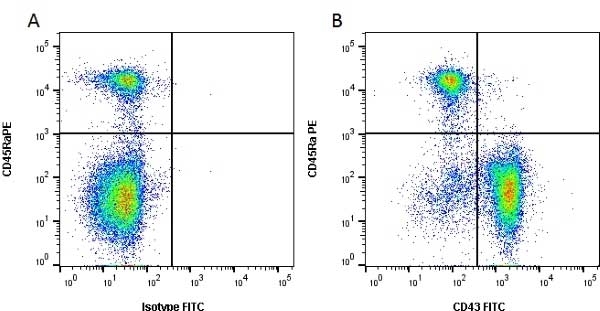
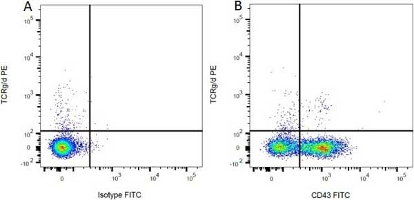
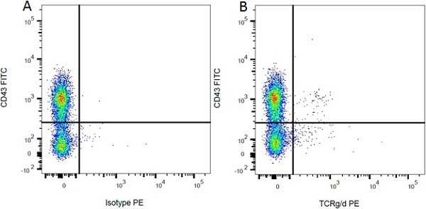
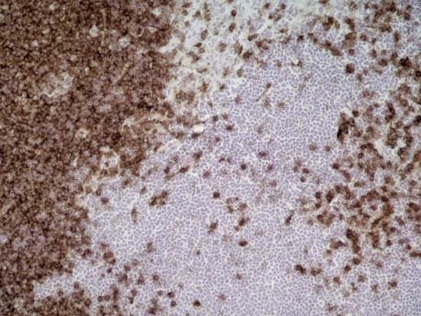
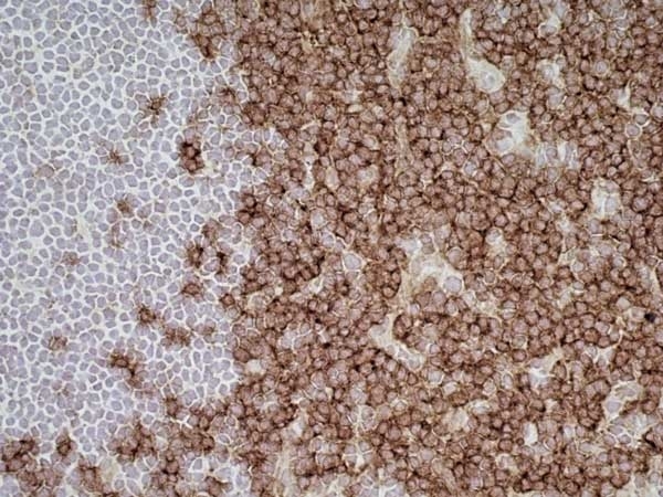
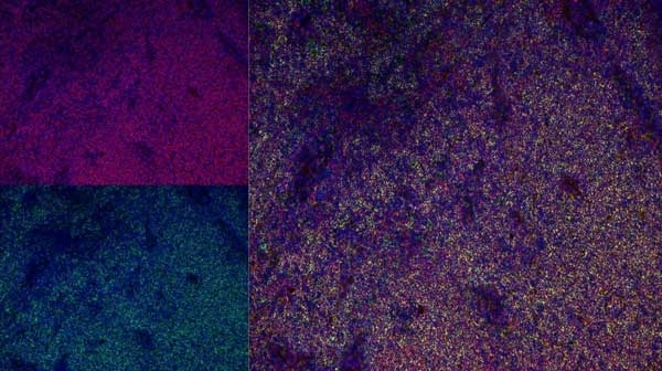
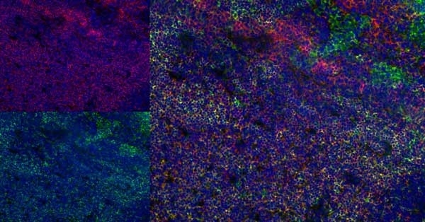
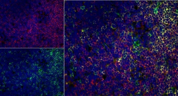
| Mouse anti Rat CD43 antibody, clone W3/13 recognizes the rat CD43 cell surface antigen, also known as leukosialin, sialophorin or W3/13 antigen. CD43 is a 371 amino acid ~95 kDa heavily glycosylated single pass type 1 transmembrane glycoprotein (Killeen et al. 1987) expressed by all leucocytes with the exception of B lymphocytes. CD43, in mice acts as a T-cell counter-receptor for CD169 (Siglec-1) suggesting a role in cell-cell interactions (van den Berg et al. 2001) Mouse anti Rat CD43 antibody, clone W3/13 is routinely tested in flow cytometry on rat splenocytes. |
- Target Species
- Rat
- Product Form
- Purified IgG conjugated to Fluorescein Isothiocyanate Isomer 1 (FITC) - liquid
- Preparation
- Purified IgG prepared by affinity chromatography on Protein G from tissue culture supernatant
- Buffer Solution
- Phosphate buffered saline
- Preservative Stabilisers
0.09% Sodium Azide 1% Bovine Serum Albumin - Immunogen
- Rat thymocyte membrane glycoproteins.
- Approx. Protein Concentrations
- IgG concentration 0.1 mg/ml
- Fusion Partners
- Spleen cells from immunised BALB/c mice were fused with cells of the mouse NS1 myeloma cell line.
- Max Ex/Em
-
Fluorophore Excitation Max (nm) Emission Max (nm) FITC 490 525 - Regulatory
- For research purposes only
- Guarantee
- 12 months from date of despatch
Avoid repeated freezing and thawing as this may denature the antibody. Storage in frost-free freezers is not recommended. This product is photosensitive and should be protected from light.
| Application Name | Verified | Min Dilution | Max Dilution |
|---|---|---|---|
| Flow Cytometry | Neat |
- Flow Cytometry
- Use 10ul of the suggested working dilution to label 106 cells in 100ul.
| Description | Product Code | Applications | Pack Size | List Price | Your Price | Quantity | |
|---|---|---|---|---|---|---|---|
| Mouse IgG1 Negative Control:FITC | MCA1209F | F | 0.1 mg |
|
Log in | ||
| List Price | Your Price | ||||||
|
|
Log in | ||||||
| Description | Mouse IgG1 Negative Control:FITC | ||||||
References for CD43 antibody
-
Brown, W.R.A. et al. (1981) Identification of a glycophorin-like molecule at the cell surface of rat thymocytes.
Nature. 289: 456-460. -
Barclay, A. N. (1981) The localization of populations of lymphocytes defined by monoclonal antibodies in rat lymphoid tissues.
Immunology. 42: 593-600 -
Forbes, R. D. C. et al. (1983) Leukocyte subsets in first-set rat cardiac allograft rejection. A serial immunohistologic study using monoclonal antibodies
Transplantation. 36: 681-686 -
Jung, S. et al. (1994) Therapeutic effect of transforming growth factor-beta 2 on actively induced EAN but not adoptive transfer EAN
Immunology. 83: 545-551. -
Bataller, R. et al. (2003) Prolonged infusion of angiotensin II into normal rats induces stellate cell activation and proinflammatory events in liver.
Am J Physiol Gastrointest Liver Physiol. 285: G642-651 -
Rice, E.K. et al. (2003) Induction of MIF synthesis and secretion by tubular epithelial cells: a novel action of angiotensin II.
Kidney Int. 63 (4): 1265-75. -
Schwab, J.M. et al. (2005) Spinal cord injury induces early and persistent lesional P2X4 receptor expression.
J Neuroimmunol. 163: 185-9. -
Conrad, S. et al. (2005) Prolonged lesional expression of RhoA and RhoB following spinal cord injury.
J Comp Neurol. 487: 166-75.
View The Latest Product References
-
Zhang, Z. et al. (2008) FTY720 ameliorates experimental autoimmune neuritis by inhibition of lymphocyte and monocyte infiltration into peripheral nerves.
Exp Neurol. 210: 681-90. -
Yamanaka, Y. et al. (2011) Immunohistochemical analysis of subcutaneous tissue reactions to methacrylate resin-based root canal sealers.
Int Endod J. 44: 669-75. -
Dort, J. et al. (2012) Beneficial effects of cod protein on skeletal muscle repair following injury.
Appl Physiol Nutr Metab. 37 (3): 489-98. -
Duchesne, E. et al. (2013) Mast cells can regulate skeletal muscle cell proliferation by multiple mechanisms.
Muscle Nerve. 48 (3): 403-14. -
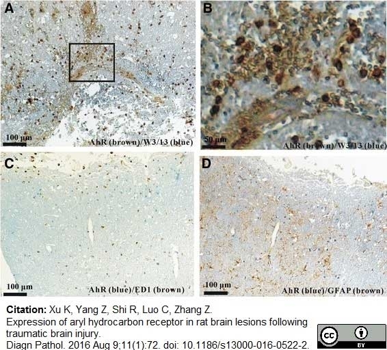
Xu, K. et al. (2016) Expression of aryl hydrocarbon receptor in rat brain lesions following traumatic brain injury.
Diagn Pathol. 11 (1): 72. -
Dort, J. et al. (2016) Shrimp Protein Hydrolysate Modulates the Timing of Proinflammatory Macrophages in Bupivacaine-Injured Skeletal Muscles in Rats.
Biomed Res Int. 2016: 5214561. -
Zhang, Z.M. et al. (2016) Lesional accumulation of CD8(+) cells in sciatic nerves of experimental autoimmune neuritis rats.
Neurol Sci. 37 (2): 199-203. -
Ornellas, F.M. et al. (2019) Mesenchymal Stromal Cells Induce Podocyte Protection in the Puromycin Injury Model.
Sci Rep. 9 (1): 19604. -
Grad, E. et al. (2019) Monocyte Modulation by Liposomal Alendronate Improves Cardiac Healing in a Rat Model of Myocardial Infarction
Regenerative Engineering and Translational Medicine. 5 (3): 280-9. -
Kami, K. et al. (2019) Role of 72-kDa Heat Shock Protein in Heat-stimulated Regeneration of Injured Muscle in Rat.
J Histochem Cytochem. 67 (11): 791-9. -
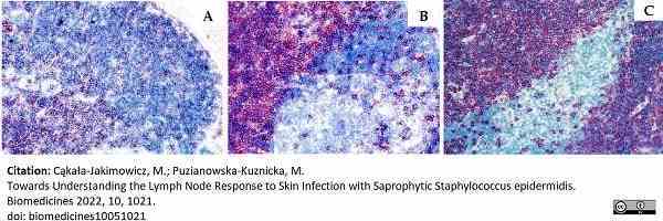
Cąkała-Jakimowicz, M. & Puzianowska-Kuznicka, M. (2022) Towards Understanding the Lymph Node Response to Skin Infection with Saprophytic Staphylococcus epidermidis..
Biomedicines. 10 (5): 1021. -
Merlini, A. et al. (2022) Distinct roles of the meningeal layers in CNS autoimmunity.
Nat Neurosci. 25 (7): 887-99.
- Synonyms
- Leukosialin
- RRID
- AB_322579
- UniProt
- P13838
- Entrez Gene
- Spn
- GO Terms
- GO:0002544 chronic inflammatory response
- GO:0016021 integral to membrane
- GO:0005902 microvillus
- GO:0009986 cell surface
- GO:0019904 protein domain specific binding
- GO:0032154 cleavage furrow
- GO:0042117 monocyte activation
- GO:0071594 thymocyte aggregation
MCA54FT
If you cannot find the batch/lot you are looking for please contact our technical support team for assistance.
Please Note: All Products are "FOR RESEARCH PURPOSES ONLY"
View all Anti-Rat ProductsAlways be the first to know.
When we launch new products and resources to help you achieve more in the lab.
Yes, sign me up