Growth Cone antibody | 2G13
Mouse anti Growth Cone
- Product Type
- Monoclonal Antibody
- Clone
- 2G13
- Isotype
- IgM
- Specificity
- Growth Cone

Filter by Application:
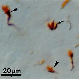
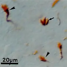
P Reset| Mouse anti Growth Cone antibody, clone 2G13 recognizes a protein originally termed 2G13P, localized to growth cones. Subsequent investigation has identified this protein to be 40S ribosomal protein SA, also known as 37 kDa laminin receptor precursor or Laminin receptor 1 (Baloui et al. 2004). 40S ribosomal protein SA is a 296 amino acid ~37 kDa membrane, cytoplasmic and nuclear protein required for the assembly and/or stability of the 40S ribosomal subunit.. In vertebrate evolution the molecule has acquired a secondary function as a laminin receptor (UniProt: P50890). In growth cones expression is notable particularly in filopodia and lamellipodia in developing rat CNS and embryonic neurons in culture (Stettler et al. 1999). 40S ribosomal protein SA interacts with the filamentous actin cytoskeleton and therefore may be involved in growth cone motility (Stettler et al. 1999). Mouse anti Growth Cone antibody, clone 2G13 has been used for the detection of growth cones by immunohistochemistry and identification of 40S ribosomal protein SA by western blotting in chicken and rat samples (Baloui et al. 2004). |
- Target Species
- Chicken
- Species Cross-Reactivity
-
Target Species Cross Reactivity Rat Mouse - N.B. Antibody reactivity and working conditions may vary between species.
- Product Form
- Tissue culture supernatant - liquid
- Preparation
- Tissue culture supernatant containing 0.2M Tris/HCl pH7.4 and 5-10% foetal calf serum
- Preservative Stabilisers
- 0.09% sodium azide (NaN3)
- Immunogen
- Embryonic chick tectal membranes.
- Fusion Partners
- Spleen cells from immunized mice were fused with cells of the mouse NS1 myeloma cell line.
- Regulatory
- For research purposes only
- Guarantee
- 12 months from date of despatch
This product is shipped at ambient temperature. It is recommended to aliquot and store at -20°C on receipt. When thawed, aliquot the sample as needed. Keep aliquots at 2-8°C for short term use (up to 4 weeks) and store the remaining aliquots at -20°C.
Avoid repeated freezing and thawing as this may denature the antibody. Storage in frost-free freezers is not recommended.
Avoid repeated freezing and thawing as this may denature the antibody. Storage in frost-free freezers is not recommended.
This product has been reported to work in the following applications. This information is derived from testing within our laboratories, peer-reviewed publications or personal communications from the originators. Please refer to references indicated for further information. For general protocol recommendations, please visit the antibody protocols page.
| Application Name | Verified | Min Dilution | Max Dilution |
|---|---|---|---|
| Flow Cytometry | |||
| Immunohistology - Frozen |  |
||
| Immunohistology - Paraffin | |
Where this product has not been tested for use in a particular technique this does not necessarily exclude its use in such procedures. Suggested working dilutions are given as a guide only. It is recommended that the user titrates the product for use in their own system using appropriate negative/positive controls.
| Description | Product Code | Applications | Pack Size | List Price | Your Price | Quantity | |
|---|---|---|---|---|---|---|---|
| Goat anti Mouse IgM:Alk. Phos.(Human Adsorbed) | STAR138A | C E P WB | 1 ml |

|
Log in | ||
| List Price | Your Price | ||||||
|
|
Log in | ||||||
| Description | Goat anti Mouse IgM:Alk. Phos.(Human Adsorbed) | ||||||
| Goat anti Mouse IgG/A/M:HRP (Human Adsorbed) | STAR87P | E | 1 mg |
|
Log in | ||
| List Price | Your Price | ||||||
|
|
Log in | ||||||
| Description | Goat anti Mouse IgG/A/M:HRP (Human Adsorbed) | ||||||
| Description | Product Code | Applications | Pack Size | List Price | Your Price | Quantity | |
|---|---|---|---|---|---|---|---|
| Mouse anti Chicken CD184 / CXCR4 | MCA6012GA | C F IF | 0.1 mg |
|
Log in | ||
| List Price | Your Price | ||||||
|
|
Log in | ||||||
| Description | Mouse anti Chicken CD184 / CXCR4 | ||||||
Source Reference
-
Stettler, O. et al. (1999) Monoclonal antibody 2G13, a new axonal growth cone marker.
J Neurocytol. 28 (12): 1035-44.
References for Growth Cone antibody
-
Penkowa, M. et al. (2003) Metallothionein-I overexpression alters brain inflammation and stimulates brain repair in transgenic mice with astrocyte-targeted interleukin-6 expression.
Glia. 42 (3): 287-306. -
Baloui, H. et al. (2004) Cellular prion protein/laminin receptor: distribution in adult central nervous system and characterization of an isoform associated with a subtype of cortical neurons.
Eur J Neurosci. 20 (10): 2605-16. -
Espejo, C. et al. (2005) Time-course expression of CNS inflammatory, neurodegenerative tissue repair markers and metallothioneins during experimental autoimmune encephalomyelitis.
Neuroscience. 132(4):1135-49. -
Kim, S.R. et al. (2011) Dopaminergic pathway reconstruction by Akt/Rheb-induced axon regeneration.
Ann Neurol. 70: 110-20. -
Nordman, J.C. & Kabbani, N. (2012) An interaction between α7 nicotinic receptors and a G-protein pathway complex regulates neurite growth in neural cells.
J Cell Sci. 125 (Pt 22): 5502-13.
- Synonyms
- 40S Ribosomal Protein Sa
- RRID
- AB_322754
- UniProt
- P50890
- Entrez Gene
- RPSA
- GO Terms
- GO:0005886 plasma membrane
- GO:0003735 structural constituent of ribosome
- GO:0004872 receptor activity
- GO:0005634 nucleus
- GO:0006412 translation
- GO:0015935 small ribosomal subunit
- GO:0043236 laminin binding
MCA1712
If you cannot find the batch/lot you are looking for please contact our technical support team for assistance.
Request a different product with this specificity
Please Note: All Products are "FOR RESEARCH PURPOSES ONLY"
View all Anti-Chicken ProductsAlways be the first to know.
When we launch new products and resources to help you achieve more in the lab.
Yes, sign me up