
Small Particle Analysis with Flow Cytometry
Overview
Small particle analysis by flow cytometry is opening up a category of biology that used to sit just out of reach. Standard flow cytometry was built for cells a few microns across, bright, and easy to set your detection threshold on, but a huge amount of important biology happens well below that size: extracellular vesicles carrying diverse biological cargo, bacterial membrane vesicles mediating infection and immunity, and lipid nanoparticles delivering mRNA therapeutics. These particles are typically tens to a few hundred nanometers across, small enough that conventional instruments struggle to distinguish real signal from optical noise.
With a small particle detector and the sensitivity to resolve events at this scale, the ZE5 Cell Analyzer can characterize these particles one at a time, not just confirming they exist, but measuring what each individual particle is made of, what it's carrying, and how it differs from its neighbors. That shift, from population averages to true single-particle resolution, is what's making small particle analysis such a fast-moving area in both diagnostics and drug development.
Why Researchers Choose the ZE5 Cell Analyzer
- Study diverse small-particle populations on a single platform
- Investigate differences between individual particles rather than population averages
- Support applications ranging from biomarker discovery to nanoparticle development
- Generate actionable insights across extracellular vesicle, exosome, membrane vesicle, and lipid nanoparticle research
Small Particle Discovery Resources
To illustrate how the ZE5 Cell Analyzer can be used to successfully detect and characterize a variety of small particles, including extracellular vesicles (EVs), lipid nanoparticles (LNPs), membrane vesicles (MVs), and exosomes, we have prepared the following resources:
Flow Cytometry-Based Exosome Detection and Analysis Using the ZE5 Cell Analyzer
 Exosomes are secreted membrane vesicles, approximately 50–100 nm in diameter, that support intercellular communication and processes such as metastasis and antigen presentation. Because they are found in major bodily fluids and carry cellular information, they are promising biomarkers, but their small size makes them difficult to study. Here, we show detection of exosome markers CD63, CD81, ALIX, and TSG101 using the ZE5 Cell Analyzer, highlighting its utility for small particle analysis.
View the Application Note
Single-Particle and Multicolor Analysis of Extracellular Vesicles (EVs)
 Extracellular vesicles (EVs) are nanosized, lipid-bilayer particles released by most cells. They lack a nucleus, cannot self-replicate, and carry biomolecules such as mRNA, miRNA, proteins, and lipids that support intercellular communication and influence processes including gene transcription and immune responses. EVs are gaining attention for their value as biomarkers and potential therapeutic agents. Using the ZE5 Cell Analyzer, this application note demonstrates single-particle, multicolor analysis of culture-cell-derived EVs.
View the Application Note
Single-Particle and mRNA-Encapsulation-Rate Analysis of Lipid Nanoparticle (LNP) Formulations
 Lipid nanoparticles (LNPs), typically 10–1,000 nm in diameter, can encapsulate and deliver active ingredients to targeted sites with controlled release. This makes them valuable drug-delivery systems, especially for nucleic-acid-based drugs that require protection to improve in vivo stability. While encapsulation efficiency is usually measured in bulk, single-particle methods can reveal which particles contain active ingredients. Using the ZE5 Cell Analyzer, we demonstrate single-particle LNP detection and mRNA encapsulation-efficiency analysis in a model formulation.
View the Application Note
Simultaneous Analysis of Bacterial Cells and Single Membrane Vesicles (MV)
 Membrane vesicles (MVs) are membrane-bound particles released by bacteria. They support key processes including cell-to-cell communication, transfer of signaling molecules and nucleic acids, delivery of virulence factors, immune evasion, and nutrient cycling in soil and aquatic environments. MV research has also improved understanding of infectious disease and supported new therapeutic and preventive approaches. This application note shows how the ZE5 Cell Analyzer can simultaneously measure bacterial cells and their released MVs.
View the Application Note
Exosome Workflows and Training Resources
The following resources illustrate how the ZE5 Cell Analyzer can help you characterize and study extracellular vesicles.
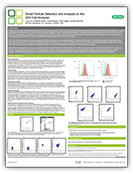
Exosome Workflow
Exosomes can be detected using surface or intravesicular staining on the ZE5 Cell Analyzer.
We share both methods, along with details of direct exosome detection and instrument setup, in this useful poster.
Download the Poster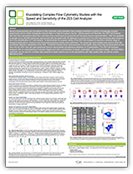
Small Particle Studies
In addition to performing small particle detection with ease, the ZE5 Cell Analyzer combines speed with enhanced electronics to perform rapid analysis without compromising data quality. Automation of crucial processes, such as washing the sample probe between samples, can reduce data variation.
The low-noise electronics of the ZE5 Cell Analyzer also improves the quality and reproducibility of your experiments. Download this poster to learn how the ZE5 Cell Analyzer can help you do more.
Download the Poster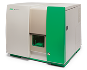
Webinar: How to Detect Exosomes Directly
In this on-demand webinar, we demonstrate how the small particle detection capability of the ZE5 Cell Analyzer can be used for exosome detection. We also present the instrument setup for direct exosome detection and for detection of exosomes captured on beads.
Watch this WebinarSmall Particle Analysis: FAQ and Glossary
General
What counts as a “small particle” in flow cytometry?
In most cytometry contexts, a small particle is anything well below the size of a typical cell, roughly 50 nm to 1 µm in diameter. This covers extracellular vesicles, bacterial membrane vesicles, lipid nanoparticles, and viral particles. Standard cytometers are built and optimized for cell-sized events (several microns and up), so particles in this smaller range often fall below their noise floor.Why is single-particle analysis better than a bulk assay?
A bulk assay reports an average across an entire population, for example, “70% encapsulation efficiency” for a batch of nanoparticles. It can't tell you whether that's 70% of particles fully loaded and 30% completely empty, or every particle sitting at 70% capacity. Single-particle analysis measures each event individually, so you get the actual distribution, not just the average.What is a small particle detector (SPD)?
An SPD is an added detection configuration on a flow cytometer that uses forward scatter and is tuned for enhanced sensitivity at sub-micron sizes. It allows a cytometer that would normally only resolve cells to also resolve much smaller biological and synthetic particles, down to roughly 50–100 nm depending on the sample.How is particle size estimated on a cytometer if there's no direct sizing detector?
By comparison to sizing bead standards. Beads of known, calibrated diameters (commonly ranging from ~100 nm to 500 nm) are run under identical instrument settings, and their scatter or fluorescence signal is used as a reference ladder against which the unknown sample's particles can be placed.Extracellular Vesicles (EVs)
What is an extracellular vesicle (EV)?
An EV is a small, membrane-bound particle released by nearly all cell types. EVs are naturally secreted, enclosed by a lipid bilayer, and, unlike cells, have no nucleus and cannot replicate. They range from roughly 30 nm to over 1 µm depending on subtype.What's the difference between an EV and an exosome?
“Exosome” refers to one specific biogenesis pathway of EV (formed inside a cell and released when a multivesicular body fuses with the plasma membrane), while “EV” is the umbrella term covering exosomes plus other subtypes, such as microvesicles that bud directly from the cell surface. Without dedicated biogenesis markers, most flow-based methods can't distinguish subtypes, so “EV” is the more accurate general term.Why are EVs of interest for diagnostics?
EVs circulate in blood and other body fluids and carry molecular cargo, proteins, mRNA, microRNA, and lipids, that reflect the cell they came from. Because they're accessible via a simple blood draw, they're being explored as a liquid-biopsy tool for non-invasive diagnostics and disease monitoring.What are EVs used for therapeutically?
Beyond diagnostics, EVs, particularly those derived from mesenchymal stem cells (MSCs), are being investigated as therapeutic agents in their own right, and as delivery vehicles for drugs or genetic material, due to their natural ability to communicate with and be taken up by other cells.How do you confirm an EV sample is real, and not just instrument noise?
By comparing against a blank (buffer-only) control and known sizing standards. A genuine EV sample should show a distinct, gated population of events that's absent in the blank, with a size distribution consistent with EVs when compared to reference beads. When using dyes, it is also important to use a dye-only control, which can be useful for distinguishing dye aggregates from EVs.Bacterial Membrane Vesicles (MVs)
What is a bacterial membrane vesicle (MV)?
An MV is a small, membrane-bound particle released by bacteria, analogous in structure to a mammalian EV but originating from a microbial membrane. MVs typically range from about 20 to 400 nm in diameter.What do bacterial MVs actually do?
MVs support several bacterial survival functions: cell-to-cell communication via transfer of signaling molecules and nucleic acids, delivery of virulence factors into host cells during infection, immune evasion by acting as decoys, and nutrient cycling in environments like soil and ocean water.Do MVs contain DNA?
Some do. MVs can carry double-stranded DNA internally, which can be detected using membrane-permeable, DNA-binding dyes in combination with a membrane stain, allowing researchers to distinguish MVs that are genuinely carrying genetic cargo from those that are not.Why analyze MVs and bacterial cells in the same sample?
Because MV production is a live process, bacteria continuously shed vesicles into their environment. Being able to resolve intact cells and the vesicles they release side by side in a single acquisition makes it possible to study the shedding process, MV uptake, and host-pathogen interactions more directly.Are MVs relevant to vaccine development?
Yes. Because MVs naturally carry immunostimulatory bacterial components, they're being investigated as a basis for MV-based vaccines and as tools for modulating immune responses, alongside applications in cancer therapy research.Instrumentation
What makes the ZE5 Cell Analyzer suitable for small particle work?
Three things in combination: a small particle detector configuration for sub-micron sensitivity, high-speed acquisition (up to 100,000 events/second) so rare small-particle populations can still be captured in statistically meaningful numbers, and robust software/hardware noise reduction to keep true signal distinguishable from background at these smaller sizes.Does temperature control matter for small particle samples?
It can. EVs, MVs, and LNPs can all be sensitive to storage conditions, so a temperature-controlled sample loader (4–37°C) helps preserve sample integrity during longer acquisition runs and supports formulation- and storage-stability studies.Have a question about small particle analysis that isn't covered here?
Speak to an ExpertRelated Topics
Bio-Rad — experts in flow cytometry — can help you discover everything you need to conduct successful flow experiments with the following additional resources:
- Discover Our Flow Cytometer Range
- Visit Our Flow Cytometry Hub
- Explore Our Flow Cytometry Resources Hub
- Improve Your Cell Analysis with the ZE5 Cell Analyzer
Be the First to Receive Updates on Products and Resources
You'll receive regular communications featuring free lab resources such as application guides, posters, mini-reviews, protocols, practical tips, webinar invitations, special offers, and updates on new research tools and products.
You'll also get our specialist newsletters—including Fab Fragments, Flow. Sorted., and RiVETing—along with news on the latest antibody innovations, such as TrailBlazer™ Antibodies and StarBright™ Dyes.
During sign-up, you can choose your areas of interest so you only receive content relevant to your research, and you can unsubscribe at any time.
