
Aldehyde Fixation
Aldehyde Fixation
Aldehyde fixation is a technique used in the preparation of biological samples for immunohistochemistry and related staining methods. Its purpose is to preserve tissue integrity by halting degenerative processes, such as autolysis, which can rapidly compromise sample quality. The fixation method relies on the chemical reactivity of aldehydes, which stabilise proteins through the formation of cross-links. However, this can inadvertently mask antigenic sites, affecting the intensity and specificity of immunological staining.
The choice of fixative, duration of fixation, and subsequent antigen retrieval procedures are all critical factors to consider when designing protocols for reliable and reproducible immunostaining. These factors can vary significantly between different antibodies, and it is important for end users to consider optimizing each antibody separately prior to staining.
Purpose of Fixation
Once tissue is separated from its blood supply or cells removed from their growth media, several degenerative processes commence. Processes like putrefaction (degradation due to the presence of microbes) or autolysis (protein digestion from the release of intracellular enzymes by the rupture of organelle membranes).
Fixation attempts to arrest or prevent these processes and preserve the sample for study and is commonly incorporated into immunohistochemistry, immunocytochemistry, immunofluorescence, and flow cytometry protocols.
Protein Cross-Linking and Antigen/Epitope Masking
Aldehydes react with amine side chains to form Schiff bases which, in turn, react with proteins and peptides to form stable methylene bridges (see Fig. 1). As a result, amino acid sequences that are recognized by antibodies in unfixed tissue may become hidden or ‘masked’ behind the induced protein folding caused by these cross-links. This reduces antigen accessibility and consequently decreases staining intensity. The extent of ‘masking’ also depends on the antigen being studied, some are more susceptible to aldehyde than others.
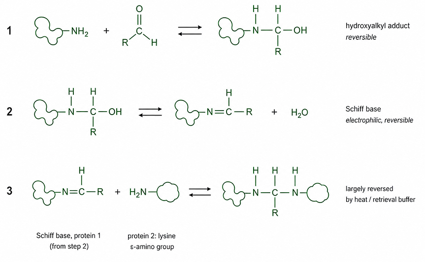
Fig. 1. Steps in the formation of protein crosslinks. 1) An aldehyde (R–CHO) adds to a nucleophilic amino group—typically the ε-amine of a lysine residue or an N-terminal α-amine—to give a hydroxyalkyl adduct (or a methylol adduct for formaldehyde, when R = H). 2) Loss of water results in a Schiff base, which at physiological pH is in equilibrium with its protonated, strongly electrophilic iminium form. Steps 1 and 2 are both readily reversible. 3) A nucleophilic group from the same or a different protein reacts with the Schiff base to form a covalent cross-link. These cross-links may reduce epitope accessibility and thereby contribute to epitope masking.
Considerations When Immunologically Staining Aldehyde-Fixed Tissue
Three key considerations for staining aldehyde-fixed tissue:
1. Type of fixative—below are a few examples of fixatives used in tissue preparation. However, it is good to keep in mind that other aldehyde-based fixatives (glutaraldehyde, glyoxal, acrolein, etc) are used in laboratories:
a. 10% neutral buffered formalin (NBF)—10% formalin is a 1:10 dilution of a saturated solution of formaldehyde in water. Because the maximum concentration of formaldehyde in water is 40%, a 1:10 dilution of a saturated solution of formaldehyde in water would contain 4% formaldehyde. Methanol is typically added to saturated solutions of formaldehyde to inhibit polymerization.
b. 4% paraformaldehyde (PFA)—paraformaldehyde is polymerized formaldehyde. Once mixed with water and heated, it depolymerizes and becomes formaldehyde. In terms of formaldehyde concentration, 4% PFA is equivalent to 10% NBF. However, since PFA solutions need to be kept refrigerated to inhibit re-polymerization, tissue penetration is slower than NBF and fixation may take longer.
c. Periodate-lysine-paraformaldehyde (PLP)—an alternative fixative first published by McLean and Nakane (1974) for the preservation of carbohydrate-rich/glycosylated cell surface antigens. Periodate oxidizes carbohydrate residues to create aldehyde groups. Paraformaldehyde reacts with the aldehyde groups on the carbohydrates and the lysine residues, creating cross-links that stabilize the tissue. Lysine cross-links periodate-oxidized molecules to other proteins, further stabilizing the tissue structure. PLP is considered a milder fixative that results in reduced levels of antigen/epitope masking and, thus, reduces the need for downstream processes like antigen/epitope retrieval.
Table 1. Fixative comparison.
Fixative |
Formaldehyde Equivalent |
Key Benefit |
Limitation |
|---|---|---|---|
|
10% NBF |
4% formaldehyde |
Rapid penetration |
Antigen masking |
|
4% PFA |
4% formaldehyde |
Widely used |
Antigen masking 4% PFA solutions may also require longer fixation times |
|
PLP |
Variable |
Reduced levels of antigen masking |
Less commonly used |
2. Fixation time.
The longer a sample is exposed to an aldehyde fixative, the greater the opportunity for more methylene bridges/cross-links to form. This increases the probability that any epitope becomes ‘masked’ and unavailable for antibody binding, leading to a decrease in staining intensity (Pollard et al. 1987). Most published guidelines recommend 24 h to 36 h fixation periods (Engel and Moore 2011).
3. Antigen/epitope retrieval.
Antigen retrieval attempts to restore antigenicity by breaking the cross-links and re-exposing the epitopes. This is commonly achieved by a combination of physical (heating) and chemical (pH) manipulation or through enzymatic (protease) methods. These processes are, by their nature, destructive, and care and attention must be taken to ensure that the tissue is not exposed to too much damage, making it unsuitable for staining.
Example IHC Images
|
|
||
|
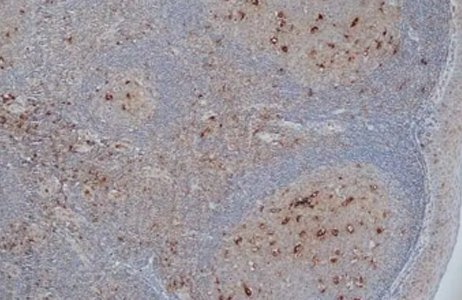
Fig. 2. Staining of formalin-fixed, paraffin-embedded human tonsil. |
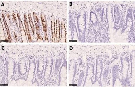
Fig. 3. Staining of formalin-fixed, paraffin-embedded rat jejunum. |
||
|
|||
|
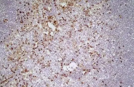
Fig. 4. Staining of a cryosection of mouse lymph node. |
|||
Frequently Asked Questions
What is aldehyde fixation?
Aldehyde fixation is a tissue preservation method that uses aldehydes such as formaldehyde to create protein cross-links, preventing degradation while maintaining tissue structure for downstream staining applications.
Why does aldehyde fixation reduce staining intensity?
Cross-linked proteins can hide antibody-binding sites, reducing antigen accessibility.
Is paraformaldehyde the same as formalin?
Paraformaldehyde is a solid, polymerized form of formaldehyde that generates formaldehyde in solution. Formalin is a formaldehyde solution often stabilized with methanol to prevent repolymerization. In terms of formaldehyde concentration, 10% neutral-buffered formalin (NBF) is equivalent to 4% paraformaldehyde (PFA).
Does fixation time affect antigen detection?
Yes. Extended fixation can increase protein cross-linking and epitope masking.
When is antigen retrieval necessary?
Antigen retrieval is commonly required when aldehyde fixation has masked epitopes needed for antibody binding.
What is epitope masking?
Epitope masking occurs when protein cross-linking caused by fixation hides antibody-binding sites, reducing antibody access and staining intensity.
References
- Engel K et al. (2011). Effects of preanalytical variables on the detection of proteins by immunohistochemistry in formalin-fixed, paraffin-embedded tissue. Arch Pathol Lab Med 135, 537–543
- McLean IW and Nakane PK (1974). Periodate-lysine-paraformaldehyde fixative. A new fixation for immunoelectron microscopy. J Histochem Cytochem 22, 1077–1083
- Pollard K et al. (1987). Fixation, processing, and immunochemical reagent effects on preservation of T-lymphocyte surface membrane antigens in paraffin-embedded tissue. J Histochem Cytochem 35, 1329–1338
Further Reading
Be the First to Receive Updates on Products and Resources
You'll receive regular communications featuring free lab resources such as application guides, posters, mini-reviews, protocols, practical tips, webinar invitations, special offers, and updates on new research tools and products.
You'll also get our specialist newsletters—including Fab Fragments, Flow. Sorted., and RiVETing—along with news on the latest antibody innovations, such as TrailBlazer™ Antibodies and StarBright™ Dyes.
During sign-up, you can choose your areas of interest so you only receive content relevant to your research, and you can unsubscribe at any time.
