Cytotrack
Cytotrack Blue 403/454 Cell Proliferation Assay Kit
- Product Type
- Accessory Reagent
- Specificity
- Cytotrack


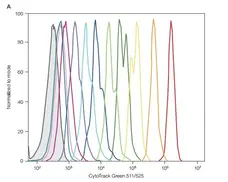
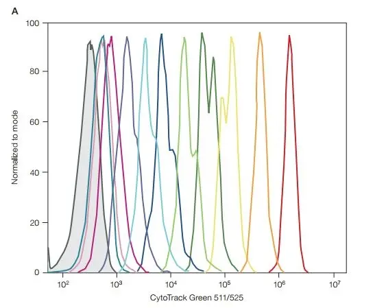
CytoTrack Green 511/525 Cell Proliferation Assay Kit (1351203) used to assess proliferation of B16-F10 mouse myeloma cells by flow cytometry.
Image caption:
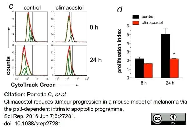
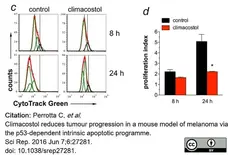
Cytotoxic and anti-proliferative properties of climacostol in melanoma cells.
(c) Flow cytometry analysis of B16-F10 proliferation at 8 and 24 h after cell treatment with climacostol (30 μg/ml) or vehicle (control). The vertical black line represents the undivided cell peak used as point of reference for the CytoTrack Green profile. The data are representative of 3 independent experiments. (d) Proliferation index for experiments shown in (c). *p <0.001 vs the respective control.
From: Perrotta C, Buonanno F, Zecchini S, Giavazzi A, Proietti Serafini F, Catalani E, Guerra L, Belardinelli MC, Picchietti S, Fausto AM, Giorgi S, Marcantoni E, Clementi E, Ortenzi C, Cervia D.
Climacostol reduces tumour progression in a mouse model of melanoma via the p53-dependent intrinsic apoptotic programme.
Sci Rep. 2016 Jun 7;6:27281.
doi: 10.1038/srep27281.
This image is from an open access article distributed under terms of a Creative Commons Attribution License.

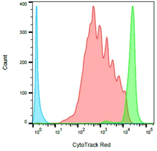
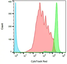
CytoTrack Red 628/643 Cell Proliferation Assay Kit (1351205) and CYTOTRACK Green 511/525 Cell Proliferation Assay Kit (1351203) used to assess proliferation in murine cells by flow cytometry.
Image caption:
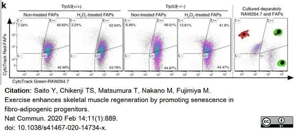
Trp53(−/−) FAP transplantation impairs muscle regeneration after acute muscle injury.
Representative CytoTrack Green-labeled RAW264.7 and CytoTrack Red-labeled FAPs plots after co-culture with RAW264.7 and FAPs.
From: Saito Y, Chikenji TS, Matsumura T, Nakano M, Fujimiya M.
Exercise enhances skeletal muscle regeneration by promoting senescence in fibro-adipogenic progenitors.
Nat Commun. 2020 Feb 14;11(1):889.
doi: 10.1038/s41467-020-14734-x.
This image is from an open access article distributed under terms of a Creative Commons Attribution License
Filter by Application:
F Reset| CytoTrack cell proliferation assay kits are available in three distinct dyes for easy multicolor cell analysis: blue, green and red. Easily incorporate a cell tracking stain into your multicolor panel. The proprietary chemistry of CytoTrack dyes enables the resolution of up to ten cell divisions. Each dye is cell permeable and comprises a fluorophore, a fluorescence blocker and a cell-retaining group. Upon entering a live cell, the fluorescence blocker is cleaved by intracellular esterases and the cell-retaining group of the fluorophore reacts with intracellular proteins to create a stable, covalent bond. As the cells divide, the fluorescence intensity is successively halved and each cell divison can be identified. |
- Reagents In The Kit
- CytoTrack Dye (4 vials, 50 assays/vial)
DMSO (1 vial, 250 μl) - Regulatory
- For research purposes only
- Guarantee
- Guaranteed until date of expiry. Please see product label.
Store at -20oC only
This product is photosensitive and should be protected from light
This product is photosensitive and should be protected from light
This product has been reported to work in the following applications. This information is derived from testing within our laboratories, peer-reviewed publications or personal communications from the originators. Please refer to references indicated for further information. For general protocol recommendations, please visit the antibody protocols page.
| Application Name | Verified | Min Dilution | Max Dilution |
|---|---|---|---|
| Flow Cytometry |  |
1/500 |
Where this product has not been tested for use in a particular technique this does not necessarily exclude its use in such procedures. Suggested working dilutions are given as a guide only. It is recommended that the user titrates the product for use in their own system using appropriate negative/positive controls.
- Instructions For Use
-
1351202
Important: Thaw all components prior to use.
1. Prepare a 500x stock solution. Add 50 μl of DMSO and mix.
2. Protocol for use in culture medium (for product 1351203) - Add 1 μl of stock solution into 500 μl of media containing 1 x 10⁶ cells of interest.
Protocol for use with buffer (for products 1351202, 1351203 and 1351205) - Prepare a 1x working solution. Add 1 μl of stock solution into 500 μl of buffer, pH 7. Add 500 μl of 1x solution to 1 x 10⁶cells.
3. Incubate at room temperature for 15 mins. Protect from light.
4. Pellet the cells by centrifugation.
5. Remove the supernatant and wash the cells using 3 ml of fresh prewarmed culture media.
6. Resuspend the cell in 500 μl of culture media.
7. Place the cells in the appropriate conditions for cells proliferation.
8. Harvest the cells and stain them for other markers if appropriate.
9. Analyze or sort cells using a flow cytometer equipped with a suitable excitation laser and emission filter.
References for Cytotrack
-
Perrotta, C. et al. (2018) Nitric Oxide Generated by Tumor-Associated Macrophages Is Responsible for Cancer Resistance to Cisplatin and Correlated With Syntaxin 4 and Acid Sphingomyelinase Inhibition.
Front Immunol. 9: 1186. -
Perrotta, C. et al. (2016) Climacostol reduces tumour progression in a mouse model of melanoma via the p53-dependent intrinsic apoptotic programme.
Sci Rep. 6: 27281. -
Zecchini, S. et al. (2019) Autophagy controls neonatal myogenesis by regulating the GH-IGF1 system through a NFE2L2- and DDIT3-mediated mechanism.
Autophagy. 15 (1): 58-77. -
Saito, Y. et al. (2020) Exercise enhances skeletal muscle regeneration by promoting senescence in fibro-adipogenic progenitors.
Nat Commun. 11 (1): 889. -
Cappello, P. et al. (2016) Anti-α-enolase antibody limits the invasion of myeloid-derived suppressor cells and attenuates their restraining effector T cell response.
Oncoimmunology. 5 (5): e1112940. -
Tario, J.J. et al. (2018) Monitoring Cell Proliferation by Dye Dilution: Considerations for Probe Selection.
Methods Mol Biol. 1678: 249-99. -
Loef, E.J. et al. (2021) Live-Cell Microscopy Reveals That Human T Cells Primarily Respond Chemokinetically Within a CCL19 Gradient That Induces Chemotaxis in Dendritic Cells.
Front Immunol. 12: 628090. -
Wang, X. et al. (2021) Activatable Biomineralized Nanoplatform Remodels the Intracellular Environment of Multidrug-Resistant Tumors for Enhanced Ferroptosis/Apoptosis Therapy.
Small. : e2102269.
Request a different product with this specificity
Please Note: All Products are "FOR RESEARCH PURPOSES ONLY"
Always be the first to know.
When we launch new products and resources to help you achieve more in the lab.
Yes, sign me up