CD86 antibody | 24F
Mouse anti Rat CD86:Alexa Fluor® 488
- Product Type
- Monoclonal Antibody
- Clone
- 24F
- Isotype
- IgG1
- Specificity
- CD86


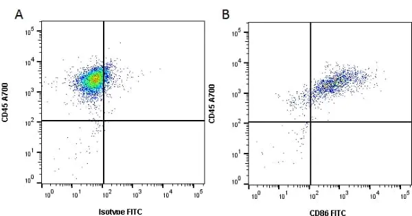
Figure B. Alexa Fluor® 700 conjugated Mouse anti Rat CD45 antibody, clone OX-1 (MCA43A700) and FITC conjugated Mouse anti Rat CD86 antibody, clone 24F (MCA2874F). All experiments performed on Rat splenocytes stimulated with LPS for 48 hours gated on live, single cells in the presence of 10% Rat serum.
Data acquired on the ZE5 Cell analyser.
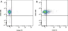
Figure B. Alexa Fluor® 488 conjugated Mouse anti Rat CD45 antibody, clone OX-1 (MCA43A488) and RPE conjugated Mouse anti Rat CD86 antibody, clone 24F (MCA2874PE). All experiments performed on red cell lysed rat blood gated on mononuclear cells.
Data acquired on the ZE5 Cell analyser.
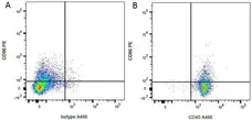
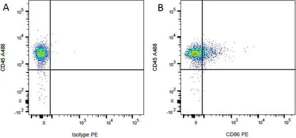
Figure B. RPE conjugated Mouse anti Rat CD86 antibody, clone 24F (MCA2874PE) and Alexa Fluor® 488 conjugated Mouse anti Rat CD45 antibody, clone OX-1 (MCA43A488). All experiments performed on red cell lysed rat blood gated on mononuclear cells.
Data acquired on the ZE5 Cell analyser.


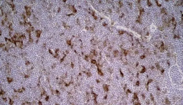
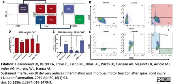
AlexaFluor® 647 conjugated Mouse anti Rat CD86 antibody, clone 24F (MCA2847A647) used as part of a panel of antibodies to assess M1 and M2 macrophages by flow cytometry.
Image caption:
Percentages of M1 and M2 macrophages. The cells determined to be CD11b+CD45HIGH macrophages were further analyzed for macrophage phenotype. Cells that were positive for the M1 markers CD68 or CD80 were characterized as “M1” macrophages and the CD68−CD80− macrophages were further analyzed with CD86 and CD163 to distinguish early stage“M2b” and later stage “M2c,” as shown in the schematic (a). The FMOs for CD68 and CD80 were used to set the gates and a representative sample of MCMs+IL-10 is shown for the CD68 vs CD80 (b). The FMOs for CD86 and CD163 were used to set the gates for distinguishing “M2b” and “M2c” cells and the CD68−CD80− “M2” macrophages were further analyzed for CD86 vs CD163 (c). Comparing across groups, MCMs+IL-10 had significantly less “M1” macrophages than the Controls, Local IL-10, and MCMs (d). MCMs+IL-10 also had significantly more “M2b” macrophages than the Controls, Local IL-10, and MCMs (e). *P <0.05 (Tukey's Test); error bars represent ± SEM; n = 3 for Controls and 4 for all other groups
From: Hellenbrand DJ, Reichl KA, Travis BJ, Filipp ME, Khalil AS, Pulito DJ, Gavigan AV, Maginot ER, Arnold MT, Adler AG, Murphy WL, Hanna AS.
Sustained interleukin-10 delivery reduces inflammation and improves motor function after spinal cord injury.
J Neuroinflammation. 2019 Apr 30;16(1):93.
doi: 10.1186/s12974-019-1479-3.
This image is from an open access article distributed under the terms of a Creative Commons Attribution License.
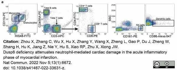
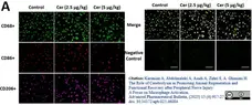
Alexa Fluor® 647 conjugated Mouse anti Rat CD86 antibody, clone 24F (MCA2874A647) used as a dendritic cell marker for rat cell samples in flow cytometry.
Image caption:
Percentage of multiple immune cell populations in peripheral leukocytes and mediastinal lymph nodes of WT and Dusp6-deficient rats.
(a) Representative gating images of flow cytometry analysis for immune cell populations using myeloid cell marker CD11b, granulocyte marker HIS48, T cell marker CD3/CD4/CD8, B cell marker CD45RA, NK cell marker CD161, and dendritic cell marker CD86.
From: Zhou X, Zhang C, Wu X, Hu X, Zhang Y, Wang X, Zheng L, Gao P, Du J, Zheng W, Shang H, Hu K, Jiang Z, Nie Y, Hu S, Xiao RP, Zhu X, Xiong JW.
Dusp6 deficiency attenuates neutrophil-mediated cardiac damage in the acute inflammatory phase of myocardial infarction.
Nat Commun. 2022 Nov 5;13(1):6672. doi:
10.1038/s41467-022-33631-z.
This image is from an open access article distributed under terms of a Creative Commons Attribution License.
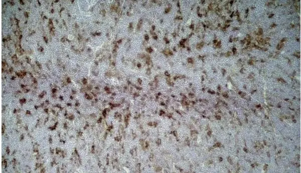
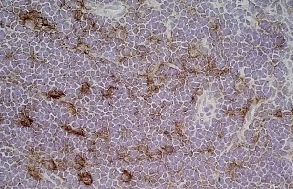
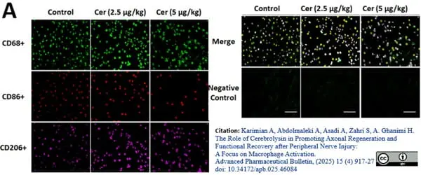
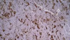
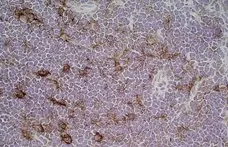
Mouse anti Rat CD86 antibody, clone 24F (MCA2874) used to assess CD68 expression in rat sciatic nerve by immunofluorreescence.
Image caption:
Impact of Cerebrolysin treatment on macrophage polarization in the sciatic nerve. (A) Triple immunofluorescent staining of resident macrophages in the sciatic nerve, highlighting the M0 marker (CD68/ green), M1 marker (CD86+/red), and M2 marker (CD206+/magenta).
From: Karimian A, Abdolmaleki A, Asadi A, Zahri S, A. Ghanimi H.
The Role of Cerebrolysin in Promoting Axonal Regeneration and Functional Recovery after Peripheral Nerve Injury: A Focus on Macrophage Activation.
Adv Pharm Bull. 2025;15(4):917-927.
doi: 10.34172/apb.025.46084
This image is from an open access article distributed under terms of a Creative Commons Attribution License.
Filter by Application:
F P C Reset| Mouse anti Rat CD86 antibody, clone 24F recognizes rat CD86, otherwise known as B7-2, a type I transmembrane protein and member of the Ig superfamily, which acts as a ligand for both CD28 and CD152 (CTLA-4), and is primarily expressed on antigen presenting cells (APCs) including dendritic cells, and also on germinal centre B cells and macrophages. Like CD80, CD86 is an accessory molecule which functions in the CD28-CD80/CD86 co-stimulatory pathway, vital for T cell activation, crosstalk between T and B cells, and Th2-mediated Ig production. Mouse anti Rat CD86 antibody, clone 24F has been shown to block the co-stimulatory activity of rat CD86 (Maeda et al. 1997). |
- Target Species
- Rat
- Product Form
- Purified IgG conjugated to Alexa Fluor 488 - liquid
- Preparation
- Purified IgG prepared by affinity chromatography on Protein G from tissue culture supernatant
- Buffer Solution
- Phosphate buffered saline
- Preservative Stabilisers
- 0.09% Sodium Azide (NaN3)
1% Bovine Serum Albumin - Immunogen
- HTLV-1 transformed Lewis-S1 cells.
- Approx. Protein Concentrations
- IgG concentration 0.05mg/ml
- Fusion Partners
- Spleen cells from immunised Balb/c mice were fused with cells of the P3U1 mouse myeloma cell line.
- Max Ex/Em
-
Fluorophore Excitation Max (nm) Emission Max (nm) Alexa Fluor®488 495 519 - Regulatory
- For research purposes only
- Guarantee
- 12 months from date of despatch
- Acknowledgements
- This product is provided under an intellectual property licence from Life Technologies Corporation. The transfer of this product is contingent on the buyer using the purchase product solely in research, excluding contract research or any fee for service research, and the buyer must not sell or otherwise transfer this product or its components for (a) diagnostic, therapeutic or prophylactic purposes; (b) testing, analysis or screening services, or information in return for compensation on a per-test basis; (c) manufacturing or quality assurance or quality control, or (d) resale, whether or not resold for use in research. For information on purchasing a license to this product for purposes other than as described above, contact Life Technologies Corporation, 5791 Van Allen Way, Carlsbad CA 92008 USA or outlicensing@thermofisher.com
This product is shipped at ambient temperature. It is recommended to aliquot and store at -20°C on receipt. When thawed, aliquot the sample as needed. Keep aliquots at 2-8°C for short term use (up to 4 weeks) and store the remaining aliquots at -20°C.
Avoid repeated freezing and thawing as this may denature the antibody. Storage in frost-free freezers is not recommended. This product is photosensitive and should be protected from light.
Avoid repeated freezing and thawing as this may denature the antibody. Storage in frost-free freezers is not recommended. This product is photosensitive and should be protected from light.
This product has been reported to work in the following applications. This information is derived from testing within our laboratories, peer-reviewed publications or personal communications from the originators. Please refer to references indicated for further information. For general protocol recommendations, please visit the antibody protocols page.
| Application Name | Verified | Min Dilution | Max Dilution |
|---|---|---|---|
| Flow Cytometry |  |
Neat | 1/10 |
Where this product has not been tested for use in a particular technique this does not necessarily exclude its use in such procedures. Suggested working dilutions are given as a guide only. It is recommended that the user titrates the product for use in their own system using appropriate negative/positive controls.
- Flow Cytometry
- Use 10ul of the suggested working dilution to label 1x10⁶ cells in 100ul.
How to Use the Spectraviewer
Watch the Tool Tutorial Video ▸- Start by selecting the application you are interested in, with the option to select an instrument from the drop down menu or create a customized instrument
- Select the fluorophores or fluorescent proteins you want to include in your panel to check compatibility
- Select the lasers and filters you wish to include
- Select combined or multi-laser view to visualize the spectra
| Description | Product Code | Applications | Pack Size | List Price | Your Price | Quantity | |
|---|---|---|---|---|---|---|---|
| Mouse IgG1 Negative Control:Alexa Fluor® 488 | MCA1209A488 | F | 100 Tests/1ml |

|
Log in | ||
| List Price | Your Price | ||||||
|
|
Log in | ||||||
| Description | Mouse IgG1 Negative Control:Alexa Fluor® 488 | ||||||
Source Reference
-
Maeda, K. et al. (1997) Characterization of rat CD80 and CD86 by molecular cloning and mAb.
Int Immunol. 9 (7): 993-1000.
References for CD86 antibody
-
Damoiseaux, J.G. et al. (1998) Costimulatory molecules CD80 and CD86 in the rat; tissue distribution and expression by antigen-presenting cells.
J Leukoc Biol. 64 (6): 803-9. -
Kano, M. et al. (1998) A crucial role of host CD80 and CD86 in rat cardiac xenograft rejection in mice.
Transplantation. 65: 837-43. -
Sacedón, R. et al. (1999) Glucocorticoid-mediated regulation of thymic dendritic cell function.
Int Immunol. 11: 1217-24. -
Hanabuchi, S. et al. (2000) Development of human T-cell leukemia virus type 1-transformed tumors in rats following suppression of T-cell immunity by CD80 and CD86 blockade.
J Virol. 74: 428-35. -
Tamatani, T. et al. (2000) AILIM/ICOS: a novel lymphocyte adhesion molecule.
Int Immunol. 12: 51-5. -
Kawai, T. et al. (2000) T(h)1 transmigration anergy: a new concept of endothelial cell-T cell regulatory interaction.
Int Immunol. 12: 937-48. -
Macphee, I.A. et al. (2002) The Th2-response in mercuric chloride-induced autoimmunity requires continuing costimulation via CD28.
Clin Exp Immunol. 129: 405-10. -
Ghiringhelli, F. et al. (2005) Tumor cells convert immature myeloid dendritic cells into TGF-beta-secreting cells inducing CD4+CD25+ regulatory T cell proliferation.
J Exp Med. 202: 919-29.
View The Latest Product References
-
MacPhee, I.A. et al. (2006) Blockade of OX40-ligand after initial triggering of the T helper 2 response inhibits mercuric chloride-induced autoimmunity.
Immunology. 117: 402-8. -
Yrlid, U. et al. (2006) A distinct subset of intestinal dendritic cells responds selectively to oral TLR7/8 stimulation.
Eur J Immunol. 36: 2639-48. -
Dilek, N. et al. (2012) Control of transplant tolerance and intragraft regulatory T cell localization by myeloid-derived suppressor cells and CCL5.
J Immunol. 188: 4209-16. -
Matsumoto, S. et al. (2015) CD200+ and CD200- macrophages accumulated in ischemic lesions of rat brain: the two populations cannot be classified as either M1 or M2 macrophages.
J Neuroimmunol. 282: 7-20. -
Patil, P.S. et al. (2016) Fluorinated methacrylamide chitosan hydrogels enhance collagen synthesis in wound healing through increased oxygen availability.
Acta Biomater. 36: 164-74. -
Hellenbrand, D.J. et al. (2019) Sustained interleukin-10 delivery reduces inflammation and improves motor function after spinal cord injury.
J Neuroinflammation. 16 (1): 93. -
Zhou, X. et al. (2022) Dusp6 deficiency attenuates neutrophil-mediated cardiac damage in the acute inflammatory phase of myocardial infarction.
Nat Commun. 13 (1): 6672. -
Karimian, A. et al. (2025) The Role of Cerebrolysin in Promoting Axonal Regeneration and Functional Recovery after Peripheral Nerve Injury: A Focus on Macrophage Activation
Adv Pharm Bull. 15 (4): 917-927 -
Ye, Y. et al. (2025) Immunomodulating red blood cell coating for mitigation of foreign body reactions
Engineered Regeneration. 6: 218-229.
- Synonyms
- B7-2
- RRID
- AB_1658078
MCA2874A488
If you cannot find the batch/lot you are looking for please contact our technical support team for assistance.
View more products with CD86 specificity
Please Note: All Products are "FOR RESEARCH PURPOSES ONLY"
View all Anti-Rat ProductsAlways be the first to know.
When we launch new products and resources to help you achieve more in the lab.
Yes, sign me up