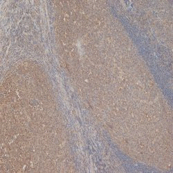
Ki67 antibody | AbD02531_h/m_IgG1

Human anti Ki67
- Product Type
- Monoclonal Antibody
- Clone
- AbD02531_h/m_IgG1
- Isotype
- HuCAL/mouse IgG1 Fc
- Specificity
- Ki67
| Human anti Ki67 antibody, clone AbD02531 recognizes the Ki67 cell-cycle associated protein. Ki67 is expressed in proliferating cells but not in quiescent cells. Expression of this antigen occurs preferentially during late G1, S, G2, and M phases of the cell cycle, while in cells in G0 phase the antigen cannot be detected. Consequently, Ki-67 antigen expression is used in tumor pathology to detect proliferating cells in neoplastic diseases. In cultured cells, Ki-67 is expressed in the nucleolus of interphase cells.
The Ki67 gene contains 15 exons. The Ki67 repeat region, within which there is a 22-amino acid Ki67 motif, is encoded by exon 13. The shorter isoform lacks exon 7. Northern blot analysis reveals multiple transcripts ranging from approximately 8.9 to 12.5 kb in proliferating but not quiescent cells. Immunoblot analysis shows expression of 320 and 359 kDa proteins. Antisense oligonucleotides inhibit cellular proliferation in a dose-dependent manner, suggesting that Ki67 protein expression may be an absolute requirement for cell proliferation. Within cells Ki67 is predominantly localized in the G1 phase in the perinucleolar region, in the later phases it is also detected throughout the nuclear interior, being predominantly localized in the nuclear matrix. In mitosis, it is present on all chromosomes. |
- Target Species
- Human
- Species Cross-Reactivity
-
Target Species Cross Reactivity Bovine Expected from Sequence Macaque Expected from Sequence Dog Expected from Sequence Chimpanzee Expected from Sequence Rhesus Monkey Expected from Sequence - N.B. Antibody reactivity and working conditions may vary between species.
- Product Form
- Chimeric human-mouse IgG1 antibody selected from the HuCAL phage display library and expressed in a human cell line. The antibody has variable regions of human origin and an Fc portion (including CH1 and CL domains) from mouse IgG1. It can be detected by anti mouse Fc specific secondary antibodies. This antibody is supplied liquid.
- Preparation
- Purified IgG prepared by affinity chromatography on Protein A.
- Source
- HKB-11
- Buffer Solution
- Phosphate buffered saline
- Preservative Stabilisers
- 0.09% Sodium Azide (NaN3)
- Immunogen
- Peptide derived from the human Ki67 protein, sequence GFKELFQTPG, coupled via a C-terminal cysteine to carrier proteins
- Approx. Protein Concentrations
- IgG concentration 0.5 mg/ml
- Regulatory
- For research purposes only
- Guarantee
- 12 months from date of despatch
- Acknowledgements
- This product and/or its use is covered by claims of U.S. patents, and/or pending U.S. and non-U.S. patent applications owned by or under license to Bio-Rad Laboratories, Inc. See bio-rad.com/en-us/trademarks for details.
- Licensed Use
- For in vitro research purposes only, unless otherwise specified in writing by Bio-Rad.
Avoid repeated freezing and thawing as this may denature the antibody. Storage in frost-free freezers is not recommended.
| Application Name | Verified | Min Dilution | Max Dilution |
|---|---|---|---|
| ELISA | 2.0 ug/ml | ||
| Immunofluorescence | |||
| Immunohistology - Frozen | |||
| Immunohistology - Paraffin 1 | 0.2 ug/ml | 1.0 ug/ml | |
| Western Blotting |
- 1This product requires antigen retrieval using heat treatment prior to staining of paraffin sections.Sodium citrate buffer pH 6.0 is recommended for this purpose.
- Technical Advice
- Recommended protocols and further information about HuCAL recombinant antibody technology can be found in the HuCAL Antibodies Technical Manual.
- Histology Positive Control Tissue
- Human tonsil
| Description | Product Code | Applications | Pack Size | List Price | Your Price | Quantity | |
|---|---|---|---|---|---|---|---|
| Goat anti Human IgG F(ab')2:HRP | 0500-0099 | E WB | 0.5 ml |
|
Log in | ||
| List Price | Your Price | ||||||
|
|
Log in | ||||||
| Description | Goat anti Human IgG F(ab')2:HRP | ||||||
References for Ki67 antibody
-
Jarutat, T. et al. (2006) Isolation and comparative characterization of Ki-67 equivalent antibodies from the HuCAL phage display library.
Biol. Chem. 387: 995-1003. -
Liu, S.K. et al. (2011) Delta-like ligand 4-notch blockade and tumor radiation response.
J Natl Cancer Inst. 103 (23): 1778-98.
- RRID
- AB_915348
- UniProt
- P46013
- Entrez Gene
- MKI67
- GO Terms
- GO:0005524 ATP binding
- GO:0005730 nucleolus
- GO:0008022 protein C-terminus binding
- GO:0008283 cell proliferation
HCA053
If you cannot find the batch/lot you are looking for please contact our technical support team for assistance.
Please Note: All Products are "FOR RESEARCH PURPOSES ONLY"
View all Anti-Human ProductsAlways be the first to know.
When we launch new products and resources to help you achieve more in the lab.
Yes, sign me up