pSIVA™ Microscopy Kit
pSIVA™ REAL-TIME Apoptosis Fluorescent Microscopy Kit
- Product Type
- Kits
- Specificity
- pSIVA™ Microscopy Kit


Filter by Application:
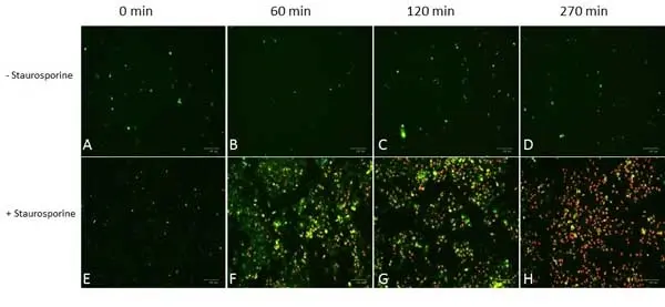
IF Reset| The process of apoptosis is undertaken in several stages defined by specific cellular morphologies. One of the earlier stages of apoptosis is a change of the plasma membrane’s phospholipid asymmetry. This rearrangement results in the translocation of phosphatidylserine (PS) from the inner to the outer plasma membrane (in non-apoptotic cells PS is exclusively located to the inner plasma membrane). However, apoptosis is reversible until reaching a certain point in the pathway and until then PS exposure can be considered as a transient event. The event defining whether the cell can be rescued and continues living is the onset of mitochondrial outer membrane permeabilization ( The pSIVA (polarity-Sensitive Indicator of Viability & Apoptosis) probe is a biosensor conjugated to the green emitting IANBD dye (excitation maximum 488 nm, emission maximum 530 nm) and only fluoresces when bound to PS in the presence of Ca2+ (Kim et al. 2010a, 2010b). The method thereby allows the analysis of kinetic apoptosis events in real time by live cell imaging and immunofluorescence / immunocytochemistry. In contrast to other PS detection based assays (e.g. annexin V) the pSIVA Real-Time Apoptosis Fluorescent Microscopy Kit does not require washing steps as you can simply add the probe and start analyzing. |
- Reagents In The Kit
- pSIVA-IANBD 200 μl
Propidium Iodide Staining Solution 500 μl - Regulatory
- For research purposes only.
- Guarantee
- 6 months from date of despatch.
- Acknowledgements
- pSIVA is a trademark of Novus Biologicals and is protected under patent no. 8.541.549.
This product is shipped at ambient temperature.
Store at +4°C. DO NOT FREEZE.
This product should be stored undiluted. This product is photosensitive and should be protected from light.
Store at +4°C. DO NOT FREEZE.
This product should be stored undiluted. This product is photosensitive and should be protected from light.
This product has been reported to work in the following applications. This information is derived from testing within our laboratories, peer-reviewed publications or personal communications from the originators. Please refer to references indicated for further information. For general protocol recommendations, please visit the antibody protocols page.
| Application Name | Verified | Min Dilution | Max Dilution |
|---|---|---|---|
| Immunocytochemistry |  |
Refer to Instructions For Use | |
| Immunofluorescence | |
Refer to Instructions For Use | |
| Live Cell Imaging | |
Refer to Instructions For Use |
Where this product has not been tested for use in a particular technique this does not necessarily exclude its use in such procedures. Suggested working dilutions are given as a guide only. It is recommended that the user titrates the product for use in their own system using appropriate negative/positive controls.
- Instructions For Use
-
APO004
Prior to commencing the microscopy experiment, please ensure that your cell culture medium contains between 1-2 mM Ca2+. Ca2+ is essential for binding of the pSIVA-IANBD probe to exposed phosphatidylserine (Kim et al. 2010b). If Ca2+ levels are insufficient, supplement the culture medium with 2 mM Ca2+.
1. Seed cells into culture plates and allow cells to adhere.
2. Optional. After 24 hours exchange the culture medium for medium containing 2 mM Ca2+, if required.
3. Optional. Induce apoptosis by treating cells with apoptosis inducing agents such as staurosporine or camptothecin.
4. Add 10–20 μl/ml* of the pSIVA-IANBD probe to cells. Mix gently by moving culture plates
backwards and forwards and side to side to ensure even distribution of the probe. DO NOT PIPETTE TO MIX.
5. Optional. If distinction between apoptotic and necrotic/dead cells is desired, add between 5–10 μl/ml* of propidium iodide (PI) to cells. Mix gently by moving plates backwards and forwards and side to side to ensure even distribution of PI. DO NOT PIPETTE TO MIX.
6. Observe cells under microscope using the green fluorescence filter for pSIVA-IANBD (excitation maximum 488 nm, emission maximum 530 nm) and the red fluorescence filter for PI (excitation maximum 535 nm, emission maximum 617 nm) visualization.
* The stated pSIVA-IANBD and PI quantities are guidelines only and may have to be optimized.
pSIVA Real-time Apoptosis Fluorescent Microscopy Kit Instructions.
How to Use the Spectraviewer
Watch the Tool Tutorial Video ▸- Start by selecting the application you are interested in, with the option to select an instrument from the drop down menu or create a customized instrument
- Select the fluorophores or fluorescent proteins you want to include in your panel to check compatibility
- Select the lasers and filters you wish to include
- Select combined or multi-laser view to visualize the spectra
| Description | Product Code | Applications | Pack Size | List Price | Your Price | Quantity | |
|---|---|---|---|---|---|---|---|
| Annexin V:APC Assay Kit | ANNEX200APC | F | 200 Tests | Log in | |||
| List Price | Your Price | ||||||
| Log in | |||||||
| Description | Annexin V:APC Assay Kit | ||||||
| Annexin V:APC Assay Kit | ANNEX50APC | F | 50 Tests | Log in | |||
| List Price | Your Price | ||||||
| Log in | |||||||
| Description | Annexin V:APC Assay Kit | ||||||
References for pSIVA™ Microscopy Kit
-
Kim, Y.E. et al. (2010) (a) Engineering a polarity-sensitive biosensor for time-lapse imaging of apoptotic processes and degeneration.
Nat Methods 7(1): 67–73. -
Kim, Y.E. et al. (2010) (b) Monitoring apoptosis and neuronal degeneration by real-time detection of phosphatidylserine externalization using a polarity-sensitive indicator of viability and apoptosis.
Nat Protoc. 5(8): 1396-405.
- Synonyms
- Annexin 12
- Annexin XII
APO004
If you cannot find the batch/lot you are looking for please contact our technical support team for assistance.
Request a different product with this specificity
Please Note: All Products are "FOR RESEARCH PURPOSES ONLY"
Always be the first to know.
When we launch new products and resources to help you achieve more in the lab.
Yes, sign me up