Cathepsin B

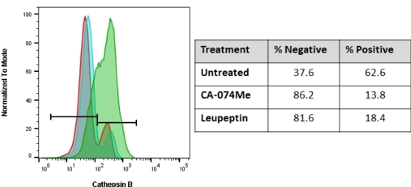
Jurkat cells were treated with 10 μM CA-074Me for 3 hours (blue histogram), 10μM Leupeptin for 1 hour (red histogram) or were untreated (green histogram). They were then stained for 1 hour with R110-(RR)2. After incubation with the R110-(RR)2 substrate, the samples were analyzed by flow cytometry using a ZE5 Cell Analyzer (488 nm excitation and a 525/35 emission filter). Treatment with CA-074Me or Leupeptin decreased the fluorescence signal detected compared to the untreated control (see table), which displayed a baseline level of cathepsin activity
Green Cathepsin B Kit
- Product Type
- Kits
- Specificity
- Cathepsin B

| Green Cathepsin B Kit enables the quantitation and monitoring of intracellular cathepsin activity over time in vitro. The Rhodamine 110 Cathepsin B substrate reagent is a non-cytotoxic and membrane permeant substrate that fluoresces green upon cleavage by active cathepsin enzymes. |
- Reagents In The Kit
- 4 vial of Rhodamine110-(RR)2 substrate - lyophilized
10X Cellular Assay Buffer, 60 ml
Hoechst Stain, 1 ml - Test Principle
- Rhodamine 110 Cathepsin B substrate utilizes the photostable green fluorophore, rhodamine 110. Rhodamine 110 cathepsin B substrate is comprised of rhodamine 110 coupled to two copies of the amino acid sequence, arginine-arginine (RR), which is the preferential target sequence for cathepsin B. When bi-substituted via amide linkage to two cathepsin B target peptide sequences, rhodamine110 is nonfluorescent. Following enzymatic cleavage at one or both arginine (R) amide linkage sites, the mono and non-substituted rhodamine 110 fluorophores generate green fluorescence when excited at 500 nm.
To use the Green Cathepsin B Assay, simply add the Rhodamine 110 Cathepsin B substrate [R110-(RR)2] directly to the cell culture media (or 1X Cellular Assay Buffer), incubate, and analyze. Because R110-(RR)2 is cell-permeant, it easily penetrates the cell membrane and the membranes of the internal cellular organelles - no lysis or permeabilization steps are required. R110-(RR)2 will enter the cell in a non-fluorescent state. If cathepsin enzymes are active, they will cleave off the two arginine-arginine cathepsin B targeting sequences and allow the rhodamine 110 fluorophore to become fluorescent upon excitation. By varying the duration and concentration of exposure to the R110-(RR)2 substrate, a picture can be obtained of the relative abundance and intracellular location of cathepsin enzymatic activity. Positive cells will fluoresce green, while negative cells will exhibit very low levels of background green fluorescence. There is no interference from pro-cathepsins forms of the enzymes. If the treatment or experimental condition stimulates cathepsin activity, cells containing elevated levels of cathepsin activity will appear brighter green than cells with lower levels of cathepsin activity. - Max Ex/Em
-
Fluorophore Excitation Max (nm) Emission Max (nm) Rhodamine110-(RR)2 525 535 - Regulatory
- For research purposes only
- Guarantee
- Guaranteed until date of expiry. Please see product label.
MULTIPLE STORAGE CONDITIONS APPLY ON ARRIVAL. Store the unopened kit (and each unopened component) according to the storage instructions on each component label. Store the Rhodamine110-(RR)2 substrate at -20oC. Once reconstituted in DMSO, use the Rhodamine110-(RR)2 substrate immediately or aliquot and store at -20oC for 6 months, protected from light. Avoid repeated freezing and thawing
This product has been reported to work in the following applications. This information is derived from testing within our laboratories, peer-reviewed publications or personal communications from the originators. Please refer to references indicated for further information. For general protocol recommendations, please visit the antibody protocols page.
| Application Name | Verified | Min Dilution | Max Dilution |
|---|---|---|---|
| Flow Cytometry | Refer to Instruction for Use | ||
| Immunofluorescence |
Where this product has not been tested for use in a particular technique this does not necessarily exclude its use in such procedures. Suggested working dilutions are given as a guide only. It is recommended that the user titrates the product for use in their own system using appropriate negative/positive controls.
- Instructions For Use
- Instructions for use can be found at https://www.bio-rad-antibodies.com/static/uploads/ifu/ict9151-2.pdf
View more products with CATHEPSIN specificity
Please Note: All Products are "FOR RESEARCH PURPOSES ONLY"
Always be the first to know.
When we launch new products and resources to help you achieve more in the lab.
Yes, sign me up