Autophagy Assay Kit, Red
Autophagy Assay, Red Detection Kit
- Product Type
- Kits
- Specificity
- Autophagy Assay Kit, Red


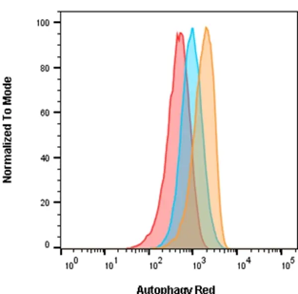
Jurkat cells were either untreated for basal conditions (red), treated with 0.5 μM Rapamycin for 18 hours (blue), or treated with 0.5 μM Rapamycin for 18 hours and 10 nM Bafilomycin A for 2 hours (orange) to induce autophagy. After staining with Autophagy Probe, Red for 30 minutes, cells were washed and analyzed on the ZE5 Cell Analyzer using the 561 nm laser and 640/20 filter

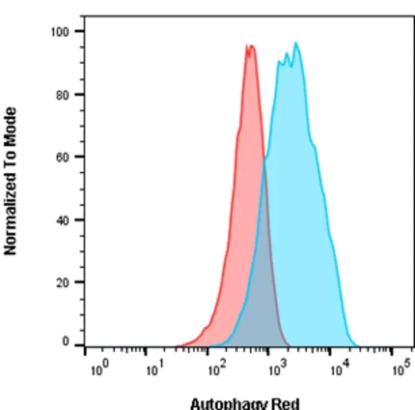
Jurkat cells were either untreated for basal conditions (red) or incubated in HBSS for 2 hours and 10 nM Bafilomycin A for 2 hours (blue) to induce autophagy. After staining with Autophagy Probe Red for 30 minutes, cells were washed and analyzed on the ZE5 Cell Analyzer using the 561 nm laser and 640/20 filter.
| Autophagy Assay, Red Detection Kit allows for the detection and monitoring of in vitro development of autophagy in living cells. Autophagy is a conserved lysosomal recycling process by which cells break down their own components such as proteins, lipids and carbohydrates. Autophagy plays a critical role in maintaining homeostasis by preventing the accumulation of damaged organelles by disassembling unnecessary or dysfunctional cells and cellular components (Mizushima et al. 2011). Autophagy occurs at low levels in the cell under normal conditions and can be rapidly upregulated during times of starvation or stress. Such degradation activities serve to provide nutrients (amino acids, nucleotides, fatty acids, etc.) and energy during periods of elevated bioenergetic demands (Mizushima et al. 2011, Levine et al. 2008). Another function of autophagy is to assist with the detection and destruction of intracellular pathogens (viruses, bacteria and parasites) (Levine et al. 2011). Dysregulation of autophagy has been associated with many disease states including cancer, infection and degenerative diseases (Levine et al. 2008). Autophagy is a dynamic process typically divided into three stages. During stage one, cytoplasmic components targeted for degradation are sequestered within a double-membrane phagopore (also called the isolation membrane). This results in the formation of a double-membrane vesicle called the autophagosome. During stage two, the autophagosome fuses with the lysosome to form the autolysosome. Degradation of the autophagosomal contents occurs during stage three (Mizushima et al. 2011, Hundeshagen et al. 2011). |
- Reagents In The Kit
-
APO010A: Autophagy Probe, Red, 1 vial - lyophilized
Fixative, 6 ml -
APO010B: Autophagy Probe, Red, 4 vials - lyophilized
Fixative, 6 ml - Test Principle
- Autophagy Probe, Red is a cell-permeant aliphatic molecule that fluoresces brightly when inserted in the lipid membranes of autophagosomes and autolysosomes. Autophagy Probe, Red can be readily detected by flow cytometry with optimal excitation at 590 nm and peak emission at 620 nm (ZE5 Cell Analyzer settings, 561 nm laser and 615/24 or 640/20 filter).
- Max Ex/Em
-
Fluorophore Excitation Max (nm) Emission Max (nm) Red Probe 590 620 - Regulatory
- For research purposes only
- Guarantee
- Guaranteed until date of expiry. Please see product label.
MULTIPLE STORAGE CONDITIONS APPLY ON ARRIVAL. Store the unopened kit (and each unopened component) according to the storage instructions on each component label.
Store the Autophagy Probe, Red at -20oC. Once reconstituted in DMSO, the Autophagy Probe, Red stock should be stored at -20oC for 6 months, protected from light. Avoid repeated freezing and thawing.
Store the Autophagy Probe, Red at -20oC. Once reconstituted in DMSO, the Autophagy Probe, Red stock should be stored at -20oC for 6 months, protected from light. Avoid repeated freezing and thawing.
This product has been reported to work in the following applications. This information is derived from testing within our laboratories, peer-reviewed publications or personal communications from the originators. Please refer to references indicated for further information. For general protocol recommendations, please visit the antibody protocols page.
| Application Name | Verified | Min Dilution | Max Dilution |
|---|---|---|---|
| Flow Cytometry |  |
Refer to Instructions For Use |
Where this product has not been tested for use in a particular technique this does not necessarily exclude its use in such procedures.
- Instructions For Use
-
APO010A, APO010B
Instructions for use can be found at www.bio-rad-antibodies.com/uploads/IFU/APO010.pdf
References for Autophagy Assay Kit, Red
-
Barghi, F. et al. (2026) Dual CDK4/6-PI3K/mTOR inhibition reinforces cytostatic programs and tumor control in preclinical models of primary and metastatic osteosarcoma.
Neoplasia. 72: 101266. -
Pandya, K. & Singh, N. (2026) MDC1 promotes nuclear localization of Beclin-1 and supports its role in ATM pathway in response to oxidative stress.
Eur J Cell Biol. 105 (3): 151540.
APO010A
APO010B
If you cannot find the batch/lot you are looking for please contact our technical support team for assistance.
Request a different product with this specificity
Please Note: All Products are "FOR RESEARCH PURPOSES ONLY"
Always be the first to know.
When we launch new products and resources to help you achieve more in the lab.
Yes, sign me up