
GATA transcription factors and their role in cancer diagnosis and prognosis
- Feb 15, 2016
- 3 min read
- Annalise Barnette, PhD
When a 22-month-old boy with trisomy 21 showing skin lesions on the head, neck and upper torso was presented for evaluation at the Children’s Hospital of Philadelphia, doctors initially suspected the root cause to be a viral or bacterial infection (Boos MD et al. 2015). However, when bacterial culture and polymerase chain reaction analyses for herpes simplex virus, varicella zoster virus, enterovirus and parechovirus were negative and antibiotic and anti-inflammatory treatments did not lessen the spread or discomfort of the lesions, other diagnoses had to be explored.
A link to acute megakaryoblastic leukemia (AMKL)
It has long been known that children with trisomy 21, which accounts for more than 90% of Down syndrome (DS) cases, have a higher chance of developing hematological disorders such as transient myeloproliferative disorder (TMD) and AMKL, a subtype of acute myeloid leukemia (AML). The incidence of AMKL among children with DS is approximately 500-fold higher than in normal children (Lange BJ et al. 1998). In addition, 10-20% of DS newborns present with TMD, which is a limited leukemia that undergoes spontaneous remission within three months. TMD is often accompanied by severe skin lesions which provide an important clue for diagnosis; however in the case presented, TMD was excluded because of the patient’s age.
It was eventually determined that the 22-month-old boy indeed suffered from AMKL, which is not known to present with any cutaneous lesions, and in fact this is the first such reported case (Boos MD et al. 2015). Immunohistochemistry (IHC) analysis of the skin lesions revealed positive staining for CD31 and CD68. These are markers of megakaryocytes (large bone marrow cells), which become highly proliferative in AMKL. The definitive diagnostic indicator of AMKL in this patient was however a positive IHC staining for the transcription factor GATA-1 in leukemic cells. The IHC staining was performed using two different antibodies, one recognizing the N-terminus and one recognizing the C-terminus. As the N-terminal staining was negative the conclusion could be made that the patient expressed a mutated, truncated form of GATA-1 called GATA-1s. This shortened GATA-1 isoform develops from a frame-shift mutation in the N-terminal activation domain of GATA-1 leading to the insertion of a premature stop codon, thus preventing synthesis of full-length GATA-1 (see image below). GATA-1s therefore lacks the N-terminal activation domain, which leads to its inability to regulate megakaryocytic target genes, ultimately contributing to disease. GATA-1s is therefore a relevant biomarker for AMKL as this is not observed in any other type of leukemia (Magalhães IQ et al. 2006, Wechsler J et al. 2002).
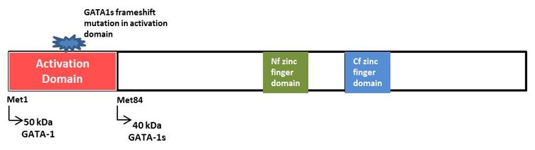
Schematic of Domain Structure of GATA-1 showing GATA-1s start site
Wechsler J et al. (2002)
You GATA get it right
GATA-1 was the first identified member of a family of six GATA transcription factors (TFs) discovered in the 1980s. These GATA factors play critical roles in homeostasis and development, and GATA knockout mice die during early embryonic development (Fujiwara Y et al. 1996). Therefore, it is unsurprising that their dysfunction would be linked to cancer development. Mutations, loss of expression or overexpression of GATA factors have all been associated with various cancers such as leukemia, breast cancer and gastrointestinal cancers (Zheng R and Blobel GA 2010), but GATA-1 and GATA-3 have been the most widely studied.
GATA-3 IHC staining is an evolving method for breast cancer diagnosis and characterization (Krings G et al. 2014). Its expression is associated with estrogen receptor (ER) negative breast cancer, specifically as a diagnostic marker for triple negative breast cancer (Krings G et al. 2014). Moreover, in vitro studies in human breast cancer cells determined a correlation between GATA-3 expression levels and metastasis formation; metastatic cells have low GATA-3 expression and non-metastatic cells have high GATA-3 expression (Kouros-Mehr H et al. 2008). Accordingly, GATA-3 expression profiles have been shown to correlate with disease outcome, where low GATA-3 expression is strongly associated with poor prognosis in patients, including reduced survival.
Altered expression of GATA-4, -5 and -6 is associated with a wide range of tumors emerging from the gastrointestinal tract, lungs, ovaries, and even the brain (Zheng R and Blobel GA 2010), but the studies linking these GATA TFs to cancer have been mostly correlative and further research is needed to fully understand the implications of these GATA family members in cancer.
Establishing new biomarkers for rapid and accurate detection of cancer is of significant interest to researchers and clinicians alike and the GATA family of transcription factors is emerging as key players in this important area of research.
References
Boos MD et al. (2015). Presentation of Acute Megakaryoblastic Leukemia Associated with a GATA-1 Mutation Mimicking the Eruption of Transient Myeloproliferative Disorder. Pediatr Dermatol 32, e204-e207.
Fujiwara Y et al. (1996). Arrested development of embryonic red cell precursors in mouse embryos lacking transcription factor GATA-1. Proc Natl Acad Sci USA 96, 12355-12358.
Kouros-Mehr H et al. (2008). GATA-3 links tumor differentiation and dissemination in a luminal breast cancer model. Cancer Cell 13, 141-152.
Krings G et al. (2014). Diagnostic utility and sensitivities of GATA3 antibodies in triple-negative breast cancer. Hum Pathol 45, 2225-2232.
Lange BJ et al. (1998). Distinctive demography, biology, and outcome of acute myeloid leukemia and myelodysplastic syndrome in children with Down syndrome: Children’s Cancer Group studies 2861 and 2891. Blood 91, 608-615.
Magalhães IQ et al. (2006). GATA-1 mutations in acute leukemia in children with Down syndrome. Cancer Genet Cytogenet 166, 112-116.
Wechsler J et al. (2002). Acquired mutations in GATA1 in the megakaryoblastic leukemia of Down syndrome. Nat Genet 32, 148-152.
Zheng R and Blobel GA (2010). GATA Transcription Factors and Cancer. Genes Cancer 1, 1178-1188.