Stain-Free vs. No-Stain — Western Blotting Mythbusting
- Jun 10, 2026
- 6 min read
- Chloe Fenton, PhD
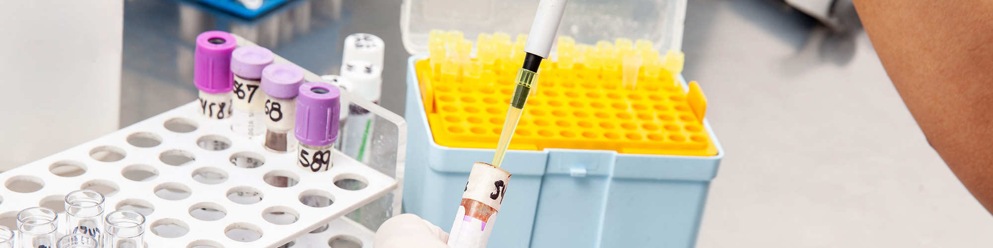
Western blotting remains a cornerstone for target protein analysis across multiple areas in academia and biopharma research. This makes data accuracy and reproducibility, as well as time and cost efficiency, critical for reliable decision-making.
However, western blotting has a reputation for being a tricky and time-consuming skill to get right. Therefore, modern advances aim to speed up and simplify the process, taking the pain out of western blotting and accelerating your workflows.
That said, with novel products, protocols, and technologies, it can sometimes be difficult to stay up to date with the ins and outs of each method.
One common misunderstanding is the mix-up between Stain-Free and No-Stain methodologies. While, by the name alone, you may assume they are pretty much the same technique, in reality, the differences are stark, and it’s important to understand the advantages and disadvantages of each.
Below, we outline the key differences between Stain-Free and No-Stain western blotting approaches, highlighting their respective strengths and limitations, to empower you to choose the right method for your needs.
Skip the background and show me the differences
Quick Answer: Is Stain-Free Better than No-Stain?
Yes. Both support total protein normalization, but the Stain-Free method provides faster workflows, built-in quality control checkpoints, and earlier verification of protein transfer and loading—making it more reliable for quantitative western blotting.
Back to Basics
Typically, the western blotting workflow consists of six main steps:
- Sample preparation — cells or tissues are lysed, and proteins are extracted, denatured, and quantified
- Electrophoresis — proteins are run on a gel and separated based on their characteristics
- Transfer — proteins are moved from the gel to a synthetic membrane
- Immunodetection — the membrane is incubated in blocking buffer to reduce nonspecific binding, followed by the primary antibody specific to the target protein, and then the secondary antibody conjugated with fluorescent molecules or enzymes for detection
- Image acquisition — the signal from the secondary antibody is acquired and converted to a digital image
- Image analysis — the signal intensity of the bands is quantified and normalized to a loading control
While traditional western blotting provides a well-established method for highly specific and sensitive protein detection, it also has multiple drawbacks. For example, it is a lengthy process with quality control opportunities often limited to the detection phase at the very end.
Additionally, conventional normalization methods typically use housekeeping proteins (HKPs) as loading controls. It is now known that using HKPs for normalization can be problematic. For one, target proteins are often in low abundance and cell lysates need to be loaded in large amounts to ensure detection. However, HKPs are generally highly expressed, so this can lead to oversaturation of the reference bands, exceeding their linear detection range (Bass et al. 2018).
Furthermore, HKPs are utilized under the assumption that they have constant expression levels. However, their expression levels can in fact vary under different experimental conditions (Patterson et al. 2025). This can introduce complications for downstream data analysis.
As a result, a growing number of publications and guidelines now recommend avoiding HKPs for normalization, or at the very least validating their compatibility for each experiment—an extra step that can add both time and cost to your workflow.
Overcoming Downstream Analysis Problems
The most appropriate way to overcome this is to use total protein normalization (TPN)—a method in which the entire protein content of a sample is stained and used as the loading control. As total protein stains are less prone to oversaturation than antibody-based detection of individual proteins, TPN allows the assessment of all proteins in the lane within their linear dynamic range. Importantly, it also minimizes bias introduced by condition-dependent changes in individual protein expression.
The problem with TPN, however, is that the stains can hinder blot development and visualization in the later steps of the workflow. Therefore, a destaining procedure is often additionally required.
So, how do more recent advances in western blotting technology compare to the conventional method?
Blot the Difference
Table 1. A summary of the key features of Stain-Free and No-Stain western blotting.
Feature |
Stain-Free |
No-Stain |
|---|---|---|
|
Speed |
Immediate visualization |
Labeling and washing |
|
Workflow |
Integrated in-gel checkpoints |
Post-transfer protocol |
|
Equipment |
Stain-Free gels No extra reagents UV excitation |
Standard gels
Labeling reagents |
|
Sensitivity |
10–25 ng |
20 ng |
|
Normalization |
TPN |
TPN |
Stain-Free
Stain-Free technology uses gels containing a UV-reactive compound that modifies tryptophan residues, enhancing their native fluorescence without additional staining. This allows instant visualization of proteins in gels and membranes with no impact on downstream steps.
No-Stain
No-Stain western blotting, on the other hand, is a post-transfer labeling method that covalently attaches a fluorophore to the lysine residues of proteins. Although the staining protocol can be applied to gels or membranes, it is recommended to use the post-transfer protocol for western blotting.
And the Winner Is…
Both approaches are advantageous in that they enable the use of TPN as a normalization method. However, while No-Stain protocols require additional staining and washing steps, Stain-Free western blotting does not, as the visualization method is directly incorporated into the gel. Therefore, the Stain-Free method allows you to capture your gels within minutes.
Not only does the immediate visualization capability of Stain-Free technology enable more rapid TPN, but it also facilitates verification and validation at various points in the western blot workflow. The gel can be activated and imaged following electrophoresis to verify sample migration, the membrane can be imaged to confirm efficient protein transfer, and the original gel can be reimaged to validate elution of the sample, all without the need for additional staining steps. This allows any problems to be detected early, rather than wasting time on a blot that you won’t find out has failed until the final steps.
Although Stain-Free western blotting requires the use of specific precast gels, they are compatible with standard Tris-Glycine buffer systems, making transition easy. Kits for hand casting gels are available for price-sensitive users as well, offering the same benefits as precast gels.
For activation and image acquisition, any Stain-Free enabled imager, such as the ChemiDoc Go Imaging System, using standard UV can be used. Fluorescent multiplexing capabilities using RGB or IR excitation are not limited using Stain-Free technology.
In contrast, the No-Stain reagent works with standard gels, but as labeling is applied post transfer, it does not have the integrated checkpoints. The image acquisition works on a variety of imagers as well, with 488 nm being the recommended excitation. This must be taken into consideration when setting up fluorescent multiplexing experiments.
Both methods streamline the western blotting process, offering more rapid protocols by eliminating the steps for HKP validation and detection. The No-Stain protocol for membranes requires just 16–20 minutes for labeling and washing, removing the need for the fixation and destaining steps required by traditional methods, such as Coomassie stains.
Stain-Free technology takes these time savings even further as gels can be activated and visualized without any incubation or washing steps. In addition, the gels enable faster separation and more efficient transfer compared to standard gels, resulting in an overall time of only 5 hours from start to finish, compared to the typical 16 hours for traditional western blotting.
Finally, both methods provide greater sensitivity than traditional TPN methods, with No-Stain protocols having a lower limit of detection of around 20 ng, and Stain-Free methods detecting proteins at levels as low as 10 ng.
Differences in a Nutshell
To summarize, both Stain-Free and No-Stain technologies enhance western blot workflows by enabling TPN and reducing reliance on HKPs.
However, Stain-Free stands out for speed, simplicity, and built-in quality control, eliminating additional staining steps while enabling earlier validation throughout the workflow.
So, why not try Stain-Free technology for streamlined, reliable, and stress-free western blotting?
Could Stain-Free Western Blotting Be the Method for You?
Our western blotting sales specialists can provide all the information you need about Stain-Free technology.
Interested in Learning More?
Browse our comprehensive four-part literature resource collection to find all you need to achieve reliable, high-quality results with Stain-Free western blotting.
References
Bass JJ et al. (2018). An overview of technical considerations for western blotting applications to physiological research. Scand J Med Sci Sports 27, 4–25.
Patterson AR et al. (2025). Expressions of commonly used housekeeping proteins are altered in asthmatic mouse lungs. Biochem Biophys Rep 42, 102018.