Secrets of the Haunted Brain: Ghost Tangles
- Oct 27, 2020
- 3 min read
- Marta González Prieto, PhD
If neurodegenerative diseases are not scary enough, you can sometimes find the remnants of dead neurons, or “ghost tangles” in patients’ brains. In this blog we discuss ghost tangles, the remains of tau (found after neuronal death), and their effects.
Tauopathies are neurodegenerative disorders characterized by abnormal deposits of tau protein in the brain, which suffer from synaptic loss and impaired connectivity. The most common tauopathy is Alzheimer’s disease. Other examples of tauopathies are frontotemporal lobar degeneration (known as Pick's disease), corticobasal degeneration, and supranuclear palsy. In Alzheimer’s disease, the accumulation of hyperphosphorylated tau protein, as well as extracellular amyloid-β plaques, is a hallmark of the condition. While all of these disorders are characterized by abnormal tau deposition, the processes are still being elucidated.
Why Do We Need Tau Protein?
In a healthy human brain, tau is expressed in neurons where one of its main roles is to maintain the axonal structure of microtubules (Figure 1). Microtubules are essential in cell division and neuronal activity and by binding to the tubulin that comprises them, Tau helps stabilize the cytoskeleton and provide flexibility.
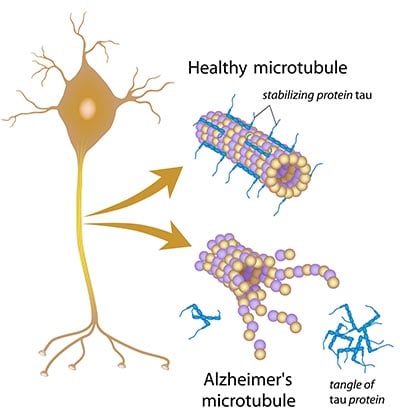
Fig. 1. Diagram illustrating the role of tau in microtubule stability. Tau protein binds to microtubules, helping to maintain their structure. When tau is not bound to axonal microtubules, it undergoes changes that lead to tau protein tangles in the cytoplasm. In this state it can no longer help stabilize neuronal microtubules leading to neuronal degradation.
However, during pathological conditions, tau protein is released and excessively phosphorylated forming insoluble aggregations in the cytoplasm, or paired helical filaments (Figure 1). Tau also undergoes multiple truncations and shifts in conformation as part of this transformation. The accumulation of the intracellular misfolded tau protein leads to cellular dysfunction and neuronal death due to perturbed microtubule function and axonal transport (Barbier et al. 2019).
The Tombstones of Dead Neurons
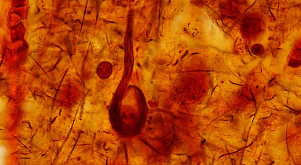
The appearance of ghost tangles starts with the formation of non-fibrillary hyperphosphorylated tau aggregates. These tau oligomers can be viewed as sparsely scattered dots. Then, the pre-tangles form groups of fibers as paired helical filaments named mature tangles. In the neuronal cell body, these fibers are known as neurofibrillary tangles. In dendrites, they are named neuropil threads. They can also be found in the axon of neurons. When these neuronal cells die, the extracellular remnants of tau proteins, called ghost tangles, are left behind.
Interestingly, during this morphological progression, tau isoforms shift from having four binding domains (found in pre-tangles) to three (observed in ghost tangles) with both three and four binding domains found in mature tangles (Uchihara 2014). The binding domains allow tau proteins to attach to tubulin; thus, four of these domains are better at stabilizing microtubules than three.
Ghost tangles have been observed in late stages of Alzheimer’s disease, where a great neuronal loss has occurred (Furcila et al. 2019), and are a spooky reminder of the neuronal death that has occurred, like a tombstone for the dead neuron.
A Toxic Legacy
Ghost tangles are toxic for neighboring cells as glial responses can contribute to further neurodegeneration. Reactive gliosis (astrocytosis and microgliosis) occurs around tau paired helical filaments and this response increases with the development of Alzheimer’s disease (Serrano-Pozo et al. 2011). However, glial cells can also damage nearby cells by spreading tau pathology. Their inflammatory response, such as the release of pro-inflammatory cytokines and reactive oxygen species, is a powerful modifier of the tau cascade. Reactive gliosis increases tau phosphorylation and accelerates tangle formation in the nearby neurons (Maphis et al. 2015) through microglial-specific fractalkine receptor (CX3CR1) (Bhaskar et al. 2010; Bolós et al. 2017).
Currently, many investigations focus on targeting tau protein pathology to try to understand the trigger for tangle formation and prevent neuronal death. Hopefully, in the future, ghost tangles and their toxic effects will be prevented through new treatments and these spooky legacies of neuronal death will be a thing of the past.
Want to Know More about Neurodegeneration?
Read our minireview on neuroinflammation and find information about antibodies for researching a range of neurodegenerative conditions.
References
Barbier P et al. (2019). Role of Tau as a microtubule-associated protein: structural and functional aspects. Front Aging Neurosci 11, 204.
Bhaskar K et al. (2010). Regulation of tau pathology by the microglial fractalkine receptor. Neuron 68, 19-31.
Bolós M et al. (2017). Absence of CX3CR1 impairs the internalization of Tau by microglia. Mol Neurodegeneration 12, 59.
Furcila D et al. (2019). Subregional density of neurons, neurofibrillary tangles and amyloid plaques in the hippocampus of patients with Alzheimer's disease. Front Neuroanat 13, 99.
Maphis N et al. (2015). Reactive microglia drive tau pathology and contribute to the spreading of pathological tau in the brain. Brain 138, 1738-1755.
Serrano-Pozo A et al. (2011). Reactive glia not only associates with plaques but also parallels tangles in Alzheimer's disease. Am J Path 179, 1373-1384.
Uchihara T (2014). Pretangles and neurofibrillary changes: similarities and differences between AD and CBD based on molecular and morphological evolution. Neuropathology 34, 571-577.