Cell Cannibalism: An Appetite for Cancer?
- Oct 24, 2025
- 3 min read
- Chloe Fenton, PhD
It’s about that time of year again when the nights get longer, the air takes on a sharper edge, and the darkness in the shadows starts to creep to the forefront of our minds. Halloween tends to bring out a morbid curiosity in us all, with graphic costumes and fake blood galore, reveling in the gore that would normally make our stomachs churn.
That spooky vibe also extends to the inner workings of the body. Previously, we’ve focused on bacterial vampirism and zombie cells, describing the mini horror movies playing out within us. This year, we’re taking a taste of something slightly different: cell cannibalism.
It’s a Cell-Eat-Cell World
Regulated cell death is a critical process in homeostasis that ensures the removal of cells that are critically damaged or mutated. At least 13 different mechanisms of cell death have been identified, differing from each other in their triggers, signaling pathways, and morphological features.
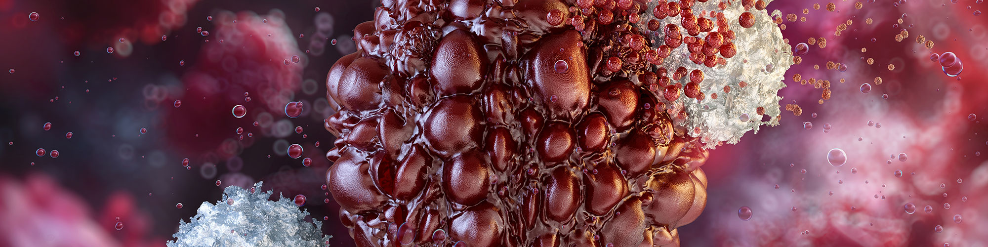
But perhaps one of the strangest of these is a process aptly known as cell cannibalism, in which one cell ruthlessly engulfs and kills another.
Phagocytosis, the process in which professional engulfers eat dead or dying cells, is probably the most well-known form of cell cannibalism (Durgan and Florey 2018). This type of cannibalism occurs in normal human physiology. For example, splenic and liver macrophages are known to be constantly devouring old and tired red blood cells to make room for fresh, new candidates (Rodriguez et al. 2025).
In contrast, in other forms of cannibalism, cells target other cells that are alive and well for their next meal. Entosis, for example, is a form of cell cannibalism occurring between seemingly healthy and viable epithelial cells. Even more intriguingly, the internalized epithelial cell, rather than being passive prey, is in fact the driver of its own destruction, suicidally invading its hungry neighbor (Durgan and Florey 2018).
But what could cause a cell to turn to such depravity?
Feeding Frenzy
The first trigger identified for this process was cell detachment from the matrix. Neighboring epithelial cells are tightly bound to one another by adherens junctions and connected to the matrix by the engagement of integrins. During matrix detachment, the stabilization provided by the integrins is lost, and so the contractile forces are off-balance, with the contraction of the adherens junctions having no counterbalance to keep them in check. This results in an unrestrained push of the detached cell into the waiting arms of the cannibal (Overholtzer et al. 2007).
Recently, however, cannibalism has been observed among epithelial cells that remain adherent, prompting research into alternative triggers.
Durgan et al. (2017) found that the structural changes associated with mitosis were able to induce cannibalism. Depletion of Cdc42, a small GTPase involved in the regulation of the cell cycle and the cytoskeleton, led to increased cell rounding and rigidity during mitosis, giving cells the strength to forcibly push themselves inside other cells to be eaten alive.
Additionally, Hamann et al. (2017) found that cells in nutrient-deprived conditions, such as in glucose withdrawal, will decide that desperate times call for desperate measures and gobble up their neighbors to avoid starvation.
A Taste for Tumors
Interestingly, these features, matrix detachment, excessive mitosis, and metabolic stress, are abundant in cancer. In fact, cannibalism is rife in the tumor microenvironment. The cell-in-cell structures produced by entosis are frequently observed in various forms of tumors, including those from breast, colon, liver, cervix, and lung cancers.
While relatively common, the precise mechanisms of entosis in cancer have unfortunately not been widely studied. Therefore, whether cannibalism is an overall beneficial or detrimental process for fighting tumors isn’t entirely known (Durgan and Florey 2018).
The fact remains that cannibalistic cells seem to preferentially eliminate cells that have characteristic tumor qualities. Additionally, Durgan et al. (2017) found that the chemotherapeutic drug Paclitaxel, which is often used to treat cancers, consistently increased cell-in-cell formations, a hallmark of entosis induction, in a breast cancer cell line. This may offer some clinical insights into the mechanism of action of the drug and point towards entosis as a tumor-suppressing function.
On the other hand, cannibalism can also occur amongst the tumor cells themselves and has been proposed as a method to promote tumor growth. In this case, the stronger tumor cell will greedily consume its weaker neighboring tumor cell to steal its nutrients, promoting clonal selection of the “better” cancer cells in a survival-of-the-fittest type of manner (Hamann et al. 2017, Sun Q et al. 2014).
The precise nature, whether good or evil, of entotic cells in cancer remains to be seen, and further research is necessary to uncover any untapped clinical potential.
But it begs the question: could we get the cannibals on our side in the fight against cancer?
Are You Interested in Studying One of the Many Mechanisms of Cell Death?
Bio-Rad offers reagents and resources to help you investigate regulated cell death in your experiments.
References
Durgan J et al. (2017). Mitosis can drive cell cannibalism through entosis. eLife 6, e27134.
Durgan J and Florey O (2018). Cancer cell cannibalism: Multiple triggers emerge for entosis. Biochim Biophys Acta Mol Cell Res 1865, 831–841.
Hamann JC et al. (2017). Entosis is induced by glucose starvation. Cell Rep 20, 201–210.
Overholtzer M et al. (2007). A nonapoptotic cell death process, entosis, that occurs by cell-in-cell invasion. Cell 131, 966–979.
Rodriguez M et al. (2025). The perils and promise of cellular cannibalism in development, homeostasis, and disease. Annu Rev Cell Dev Biol 41, 281–306.
Sun Q et al. (2014). Competition between human cells by entosis. Cell Res 24, 1299–1310.