A Guide to IHC-paraffin Success
- Feb 20, 2020
- 5 min read
- Kimberley Bryon-Dodd, PhD
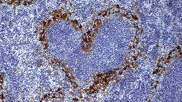
Immunohistochemistry (IHC) provides invaluable information about the localization of proteins in a tissue. However, to ensure accurate results it is important to avoid errors that could cause artefacts or reduced staining. Your results are only as good as your samples, and this blog is written to help you perfect your IHC-paraffin technique. It focuses on some of the key stages so that you can achieve publication-quality results.
Antigen Retrieval
One of the initial steps of sample preparation, fixation, can result in protein-cross linking, which impedes the antibody binding by masking the epitope. This can result in greatly reduced staining, so it is important to include a step to enable epitope recognition by your antibodies.
There are two ways of performing antigen retrieval – heat-induced antigen retrieval (HIER) and proteolytic-induced antigen retrieval (PIER). As the name suggests, HIER uses a heating step to retrieve antigens and is considered a gentler method than PIER, which requires the use of enzymes such as proteinase K, trypsin or pepsin. As well as the method of heating, the pH of the buffer used for HIER also impacts on the success of antigen retrieval. Optimal recovery for many epitopes occurs in alkaline buffers (pH~8-10) and this range is a good starting point when you are establishing a protocol.
The use of enzymes means that PIER can sometimes result in changes to specimen morphology, with fewer available antibodies working with PIER. Antibody datasheets often provide information about the recommended antigen retrieval steps for that antibody and you should use the recommended method for best results.
Including a control sample where HIER/PIER was not performed will enable you to eliminate the possibility of observed staining resulting from artefacts caused by the antigen retrieval step and give you confidence in your results.
Blocking – Non-specific Binding
Non-specific binding can also cause staining artefacts that can confound interpretation of results. The blocking step helps to reduce false positives and background signal. Where possible, block with normal (unchallenged) serum from the same species as the tissue being stained, as the serum will contain antibodies that bind to non-specific epitopes in your sample.
Alternatively, you can also use normal serum from the same species as the secondary antibody used. However, you should never use normal serum from the same species as the primary antibody as this would increase background as the secondary antibody may recognize and bind proteins in the serum.
More economical protein buffers such as bovine serum albumin (BSA), non-fat dry milk or gelatine can also be used for the blocking step but may have higher background than other blocking methods.
Not all blocking reagents and buffers are compatible with every detection method. For example, alkaline phosphatase (AP) conjugates should not be used with buffers containing phosphate. Milk should not be used with biotin-avidin detection as milk contains biotin. Make sure that you check compatibility if you are establishing a protocol!
A more specialist option that may be helpful is to use a commercial blocking reagent; they have been designed to block non-specific binding and can be more effective than the more traditional BSA/ milk methods and cheaper than normal serum.
Blocking – Optimizing Detection Methods
When using enzymatic detection methods like horseradish peroxidase (HRP) you might need to block endogenous enzymes in order to prevent high background. You can use indicators to detect endogenous enzyme activity and determine whether you should include an additional blocking step (Table 1).
Table 1: Detection methods and blocking steps
Detection Method |
Endogenous Enzymes/Proteins in Tissues |
Example Indicator |
Blocking |
|---|---|---|---|
|
HRP |
Peroxide e.g. liver and kidney |
DAB (turns brown) |
|
|
AP |
Alkaline phosphatase e.g. bone, intestine and placenta |
5-bromo-4-chloro-3-indolyl-phosphate/nitro blue tetrazolium (BCIP/NZT) (turns blue) |
Levamisole |
|
Avidin-biotin |
Biotin e.g. kidney, liver and spleen |
|
Avidin, then biotin (order is crucial) |
Primary Antibody
Ensuring that you are using a suitable primary antibody will give you the best results. If you are using a new antibody, always check whether it has been verified to work in IHC-paraffin. The antibody datasheet will have information about which applications it has been tested in and any special conditions, including recommended antigen retrieval methods. You can also review the literature to find antibodies that have been used successfully in your desired tissue type and species and get information about the experimental protocol used. This information will help you hone your protocol and avoid any basic errors.
For uncharacterized antigens, polyclonal primary antibodies can be advantageous as they bind to multiple epitopes of your antigen, which can help you overcome some epitopes remaining inaccessible after sample prep. Polyclonals can give higher background though than monoclonal antibodies, due to non-specific binding, and they also vary in lot-to-lot consistency. When you are repeating experiments remember to ask for antibodies with the same lot number if you need to reorder so that you get consistent results between experiments.
To ensure that you are confident about your staining patterns, always check where you are expecting to see protein expression in your tissue. The antibody datasheet, published literature and resources like the Human Protein Atlas all contain information about where proteins are localized and can be used to help you predict where you should (or shouldn’t) expect to see staining.
Monoclonal antibodies are often used in IHC due to their lot-to-lot reproducibility and consistent specificity, which can mean lower background staining.
Detection System
Ensure that you use the correct detection system for your secondary antibody to get clear results. You can use directly conjugated primary antibodies, but since the secondary antibody assists in signal amplification (as multiple secondaries bind to a single primary antibody) directly conjugated antibodies are only really recommended when you are detecting really abundant proteins such as beta-actin.
When you have an antigen expressed at really low levels, consider using a detection method that amplifies the signal. As a single avidin molecule can bind up to four biotin molecules, biotinylated secondaries used together with avidin/streptavidin can prove a useful method.
Controls
To give you confidence that you are interpreting your data correctly, you should ensure that you use all relevant controls. You should always include a positive control and a negative control to verify that the assay has worked. For the positive control, use a section of tissue known to express the marker that you are looking for. The negative control should use a section of tissue where you know the marker is not expressed.
Depending on your experimental set-up, you may also wish to consider secondary antibody only, background staining, absorption and isotype controls.
Need More Assistance with IHC?
Access detailed protocols, tips and troubleshooting advice on our dedicated immunohistochemistry page.