Seeing SARS – the Remarkable Story of June Almeida
- Aug 27, 2020
- 3 min read
- Rachael Preston, PhD
In this blog we continue our celebration of women in science by looking back at the career of June Almeida, the internationally renowned Scottish virologist who became a pioneer in the imaging and identification of viruses
Development of a Novel Microscopy Technique
Early on in her career, Almeida worked as an electron microscopy technician at the Ontario Cancer Institute in Canada. There she developed a novel imaging technique for the detection of viruses, known as immune electron microscopy (IEM) (Almeida 2008). This technique involves the detection of molecules at the ultrastructural level by labeling them with specific antibodies attached to colloidal gold particles. When a beam of electrons encounters the colloidal gold particles on an antibody bound to its antigen an electron-dense label is produced. The electron beam is then scanned and the position of the beam, combined with the intensity of the signal, is used to produce an image, referred to as an electron micrograph. This allows detection and visualization of the colloidal gold particles and therefore the target molecule. If the antigen is located on the surface of the cell, scanning electron microscopy (SEM), where the beam of electrons is streamed across the surface of a sample can be used to visualize it.
When the antigen is located inside a cell, transmission electron microscopy (TEM) is required. With TEM, the beam of electrons is transmitted through a specimen, rather than across the surface, in order to form an image.
The use of antibodies in combination with electron microscopy enables researchers to distinguish between different microscopic organisms, which would have previously been impossible to achieve with electron microscopy alone. The specificity of antibodies means that antibody-bound colloidal gold particles will recognize the exact target, like a molecule found on a virus. This enables precise detection and positive identification of a virus within a sample.
Colloidal gold particles of different sizes can also easily be distinguished in electron micrographs, allowing specimens to be labeled with multiple antibodies attached to different sized colloidal gold particles at the same time.
Powering Vaccine Discovery
Almeida used this IEM technique to study several viruses including rubella and hepatitis B. This led to the identification of two distinct components of the hepatitis B virus: an outer lipid envelope and an icosahedral nucleocapsid core made of protein that encloses the viral DNA. The hepatitis B vaccine, which stimulates the production of antibodies in response to the surface component of the virus, was based on this discovery and is a key component of the hepatitis B vaccine still in use today (Almeida 2008, Dienstag and Purcell 1977).
Later in her career, while working at St Thomas's Hospital Medical School, Almeida collaborated with Dr David Tyrrell, Director of the Common Cold Research Centre, who at the time was investigating rhinoviruses. Tyrrell had developed a new technique of organ culture, whereby parts of an organ or a whole organ are cultured in vitro. Organ culture allows the structure of organ tissue to be maintained and directed towards normal development. The organ tissue can then be inoculated with a virus, allowing the virus to be cultivated, eliminating the need for human volunteers in the study of viruses. Tyrell’s organ culture technique combined with Almeida’s IEM technique eventually resulted in the successful detection of rhinoviruses (Almeida 2008).
Identification of a New Type of Virus
Tyrrell’s team of researchers had collected samples of a flu-like virus from a sick schoolboy. However, they were having trouble cultivating this virus using traditional methods, leading them to suspect that this might be a completely new type of virus (Broklehurst 2020).
Tyrrell sent some of these samples to Almeida in the hopes that she would be able to identify the virus using the IEM technique. Almeida was able to successfully produce clear images of the virus and noted that it resembled two similar viruses she had previously imaged while studying bronchitis in chickens and murine hepatitis (Brocklehurst 2020). These had previously been dismissed as poor images of influenza viruses. However, the images of the virus in the samples from the schoolboy combined with the images from Almeida’s previous work convinced Almeida and Tyrrell that these were in fact a new family of viruses.
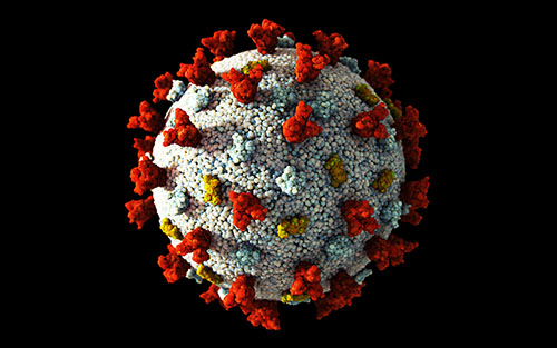
Inspired by the virus’ halo-like structure, Alemeida and Tyrrell decided to name the new family of viruses after the Latin word for crown, corona. This family of viruses, referred to as coronaviruses, is known to cause respiratory tract infections in mammals and birds and includes severe acute respiratory syndrome coronavirus (SARS) and SARS-CoV-2, also known as COVID-19 (Almeida et al. 1968, Brocklehurst 2020).
Almeida also taught other virologists how to perform IEM, including Albert Kapikian, who used the technique to identify Norovirus, the cause of ‘winter vomiting disease’. This approach also enabled healthcare workers to visualize the hepatitis A virus for the first time (Almeida 2008, Banatvala 2011).
Today, IEM is still one of the most sensitive methods available for the detection and diagnosis of viruses and June Almeida’s pioneering approach revolutionized the virology field. Her contribution to the field of virology is monumental with many of her electron micrographs still published in textbooks many decades later and IEM continuing to enable virologists to study new viruses like SARS-CoV-2. It is likely she will remain an inspiration for many generations of scientists.
References
Almeida J (2008). June Almeida (née Hart), BMJ 336, 1511.
Almeida JD et al. (1968). Coronaviruses. Nature 220, 650.
Banatvala JE (2011). Almeida [née Hart], June Dalziel, Oxford DNB https://doi.org/10.1093/ref:odnb/99332 Accessed 05/13/20202
Broklehurst S (2020). The woman who discovered the first coronavirus. https://www.bbc.co.uk/news/uk-scotland-52278716 Accessed 05/13/202
Dienstag JL and Purcell RH (1977). Recent advances in the identification of hepatitis viruses*. PostgradMedJ 53, 364-373.