Measuring the inevitable divide - tools for assessing cell proliferation
- Sep 04, 2015
- 2 min read
- Bio-Rad
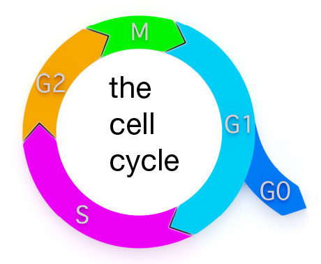
Schematic representation of the cell cycle. Ki-67 is expressed during G1, S, G2 and M phases. PCNA is present from G1 through to G2. MCM 2 is critical for replication initiation while the BrdU method recognizes replicating cells during S phase.
It was in the 1900s that scientists first discovered cells in the process of dividing, however they did not know then how this was happening and what the process should be called. Since then, the term cell proliferation came to be defined as the increase in cell number due to cell growth and division.
Further research demonstrated that cell proliferation has to be carefully regulated as the smallest alterations can result in an uncontrolled increase in cell numbers, and eventually cancer. It is inherently clear that cancer is bad but determining just how bad is partly based on measuring the rate of cell proliferation.
Our understanding of the cell cycle has led to the development of useful reagents to assess cell proliferation by measuring the number of cells in growth phases of the cell cycle.
The eukaryotic cell cycle
In eukaryotes the cell cycle can be grouped into three major stages:
- Interphase (a growth period, comprising of G1, S and G2 phase, in which the cell duplicates its DNA)
- Mitotic phase (the chromosomes get separated into two sets in preparation for division. M phase consists of 4 stages: prophase, metaphase, anaphase, and telophase)
- Cytokinesis (division to form two daughter cells)
The transition of cells through these stages of the cell cycle is regulated by checkpoint proteins. So it should come as no surprise that cell cycle misregulation and impaired cell cycle checkpoints are two of the most common causes of aberrant cell proliferation.
One of the main strategies for measuring cell proliferation is to quantify protein levels of proliferation markers such as Ki-67, proliferating cell nuclear antigen (PCNA), and minichromosome maintenance 2 (MCM 2). Detection and quantification of these proteins provides information on cell cycle progression. Ki-67 for example is present in all stages of the cell cycle, but is absent in cells in the G0 resting phase. During interphase, it is exclusively expressed in the nucleus, whereas during the mitotic phase it is predominantly located on the surface of chromosomes. This makes it an excellent growth marker, particularly in disease contexts such as cancer where identifying the growing fraction of a cell population is critical (Scholzen and Gerdes, 2000).
Another strategy for determining cell proliferation is through the use of a thymidine analog called Bromodeoxyuridine (BrdU). BrdU can be added to the culture media of cells, where it incorporates into newly synthesized DNA of proliferating cells instead of thymidine. The incorporated BrdU can then be detected and measured with the help of BrdU specific antibodies.
References
Scholzen T, Gerdes J. The Ki-67 protein: from the known and the unknown. J Cell Physiol. 182:311-322, 2000.