"Fin"tech: Zebrafish Models
- Jul 14, 2021
- 5 min read
- Keira Lee Rice
2021 Bio-Rad Science Writing Competition Winner
Keira’s enthusiasm about her PhD topic and model organism, zebrafish, is clearly conveyed in her article through the warmth of her tone. The judges were impressed by her writing style and her hand drawn illustrations that accompany her article, adding a touch of humor to the discussion of her PhD subject.
Keira is a third year PhD Candidate at the Hong Kong University of Science and Technology (HKUST). She works in an imaging lab that uses zebrafish to study calcium signaling in embryonic development. We are delighted to publish Keira’s winning entry below.
“Give a man a fish and you feed him for a day; teach a man to [work with zebra-] fish and you feed [his mind] for a lifetime”.
I am on “fish duty” two days a week, which is a rotational duty assigned to our lab members to feed and maintain our zebrafish facility. I usually have my earphones on, singing along to Hamilton while I shuffle from tank to tank, feeding the zebrafish that hold the fate of my PhD in their fins. Specifically, their tail fins (you’ll see why shortly).
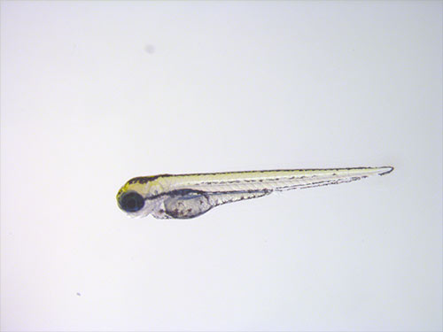
The zebrafish are a small freshwater fish that have made their way into aquariums and laboratories around the world, including the Calcium Aequorin Imaging Lab where I work. These stripey swimmers (pale yellow with dark blue stripes (Singh and Nüsslein-Volhard 2015), I should add) are a popular vertebrate model in a variety of research fields, stretching from basic research in neuroscience, to preclinical studies in drug screening and development. The short generation time, optical clarity, and genetic tractability of zebrafish are just some of many characteristics that make them an ideal in vivo system to dive into the “hows” and “whys” of biological phenomena or to “fish out” the function of certain genes...
And that is exactly how I came to use zebrafish within my own research. Namely, I started generating my own zebrafish mutant to test a hypothesis about one particular gene. Despite a few setbacks, the availability of genetic tools and a sea of existing protocols streamlined the process significantly. Eventually, I settled with the CRISPR/Cas9 mutagenesis system (Varshney et al. 2015) and a High-Resolution Melt Analysis (HRMA)-based genotyping method (Samarut et al. 2016) (Figure 1). Now, I am working to screen second generation adult zebrafish to identify homozygotes, which will allow me to propagate a stable mutant line.
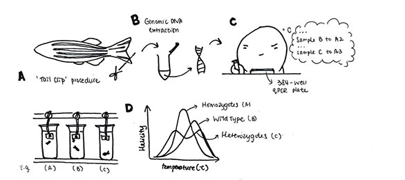
Fig. 1. Schematic illustrating tail clip procedure in zebrafish for genotyping.
To collect embryos to raise the progeny of my mutants, I need to scoop pairs of fish with my net to prepare a cross of male and female fish in a breeding trap (Figure 2). This tank has a transparent divider to encourage visual contact, and a porous bottom to enable egg collection and allow male-released pheromones (Li et al. 2018) to dissipate in the water overnight.
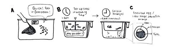
Fig. 2. Illustration of zebrafish embryo collection highlighting transparent divider and porous bottom.
Together, this forms the setting for “zebrafish speed-dating", which induces spawning and allows me to obtain fertilized eggs for my experiments (Figure 2C). Actually, it is by providing an experimentally accessible system to study social traits that zebrafish have emerged as a valuable vertebrate model in behavioral neuroscience. When I attended my first International Zebrafish Conference in 2019 (“It sounds fishy” was the response I got from my Dad when I tried to tell him that “zebrafish conferences are a real thing”), there was a fascinating talk by Prof. Judith Eisen about the requirements for microbiota colonization on the normal development of social behaviors in zebrafish (Bruckner et al. 2020).
Apart from the ease of genetic manipulation and the adaptation of zebrafish to laboratory conditions, the suitability of transparent zebrafish for microscopy enables in vivo imaging studies that are unparalleled in other vertebrate models. In fact, if I had performed live imaging of transgenic zebrafish, that selectively express fluorescent proteins in their macrophages and neutrophils, I would have been able to see these dynamic cells migrating (Li et al. 2012) to the site of where I performed the tail fin amputation I described earlier (Figure 3).
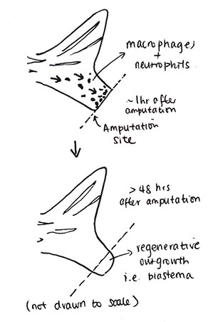
Fig. 3. Illustration showing macrophage and neutrophil migration after tail fin amputation and regeneration at wound site.
Upon arrival, these macrophages mediate an inflammatory response that also contributes to the remarkable regenerative ability of zebrafish (Petrie et al. 2014). When I check on my fish each day, I’m always amazed at how fast their tail fins grow back.
These are just some of the stories I tell my friends when they ask, “How are the fish?”, who at this point, just think of me as the “zebrafish lady”. I make sure to keep them updated with my fish tales (I have several “Finding Nemo” stories too, but that’s for another time).
Given the significance of daylight hours on biological processes, it is important that our fish are kept under a strict lighting regime, and I make sure to check the lights in our fish room before tucking the fish to bed (zebrafish do sleep (Leung et al. 2019))! It probably comes as no surprise that zebrafish are also used to study the biological basis and effects of circadian rhythms. Notably, a recent study showed that muscle growth is regulated by the biological clock, where muscle grows more in the day than at night irrespective of nutrition and muscle activity (Kelu et al. 2020). Who would have thought that fish could help to inform us on the best time of day to go to the gym?
I, too, am studying muscle biology, and my research revolves around the development of the embryonic trunk musculature. I typically conduct my studies ~24 or ~48 hours after collecting embryos, when muscle differentiation and the formation of tendon-like junctions are mostly complete (Figure 4).
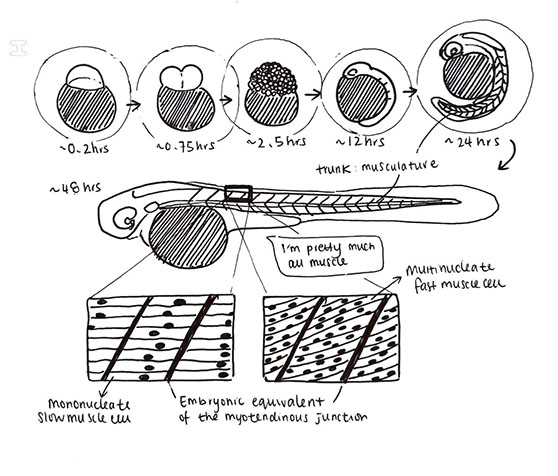
Fig. 4. Illustration of embryo development in zebrafish, highlighting muscle fiber formation.
Unlike in humans where slow and fast muscle fibers are intermixed, in zebrafish they are spatially distinct, which is particularly convenient for me to perform cell-type focused studies. Moreover, the remarkable genetic and structural similarity between zebrafish and mammalian muscle has established zebrafish as a preclinical platform for degenerative muscle diseases (Farr GH 3rd et al. 2020). Duchenne’s Muscular Dystrophy is one well-known example.
All in all, I believe that the ideal characteristics of zebrafish as a vertebrate model will help the zebrafish research community continue to make waves in the life sciences.
As for me, I will “just keep swimming” till I defend my thesis. Fins and fingers crossed.
Bio-Rad's Science Writing Competition Results
We were delighted to receive entries from PhD/Grad Students from all around the world. The judges were impressed by the high standard of submitted articles across a breadth of different topics. Keira is the winner of a commemorative trophy, a copy of ’The Scientist’s Guide to Writing: How to Write More Easily and Effectively throughout Your Scientific Career’, and Science Writing Mentorship from our Lab Crunches Editor.
"I had a wonderful time creating my 'zebrafish blog' and I'm so happy to be sharing it with a wider audience. I am especially grateful because science communication is a skill I hope to build for my career, and I would like to thank all the judges and readers for your support." - Keira Lee Rice
View the Full AnnouncementReferences
Bruckner JJ et al. (2020). The microbiota promotes social behavior by neuro-immune modulation of neurite complexity. biorxiv https://doi.org/10.1101/2020.05.01.071373.
Farr GH 3rd et al. (2020). A novel chemical-combination screen in zebrafish identifies epigenetic small molecule candidates for the treatment of Duchenne muscular dystrophy. Skeletal Muscle 10, 29.
Kelu JJ et al. (2020). Circadian regulation of muscle growth independent of locomotor activity. PNAS 117, 31,208–31,218.
Leung LC et al. (2019). Neural signatures of sleep in zebrafish. Nature 571, 198–204.
Li L et al. (2012). Live imaging reveals differing roles of macrophages and neutrophils during zebrafish tail fin regeneration. Immunology 287, 25,353–25,360.
Li J et al. (2018). Male zebrafish (Danio rerio) odorants attract females and induce spawning. Aquaculture and Fisheries 3, 139–144.
Petrie et al. (2014). Macrophages modulate adult zebrafish tail fin regeneration. Development 141, 2,581–2,591.
Samarut E et al. (2016). A simplified method for identifying early CRISPR-induced indels in zebrafish embryos using high resolution melting analysis. BMC Genomics 17, 547.
Singh AP and Nüsslein-Volhard C (2015). Zebrafish stripes as a model for vertebrate colour pattern formation. Curr Biol 25, R81–R92.
Varshney GK et al. (2015). High-throughput gene targeting and phenotyping in zebrafish using CRISPR/Cas9. Genome Res 25, 1,030–1.042.