Erebosis: Fly Guts Die in Darkness
- Mar 22, 2023
- 4 min read
- Bio-Rad
Over 200 billion cells die in our body each day (Arandjelovic and Ravichandran 2015). Cell death helps our body remove diseased or damaged cells, which are replaced constantly in our skin, gut, and other parts of the body.
Until April 2022, scientists had identified 13 pathways of cell death; the three most well-known of which are apoptosis, necrosis, and autophagy (Sasabe 2022, Caballero-Díaz and Valcárcel 2014, Galluzzi et al. 2018, Hewings-Martin 2017).
However, earlier last year scientists from Japan discovered, completely by accident, a different way for cells to die.
The new cell death mechanism, which the researchers have named ‘erebosis’, overturns the long-standing dogma about how cells regenerate and renew in the gut of flies, explains lead researcher Sa Kan Yoo. He’s now keen to discover whether cells die the same way in humans too (Sasabe 2022).
“In both humans and flies, it’s been dogma that apoptosis is important for the turnover of gut cells, and in our PLoS Biology paper, we show that apoptosis isn’t important, and erebosis is important instead,” he says.
Different Ways to Die
Mammalian cells typically die by apoptosis or necrosis. Apoptosis, or programmed cell suicide, begins when a cell loses survival signals or its DNA is damaged. The cell shrinks and changes shape, its DNA fragments, and – eventually – it, is devoured by specialized immune cells (Shimogonya et al. 2018).
Necrosis is often seen as an ‘accidental’ form of cell death, unlike apoptosis where the cell deliberately commits suicide (Galluzzi et al. 2018). When necrosis is triggered by high temperatures or pressures, cells swell and rupture, spilling out their contents like a leaky tire. The leakage of cellular components triggers a strong immune reaction.
A third process, autophagy, has also been associated with non-programmed cell death, although its main function is as a survival mechanism. When a cell is facing starvation, it can digest parts of itself to generate energy. Autophagy can also protect cells from disease by removing non-functional proteins (Glick et al. 2010).
Dying into Darkness
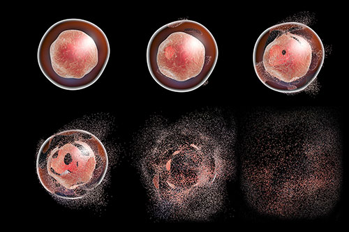
Erebosis seems to be a new type of programmed cell death, which occurs more slowly than apoptosis. Yoo and colleagues speculate that erebosis helps maintain the gut lining, which naturally renews itself over a few days or weeks (Ciesieski et al. 2022).
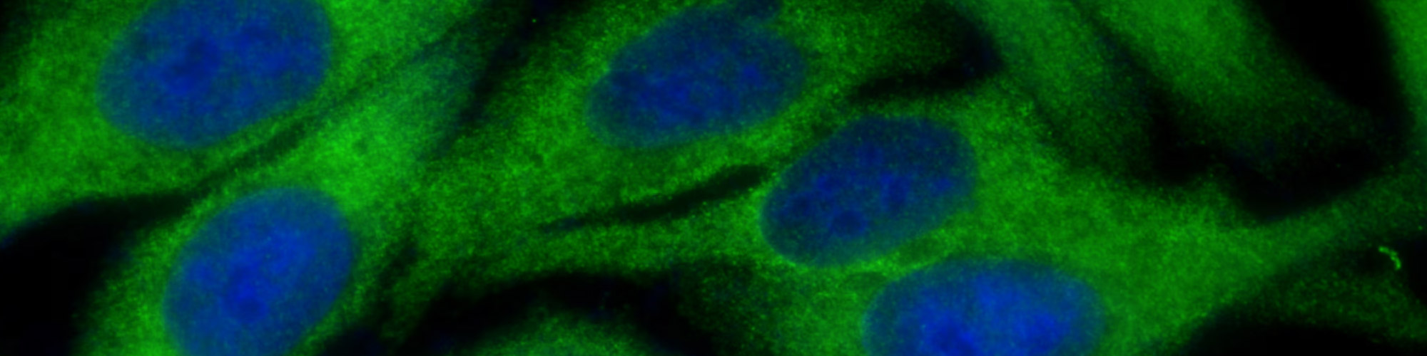
The team discovered erebosis in fruit flies, entirely by accident, while studying ANCE, an enzyme that helps lower blood pressure. After immunohistochemical staining of ANCE, they noticed that its expression in enterocytes of the fly gut was patchy, and the cells containing it looked strange when viewed under an electron microscope (Sasabe 2022).
“It took us a long time to understand these were dying cells,” says Yoo. “At first, we thought they were a weird new cell type.”
The weird cells they observed had flat nuclei. Further immunohistochemical staining for Phalloidin and DAPI revealed a loss of cytoskeleton and DNA content, respectively, and immuno-electron microscopy showed these cells lacked mitochondria, nuclear membranes and other contents of cells needed to keep them alive (Sasabe 2022, Ciesieski et al. 2022).
The team named the process erebosis after the Greek ‘erebos’, meaning ‘darkness’, because the cells appeared dark on microscope images (Sasabe 2022).
It seemed unlikely the cells could lack organelles, cytoskeleton and, even, possibly DNA and remain alive, therefore Yoo and colleagues speculated that the cells were undergoing some type of death. Analysis of a general cell death marker, TUNEL, revealed that some of the cells late in the erebotic process also showed signs of cell death (Ciesieski et al. 2022).
Searching for Novelty
The team realized the cells were dying due to a previously unknown cell-death mechanism. The erebotic cells didn’t show activation of caspases, an important enzyme in apoptosis, and inhibiting caspase activation didn’t stop erebosis.
A necrosis marker, propidium iodide, which enters cells when the membrane is breached, didn’t enter the erebotic cells. The flies were unlikely to have an infection because they’d been sterilized with bleach as embryos, and cultured in antibiotics, and their gut cells didn’t show any features common in infection-induced cell shedding.
Preventing expression of Atg genes involved in autophagy didn’t prevent erebosis either. Neither did the erebotic cells show up on a microscope image when flies expressing mCherry-tagged Atg8, labeling a gene sensitive to autophagy, were used (Ciesieski et al. 2022).
“The cell death we found didn’t have any features of the classic three types of cell death,” he says.
The Purpose of Erebosis
Yoo and his colleagues found small stem-cell-like enterocytes close to the erebotic cells, leading them to speculate that erebosis is a natural process for regenerating the gut lining.
According to them, as the erebotic cells die slowly, new enterocytes may grow to replace them, allowing a healthy turnover of nutrient-gathering cells in the fly gut (Ciesieski et al. 2022). Because the erebotic cells die slowly, the gut lining isn’t compromised.
The team now hope to investigate the mechanisms of erebosis and whether it exists in mammals.
“We’ve found this new phenomenon, but know almost nothing about its molecular mechanism, so we’re putting effort into understanding that,” he says.
Yoo and his colleagues believe erebosis may be important in the mammalian gut after a paper published earlier this year, which – he says - showed apoptosis was not important for gut turnover in mice (Ghazavi et al. 2022).
The paper didn’t suggest an alternative mechanism for gut cell turnover, he explains, and speculates it may turn out to be erebosis.
Studying Regulated Cell Death?
Bio-Rad has products and resources to support your research, including downloadable posters and minireviews.
References
Arandjelovic S and Ravichandran K. (2015). Phagocytosis of apoptotic cells in homeostasis. Nat Immunol 16, 907-917.
Caballero-Díaz E and Valcárcel M (2014) Chapter 5 - Toxicity of gold nanoparticles in Analytical Chemistry, Volume 66 of Comprehensive Analytical Chemistry, Valcárcel M and López-Lorente AI ed, pp. 207-254.
Ciesielski HM et al. (2022) Erebosis, a new cell death mechanism during homeostatic turnover of gut enterocytes. PLoS Biol 20, e3001586.
Galluzzi L et al. (2018). Molecular mechanisms of cell death: recommendations of the Nomenclature Committee on Cell Death 2018. Cell Death Differ 25, 486-541.
Ghazavi F et al (2022) Executioner caspases 3 and 7 are dispensable for intestinal epithelium turnover and homeostasis at steady state PNAS 119, e2024508119
Glick D et al. (2010) Autophagy: Cellular and molecular mechanisms. J Pathol 221, 3-12.
Hewings-Martin Y (2017) Cell death: Is our health at risk? Medical News Today. https://www.medicalnewstoday.com/articles/318927 ACCESSED 01/27/23
Sasabe M (2022) A new type of cell death discovered in fly guts RIKEN Press Release https://www.riken.jp/en/news_pubs/research_news/pr/2022/20220426_2/index.html ACCESSED 01/30/23
Shimogonya S et al (2018) Biomechanics for pathology and treatment in integrated nano-biomechanics, Yamaguchi T et al, eds. (Elsevier) pp. 101-146.