CD279 antibody


Rabbit anti Human CD279
- Product Type
- Polyclonal Antibody
- Isotype
- Polyclonal IgG
- Specificity
- CD279

| Rabbit anti Human CD279 antibody detects CD279, a co-stimulatory molecule also known as programmed cell death-1 (PD-1). CD279 is a single-pass type 1 membrane protein belonging to the CD28 family, and functions mainly as a negative regulator of T-cell activation. CD279 has two specific ligands; CD274 (PD-L1) and CD273 (PD-L2), and their interaction is key in the balance between stimulatory and inhibitory signals needed for effective immune responses to microbes and self-tolerance. CD279 is inducibly expressed by T-cells, B-cells, NK-T-cells and monocytes upon activation. Loss of CD279 function has been associated with a number of autoimmune diseases, including rheumatoid arthritis, type I diabetes and ankylosing spondylitis. CD279 may be targeted therapeutically in the treatment of HIV infection to reduce T-cell exhaustion (Freeman et al. 2006). |
- Target Species
- Human
- Species Cross-Reactivity
-
Target Species Cross Reactivity Mouse Rat - N.B. Antibody reactivity and working conditions may vary between species.
- Product Form
- Purified IgG - liquid
- Antiserum Preparation
- Antisera to Human CD279 were raised by repeated immunisation of rabbits with highly purified antigen. Purified IgG prepared from whole serum by affinity chromatography.
- Buffer Solution
- Phosphate buffered saline
- Preservative Stabilisers
- 0.02% Sodium Azide (NaN3)
- Immunogen
- A peptide corresponding to a 16 amino acid sequence from near the centre of Human CD279.
- Approx. Protein Concentrations
- IgG concentration 1.0mg/ml
- Regulatory
- For research purposes only
- Guarantee
- 12 months from date of despatch
This product is shipped at ambient temperature. It is recommended to aliquot and store at -20°C on receipt. When thawed, aliquot the sample as needed. Keep aliquots at 2-8°C for short term use (up to 4 weeks) and store the remaining aliquots at -20°C.
Avoid repeated freezing and thawing as this may denature the antibody. Storage in frost-free freezers is not recommended.
Avoid repeated freezing and thawing as this may denature the antibody. Storage in frost-free freezers is not recommended.
This product has been reported to work in the following applications. This information is derived from testing within our laboratories, peer-reviewed publications or personal communications from the originators. Please refer to references indicated for further information. For general protocol recommendations, please visit the antibody protocols page.
| Application Name | Verified | Min Dilution | Max Dilution |
|---|---|---|---|
| Immunohistology - Paraffin 1 | 5.0ug/ml | ||
| Western Blotting | 0.5 | 1.0ug/ml |
- 1This product requires antigen retrieval using heat treatment prior to staining of paraffin sections.Sodium citrate buffer pH 6.0 is recommended for this purpose.
Where this product has not been tested for use in a particular technique this does not necessarily exclude its use in such procedures. Suggested working dilutions are given as a guide only. It is recommended that the user titrates the product for use in their own system using appropriate negative/positive controls.
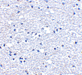
- Histology Positive Control Tissue
- Human brain tissue
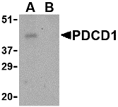
- Western Blotting
- AHP1706 detects a band of approximately 45kDa in THP-1 cell lysate
| Description | Product Code | Applications | Pack Size | List Price | Your Price | Quantity | |
|---|---|---|---|---|---|---|---|
| Antigen Retrieval Buffer, pH8.0 | BUF025A | P | 500 ml | Log in | |||
| List Price | Your Price | ||||||
| Log in | |||||||
| Description | Antigen Retrieval Buffer, pH8.0 | ||||||
| TidyBlot Western Blot Detection Reagent:HRP | STAR209P | WB * | 0.5 ml | Log in | |||
| List Price | Your Price | ||||||
| Log in | |||||||
| Description | TidyBlot Western Blot Detection Reagent:HRP | ||||||
Further Reading
-
Ishida, Y. et al. (1992) Induced expression of PD-1, a novel member of the immunoglobulin gene superfamily, upon programmed cell death.
EMBO J. 11 (11): 3887-95. -
Zhong, X. et al. (2004) Suppression of expression and function of negative immune regulator PD-1 by certain pattern recognition and cytokine receptor signals associated with immune system danger.
Int Immunol. 16 (8): 1181-8. -
Nishimura, H. et al. (1999) Development of lupus-like autoimmune diseases by the disruption of the PD-1 gene encoding an ITIM motif-carrying immunoreceptor.
Immunity 11:141-51.
- Synonyms
- PD-1
- RRID
- AB_2159158
- UniProt
- Q15116
- Entrez Gene
- PDCD1
- GO Terms
- GO:0006915 apoptosis
- GO:0016021 integral to membrane
- GO:0004725 protein tyrosine phosphatase activity
- GO:0004871 signal transducer activity
- GO:0006959 humoral immune response
- GO:0007275 multicellular organismal development
- GO:0031295 T cell costimulation
AHP1706
If you cannot find the batch/lot you are looking for please contact our technical support team for assistance.
View more products with CD279 specificity
Please Note: All Products are "FOR RESEARCH PURPOSES ONLY"
View all Anti-Human ProductsAlways be the first to know.
When we launch new products and resources to help you achieve more in the lab.
Yes, sign me up