Mitochondrial Membrane Potential MitoPT™ TMRE Kit


Mitochondrial Membrane Potential Kit
- Product Type
- Kits
- Specificity
- Mitochondrial Membrane Potential MitoPT™ TMRE Kit

| The MitoPT TMRE kit uses a quick and easy staining method to clearly differentiate between non-apoptotic and apoptotic cells through mitochondrial functionality. |
- Reagents In The Kit
- MitoPT TMRE Reagent - lyophilized - to make up 1mM stock
10x Assay Buffer, 125ml (x2)
600uL 50mM carbonyl cyanide m-chlorophenylhydrazone (CCCP) concentrate in DMSO. - Test Principle
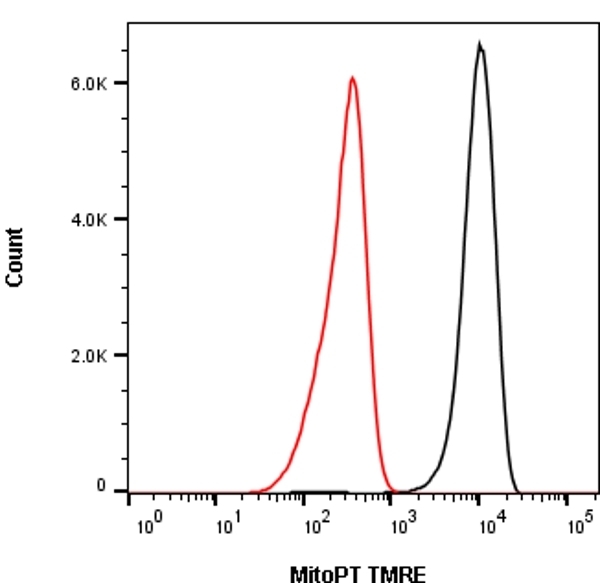
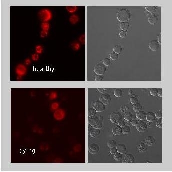
- An early indication of apoptosis involves a collapse in the electrochemical gradient across the mitochondrial membrane. Loss of mitochondrial membrane potential can be detected by a unique fluorescent cationic dye known as TMRE (tetramethylrhodamine ethyl ester), that has been incorporated into the MitoPT TMRE kit.
The MitoPT TMRE reagent easily penetrates cells and enters the mitochondria. It aggregates in the mitochondria of non-apoptotic cells and fluoresces bright orange/red, whilst in apoptotic cells it diffuses throughout the cell. Once dispersed, the reagent assumes a monomeric form and exhibits a reduced orange/red fluorescence level. This allows an easy distinction between apoptotic and non-apoptotic fluorescent cells which can be read with a flow cytometer, fluorescence microscope or a fluorescence plate reader using black microtitre plates. - Regulatory
- For research purposes only.
- Guarantee
- Guaranteed until date of expiry. Please see product label.
- Acknowledgements
- MitoPT is a trademark of Immunochemistry Technologies, LLC.
Store the unopened kit at -20°C. Once open, the 10x Assay Buffer can be stored at 2-8°C until the expiry date shown.
Protect the MitoPT TMRE Reagent from light at all times. Once reconstituted, the MitoPT TMRE stock should be stored at -20°C, protected from light and thawed no more than twice.
This product has been reported to work in the following applications. This information is derived from testing within our laboratories, peer-reviewed publications or personal communications from the originators. Please refer to references indicated for further information. For general protocol recommendations, please visit the antibody protocols page.
| Application Name | Verified | Min Dilution | Max Dilution |
|---|---|---|---|
| Flow Cytometry | |||
| Immunofluorescence |
Where this product has not been tested for use in a particular technique this does not necessarily exclude its use in such procedures. Suggested working dilutions are given as a guide only. It is recommended that the user titrates the product for use in their own system using appropriate negative/positive controls.
- Instructions For Use
- Instructions for use can be found at www.bio-rad-antibodies.com/uploads/IFU/IFUICT946.pdf
How to Use the Spectraviewer
Watch the Tool Tutorial Video ▸- Start by selecting the application you are interested in, with the option to select an instrument from the drop down menu or create a customized instrument
- Select the fluorophores or fluorescent proteins you want to include in your panel to check compatibility
- Select the lasers and filters you wish to include
- Select combined or multi-laser view to visualize the spectra
Request a different product with this specificity
Please Note: All Products are "FOR RESEARCH PURPOSES ONLY"
Always be the first to know.
When we launch new products and resources to help you achieve more in the lab.
Yes, sign me up