FGF Basic

Recombinant Human FGF Basic (PHP105) used to supplement tissue cultuure medium supporting growth of human and canine chondrocyte like cells (CLCs) in vitro.
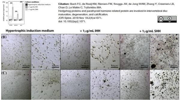
Image caption:
Indian and Sonic hedgehog facilitate calcification in human and canine chondrocyte‐like cells in vitro. (A) Hedgehog and PTHrP signaling‐related mRNA expression in canine CLC monolayers treated with hypertrophic induction medium supplemented with/without 1 μg/mL IHH or SHH for 7 days. n = 4. **P <.01. ALP, alkaline phosphatase; IHH, Indian hedgehog; PTHR1, PTHrP receptor 1, RUNX2, Runt‐related transcription factor 2; SHH, Sonic hedgehog. Alizarin Red S staining on one representative canine (B) and human (C) CLC donor. CLC monolayers were treated with hypertrophic induction medium supplemented with/without 1 μg/mL IHH or SHH for 7 days. n = 4 per species
From: Bach FC, de Rooij KM, Riemers FM, Snuggs JW, de Jong WAM, Zhang Y, Creemers LB, Chan D, Le Maitre C, Tryfonidou MA.
Hedgehog proteins and parathyroid hormone-related protein are involved in intervertebral disc maturation, degeneration, and calcification.
JOR Spine. 2019 Nov 19;2(4):e1071.
doi: 10.1002/jsp2.1071.
This image is from an open access article distributed under terms of a Creative Commons Attribution License.
Recombinant Human FGF Basic
- Product Type
- Recombinant Protein
- Specificity
- FGF Basic

| Recombinant Human FGF basic represents the C-terminal protion of human fibroblast growth factor 2 (A135 - S288). Fibroblast growth factor basic (FGF basic), also known as FGF 2, is a heparin binding growth factor which has stimulatory activity on a range of cells of mesenchymal, neuroectodermal and endothelial origin. Note: FGF basic is sensitive to acidic conditions. |
- Target Species
- Human
- Product Form
- Purified recombinant protein expressed in E. coli - lyophilized
- Reconstitution
- Reconstitute with 0.5 ml Tris (5mM, pH7.6).
Care should be taken during reconstitution as the protein may appear as a film at the bottom of the vial. Bio-Rad recommend that the vial is gently mixed after reconstitution. Further dilutions may be prepared in a buffer containing a carrier protein (eg 0.1% BSA). - Source
- E.coli
- Buffer Solution
- TRIS buffered saline.
- Preservative Stabilisers
- None present
- Carrier Free
- Yes
- Activity
- 2 x 10⁶ units/mg
- Purity
- >95% by SDS PAGE and HPLC analysis
- Approx. Protein Concentrations
- Total protein concentration 0.1 mg/ml after reconstitution.
- Protein Molecular Weight
- 17.2 kD (154 amino acid sequence)
- Endotoxin Level
- < 0.1 ng/ug
- Regulatory
- For research purposes only
- Guarantee
- Guaranteed for 3 months from the date of reconstitution or until the date of expiry, whichever comes first. Please see label for expiry date.
Prior to reconstitution store at -20oC. Following reconstitution store at -20oC.
This product should be stored undiluted.
Storage in frost-free freezers is not recommended. Avoid repeated freezing and thawing as this may denature the protein. Should this product contain a precipitate we recommend microcentrifugation before use.
This product should be stored undiluted.
Storage in frost-free freezers is not recommended. Avoid repeated freezing and thawing as this may denature the protein. Should this product contain a precipitate we recommend microcentrifugation before use.
This product has been reported to work in the following applications. This information is derived from testing within our laboratories, peer-reviewed publications or personal communications from the originators. Please refer to references indicated for further information. For general protocol recommendations, please visit the antibody protocols page.
| Application Name | Verified | Min Dilution | Max Dilution |
|---|---|---|---|
| ELISA | 0.2 | 0.4 ng/well | |
| Functional Assays | 0.1 | 10 ng/ml | |
| Western Blotting | 1.5 | 3.0 ng/lane |
Where this protein has not been tested for use in a particular technique this does not necessarily exclude its use in such procedures. Suggested working dilutions are given as a guide only. It is recommended that the user titrates the protein for use in their own system using appropriate negative/positive controls.
References for FGF Basic
-
Svendsen, C.N. et al. (1997) Long-term survival of human central nervous system progenitor cells transplanted into a rat model of Parkinson's disease.
Exp Neurol. 148: 135-46. -
Dimitrellos, V. et al. (2003) Capillary electrophoresis and enzyme solid phase assay for examining the purity of a synthetic heparin proteoglycan-like conjugate and identifying binding to basic fibroblast growth factor.
Biomed Chromatogr. 17 (1): 42-7. -
Kim, T.H. et al. (2005) Recombinant human prothrombin kringle-2 induces bovine capillary endothelial cell cycle arrest at G0-G1 phase through inhibition of cyclin D1/CDK4 complex: modulation of reactive oxygen species generation and up-regulation of cyclin-dependent kinase inhibitors.
Angiogenesis. 8: 307-14. -
van Beuningen, HM et al. (2014) Inhibition of TAK1 and/or JAK can rescue impaired chondrogenic differentiation of human mesenchymal stem cells in osteoarthritis-like conditions.
Tissue Eng Part A. 20 (15-16): 2243-52. -
Pleumeekers, M.M. et al. (2014) The in vitro and in vivo capacity of culture-expanded human cells from several sources encapsulated in alginate to form cartilage.
Eur Cell Mater. 27: 264-80. -
Narcisi R et al. (2015) Long-term expansion, enhanced chondrogenic potential, and suppression of endochondral ossification of adult human MSCs via WNT signaling modulation.
Stem Cell Reports. 4 (3): 459-72. -
Quang Le, B. et al. (2015) High-Throughput Screening Assay for the Identification of Compounds Enhancing Collagenous Extracellular Matrix Production by ATDC5 Cells.
Tissue Eng Part C Methods. 21 (7): 726-36. -
Willems, N. et al. (2015) Intradiscal application of rhBMP-7 does not induce regeneration in a canine model of spontaneous intervertebral disc degeneration.
Arthritis Res Ther. 17: 137.
View The Latest Product References
-
Pleumeekers, M.M. et al. (2015) Cartilage Regeneration in the Head and Neck Area: Combination of Ear or Nasal Chondrocytes and Mesenchymal Stem Cells Improves Cartilage Production.
Plast Reconstr Surg. 136 (6): 762e-774e. -
Lolli, A. et al. (2016) Silencing of Antichondrogenic MicroRNA-221 in Human Mesenchymal Stem Cells Promotes Cartilage Repair In Vivo..
Stem Cells. 34 (7): 1801-11. -
Cleary, M.A. et al. (2016) Expression of CD105 on expanded mesenchymal stem cells does not predict their chondrogenic potential.
Osteoarthritis Cartilage. 24 (5): 868-72. -
Grotenhuis, N. et al. (2016) Biomaterials Influence Macrophage-Mesenchymal Stem Cell Interaction In Vitro.
Tissue Eng Part A. 22 (17-18): 1098-107. -
Kroon, L.M.G. et al. (2017) Activin and Nodal Are Not Suitable Alternatives to TGFβ for Chondrogenic Differentiation of Mesenchymal Stem Cells.
Cartilage. 8 (4): 432-8. -
Rodrigues, A.I. et al. (2017) Calcium phosphates and silicon: exploring methods of incorporation.
Biomater Res. 21: 6. -
Quang Le, B. et al. (2017) An Approach to In Vitro Manufacturing of Hypertrophic Cartilage Matrix for Bone Repair.
Bioengineering (Basel). 4 (2): 35. -
Bach, F.C. et al. (2017) Link-N: The missing link towards intervertebral disc repair is species-specific.
PLoS One. 12 (11): e0187831. -
Pleumeekers, M.M. et al. (2018) Trophic effects of adipose-tissue-derived and bone-marrow-derived mesenchymal stem cells enhance cartilage generation by chondrocytes in co-culture.
PLoS One. 13 (2): e0190744. -
Bach, F.C. et al. (2019) Hedgehog proteins and parathyroid hormone-related protein are involved in intervertebral disc maturation, degeneration, and calcification.
JOR Spine. 2 (4): e1071. -
Lolli, A. et al. (2019) Hydrogel-based delivery of antimiR-221 enhances cartilage regeneration by endogenous cells.
J Control Release. 309: 220-30. -
Sivasubramaniyan, K. et al. (2019) Cell-surface markers identify tissue resident multipotential stem/stromal cell subsets in synovial intimal and sub-intimal compartments with distinct chondrogenic properties.
Osteoarthritis Cartilage. 27 (12): 1831-40. -
Vainieri, M.L. et al. (2020) Evaluation of biomimetic hyaluronic-based hydrogels with enhanced endogenous cell recruitment and cartilage matrix formation.
Acta Biomater. 101: 293-303. -
Khatab, S. et al. (2020) MSC encapsulation in alginate microcapsules prolongs survival after intra-articular injection, a longitudinal in vivo cell and bead integrity tracking study.
Cell Biol Toxicol. 36 (6): 553-570. -
Narcisi, R. et al. (2021) Expansion and Chondrogenic Differentiation of Human Bone Marrow-Derived Mesenchymal Stromal Cells.
Methods Mol Biol. 2221: 15-28. -
Tellegen, A. et al. (2021) Intra-Articular Slow-Release Triamcinolone Acetonide from Polyesteramide Microspheres as a Treatment for Osteoarthritis
Pharmaceutics. 13 (3): 372. -
Teunissen, M. et al. (2021) The lower in vitro. chondrogenic potential of canine adipose tissue-derived mesenchymal stromal cells (MSC) compared to bone marrow-derived MSC is not improved by BMP-2 or BMP-6.
Vet J. 269: 105605. -
Du, J. et al. (2022) Intradiscal injection of human recombinant BMP-4 does not reverse intervertebral disc degeneration induced by nuclectomy in sheep.
J Orthop Translat. 37: 23-36. -
Basatvat, S. et al. (2023) Harmonization and standardization of nucleus pulposus cell extraction and culture methods
JOR SPINE. Jan 10 [Epub ahead of print]. -
Schwab, A. 41:42-53. (2023) Modulating design parameters to drive cell invasion into hydrogels for osteochondral tissue formation
J Orthop Translat. -
Tryfonidou, M. et al. (2023) Notochordal cell-derived matrix inhibits MAPK signaling in the degenerative disc environment
European Cells and Materials. 46: 57-90. -
Kearney, C.M. et al. (2022) Treatment Effects of Intra-Articular Allogenic Mesenchymal Stem Cell Secretome in an Equine Model of Joint Inflammation.
Front Vet Sci. 9: 907616.
PHP105
If you cannot find the batch/lot you are looking for please contact our technical support team for assistance.
View more products with FGF BASIC specificity
Please Note: All Products are "FOR RESEARCH PURPOSES ONLY"
View all Anti-Human ProductsAlways be the first to know.
When we launch new products and resources to help you achieve more in the lab.
Yes, sign me up