CD4 antibody | Du CD4-2


Mouse antiDuck CD4 antibody, clone Du CD4-2 used for the identification of T-Helper cells in goose organs by immunohistochemistry on formalin fixed, paraffin embedded tissue sections.
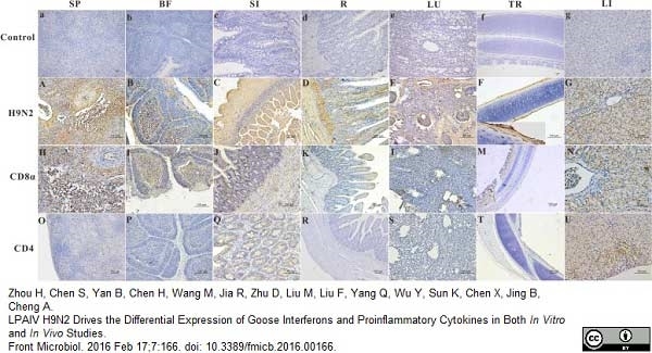
Image caption:
The location of the AIV H9N2 antigen and goose CD4 and CD8α molecules in several tissues at 5 dpi. The protein locations in the different tissues from AIV H9N2-infected birds were detected by IHC assay (Negative control group: a–g). Viral positive signals and cells positive for CD4 or CD8α antigen appeared dark brown using immunohistochemical staining. SP: SP (A,H,O), BF: BF (B,I,P), SI: SI (C,J,Q), R: R (D,K,R), LU: lung (E,L,S), TR: TR (F,M,T), LI: LI (G,N,U).
From: Zhou H, Chen S, Yan B, Chen H, Wang M, Jia R, Zhu D, Liu M, Liu F, Yang Q, Wu Y, Sun K, Chen X, Jing B, Cheng A.
LPAIV H9N2 Drives the Differential Expression of Goose Interferons and Proinflammatory Cytokines in Both In Vitro and In Vivo Studies. Front Microbiol. 2016 Feb 17;7:166.
This image is from an open access article distributed under terms of a Creative Commons Attribution License.
Mouse anti Duck CD4 antibody, clone Du CD4-2 (MCA2478) used along with Mouse anti Duck CD8&aplha; antibody, clone Du CD8-1 (MCA2479) for the demonstration of lymphocyte subsets in goose spleen sections by immunohistochemistry using formalin fixed, paraffin embedded tissue sections.
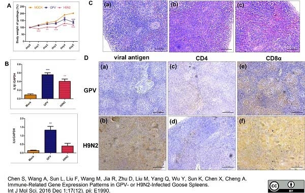
Image caption:
Global characteristics of goslings after infection with GPV and H9N2. (A) Body weight change of GPV- and H9N2-infected goslings from day 0 to 5. ** p≤0.01, *** p≤0.001; (B) the expression level of IL-1β and IL-6 in the spleen tissues of GPV- and H9N2-infected goslings. ** p≤0.01, *** p≤0.001; (C) histological changes in goose spleens infected with GPV and H9N2 at 5 dpi. The spleen tissues from (a) mock-, (b) GPV-, and (c) H9N2-infected goslings were stained with H &E, and diffuse hemorrhage was observed in either of virus-infected spleen sections, scale bar = 100 μm; and (D) Detection of (a) GPV and (b) H9N2 antigen, as well as (c,d) CD4- and (e,f) CD8α-positive cells by immunohistochemical analysis. The dark brown represents positive signs for viral antigen, CD4, or CD8α molecules, scale bar = 100 μm.
From: Chen S, Wang A, Sun L, Liu F, Wang M, Jia R, Zhu D, Liu M, Yang Q, Wu Y, Sun K, Chen X, Cheng A.
Immune-Related Gene Expression Patterns in GPV- or H9N2-Infected Goose Spleens.
Int J Mol Sci. 2016 Dec 1;17(12). pii: E1990.
This image is from an open access article distributed under terms of a Creative Commons Attribution License.
Mouse anti Duck CD4 antibody, clone Du CD4-2 (MCA2478) used for the identification of T lymphocytes in goose tissues by immunohistochemistry on formalin fixed, paraffin embedded tissue sections.
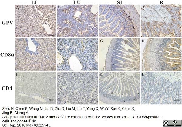
Image caption:
The location and density of GPV antigen, CD4 and CD8α molecules in the liver (LI), lung (LU), small intestine (SI), and rectum (R). Geese were humanly killed 5 days post infection by viruses. The protein locations in the different tissues of GPV-infected birds were detected by IHC assay. Positive virus signals were detected, cells positive for CD4 or CD8α antigen appeared dark brown using immunohistochemical staining, and sections were counterstained with haematoxylin. Mouse polyclonal antibody against GPV was prepared by our laboratory. The dilution folds of mouse anti-duck monoclonal CD4 antibodies (AbD Serotec MCA2478) and mouse anti-goose monoclonal CD8α (provided by our laboratory) antibodies were 1:100, respectively. Incubation with goat anti-mouse or goat anti-rabbit secondary antibody was performed according to the protocols of the immunoassay kit. Liver (A,E,I), lung (B,F,J), small intestine (C,G,K) and rectum (D,H,L).
From: Zhou H, Chen S, Wang M, Jia R, Zhu D, Liu M, Liu F, Yang Q, Wu Y, Sun K, Chen X, Jing B, Cheng A.
Antigen distribution of TMUV and GPV are coincident with the expression profiles of CD8α-positive cells and goose IFNγ. Sci Rep. 2016 May 6;6:25545.
This image is from an open access article distributed under terms of a Creative Commons Attribution License.
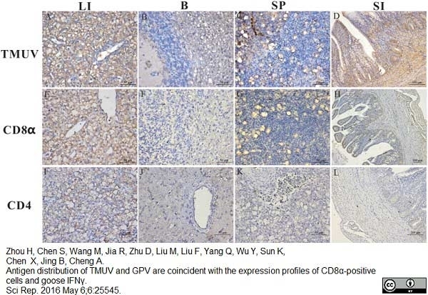
Mouse anti Duck CD4 antibody, clone Du CD4-2 (MCA2478) used for the identification of T lymphocytes in goose tissues by immunohistochemistry on formalin fixed, paraffin embedded tissue sections.
Image caption:
The location and density of TMUV antigen, CD4 and CD8α molecule in the liver (LI), brain (B), spleen (SP), and small intestine (SI). Geese were humanly killed 5 days post infection by viruses. These protein locations in the different tissues of TMUV-infected birds were detected by IHC assay. Positive virus signals were detected, cells positive for CD4 or CD8α antigen appeared dark brown using immunohistochemical staining, and sections were counterstained with haematoxylin. Rabbit polyclonal antibody against TUMV E protein was prepared by our laboratory. The dilution folds of mouse anti-duck monoclonal CD4 antibodies and mouse anti-goose monoclonal CD8α antibodies were both 1:100. Incubation of goat anti-mouse or goat anti-rabbit secondary antibody was performed by the protocols of the immunoassay kit. Liver (A,E,I), brain (B,F,J), spleen (C,G,K) and small intestine (D,H,L).
From: Zhou H, Chen S, Wang M, Jia R, Zhu D, Liu M, Liu F, Yang Q, Wu Y, Sun K, Chen X, Jing B, Cheng A.
Antigen distribution of TMUV and GPV are coincident with the expression profiles of CD8α-positive cells and goose IFNγ. Sci Rep. 2016 May 6;6:25545.
This image is from an open access article distributed under terms of a Creative Commons Attribution License.
Mouse anti Duck CD4 antibody, clone Du CD4-2 (MCA2478) used for the evaluation of CD4 expression on Pekin duck lymphocytes by flow cytometry.
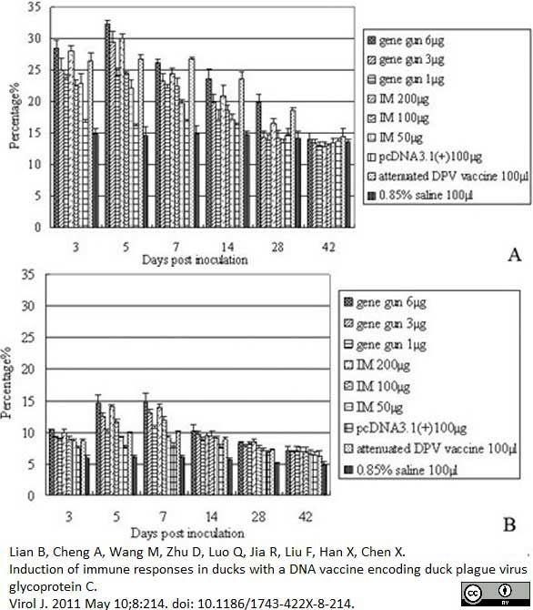
Image caption:
T lymphocytes in PBLs following DPV gC DNA vaccination. 3, 5, 7, 14, 28, 42 days after vaccination, the isolated PBLs were stained with monoclonal antibodies against duck CD4 (A), and CD8 (B). The results presented are the mean of all specimens of each group ± SD.
From: Lian B, Cheng A, Wang M, Zhu D, Luo Q, Jia R, Liu F, Han X, Chen X.
Induction of immune responses in ducks with a DNA vaccine encoding duck plague virus glycoprotein C.
Virol J. 2011 May 10;8:214.
This image is from an open access article distributed under the terms of the Creative Commons Attribution License.
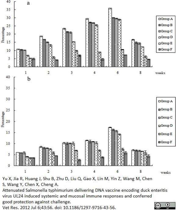
Mouse anti Duck CD4 antibody, clone Du CD4-2 (MCA2478) used for the evaluation of lymphocyte populations in PBLs following vaccination by flow cytometry.
Image caption:
T lymphocytes in PBL following DEV UL24 DNA vaccination. 1, 2, 3, 4, 6, 8 weeks after vaccination, the isolated PBL were stained with monoclonal antibodies against duck CD4 (A) and CD8 (B). The results presented are the mean of all specimens of each group. Statistical significance was determined using a student's t-test.
From: Yu X, Jia R, Huang J, Shu B, Zhu D, Liu Q, Gao X, Lin M, Yin Z, Wang M, Chen S, Wang Y, Chen X, Cheng A. Attenuated Salmonella typhimurium delivering DNA vaccine encoding duck enteritis virus UL24 induced systemic and mucosal immune responses and conferred good protection against challenge. Vet Res. 2012 Jul 6;43:56.
This image is from an open access article distributed under the terms of the Creative Commons Attribution License.
Mouse anti Duck CD4
- Product Type
- Monoclonal Antibody
- Clone
- Du CD4-2
- Isotype
- IgG2a
- Specificity
- CD4
| Mouse anti Duck CD4 antibody, clone Du CD4-2 recognizes Pekin duck CD4, shown to be expressed by thymocytes, splenocytes and peripheral lymphoid cells. Most avian immune research has been carried out on chickens, relatively little is known about the immune system of ducks, though there is a resemblance between the main lymphoid organs, the spleen, thymus and bursa of Fabricius. At the cellular level, like mammalian T cells, duck lymphocytes are responsive to phytohaemagglutinin, and all cells reacting with clone Du CD4-2 have been identified as CD3+ T cells (Kothlow et al. 2005). Clone Du CD4-2 can be used to identify duck T helper cells. Mouse anti Duck CD4 antibody, clone Du CD4-2 does not appear to react with Mallard. |
- Target Species
- Duck
- Species Cross-Reactivity
-
Target Species Cross Reactivity Chicken Goose - N.B. Antibody reactivity and working conditions may vary between species.
- Product Form
- Purified IgG - liquid
- Preparation
- Purified IgG prepared by affinity chromatography on Protein G from tissue culture supernatant
- Buffer Solution
- Phosphate buffered saline
- Preservative Stabilisers
- <0.1% Sodium Azide (NaN3)
- Immunogen
- 293T cells expressing Pekin duck CD4.
- Approx. Protein Concentrations
- IgG concentration 1.0mg/ml
- Fusion Partners
- Spleen cells from immunised Balb/c mice were fused with cells of the SP2/0 mouse myeloma cell line.
- Regulatory
- For research purposes only
- Guarantee
- 12 months from date of despatch
This product is shipped at ambient temperature. It is recommended to aliquot and store at -20°C on receipt. When thawed, aliquot the sample as needed. Keep aliquots at 2-8°C for short term use (up to 4 weeks) and store the remaining aliquots at -20°C.
Avoid repeated freezing and thawing as this may denature the antibody. Storage in frost-free freezers is not recommended.
Avoid repeated freezing and thawing as this may denature the antibody. Storage in frost-free freezers is not recommended.
This product has been reported to work in the following applications. This information is derived from testing within our laboratories, peer-reviewed publications or personal communications from the originators. Please refer to references indicated for further information. For general protocol recommendations, please visit the antibody protocols page.
| Application Name | Verified | Min Dilution | Max Dilution |
|---|---|---|---|
| Flow Cytometry | 1 | 10 ug/ml | |
| Immunohistology - Paraffin | |||
| Immunoprecipitation |
Where this product has not been tested for use in a particular technique this does not necessarily exclude its use in such procedures. Suggested working dilutions are given as a guide only. It is recommended that the user titrates the product for use in their own system using appropriate negative/positive controls.
- Flow Cytometry
- Use 10ul of the suggested working dilution to label 106 cells in 100ul.
| Description | Product Code | Applications | Pack Size | List Price | Your Price | Quantity | |
|---|---|---|---|---|---|---|---|
| Goat anti Mouse IgG (H/L):FITC (Multi Species Adsorbed) | STAR117F | F | 0.5 mg |

|
Log in | ||
| List Price | Your Price | ||||||
|
|
Log in | ||||||
| Description | Goat anti Mouse IgG (H/L):FITC (Multi Species Adsorbed) | ||||||
References for CD4 antibody
-
Kothlow, S. et al. (2005) Characterization of duck leucocytes by monoclonal antibodies.
Dev Comp Immunol. 29 (8): 733-48. -
Yu, X. et al. (2012) Attenuated Salmonella typhimurium delivering DNA vaccine encoding duck enteritis virus UL24 induced systemic and mucosal immune responses and conferred good protection against challenge.
Vet Res. 43: 56. -
Shanmugasundaram, R. and Selvaraj, R.K. (2012) Regulatory T cell properties of thymic CD4(+)CD25(+) cells in ducks.
Vet Immunol Immunopathol. 149: 20-7. -
Lian, B. et al. (2011) Induction of immune responses in ducks with a DNA vaccine encoding duck plague virus glycoprotein C.
Virol J. 8: 214. -
Huang, J. net al. (2014) An attenuated duck plague virus (DPV) vaccine induces both systemic and mucosal immune responses to protect ducks against virulent DPV infection.
Clin Vaccine Immunol. 21: 457-62. -
Chen, S. et al. (2015) Age-related development and tissue distribution of T cell markers (CD4 and CD8a) in Chinese goose.
Immunobiology. 220 (6): 753-61. -
Zhou, H. et al. (2016) LPAIV H9N2 Drives the Differential Expression of Goose Interferons and Proinflammatory Cytokines in Both In Vitro and In Vivo Studies.
Front Microbiol. 7: 166. -
Chen, S. et al. (2016) Immune-Related Gene Expression Patterns in GPV- or H9N2-Infected Goose Spleens.
Int J Mol Sci. 17 (12): pii: E1990.
View The Latest Product References
-
Zhou H et al. (2016) Antigen distribution of TMUV and GPV are coincident with the expression profiles of CD8α-positive cells and goose IFNγ.
Sci Rep. 6: 25545. -
Cornelissen, J.B. et al. (2013) Differences in highly pathogenic avian influenza viral pathogenesis and associated early inflammatory response in chickens and ducks.
Avian Pathol. 42 (4): 347-64. -
Wu, Y. et al. (2019) Changes in the small intestine mucosal immune barrier in Muscovy ducklings infected with Muscovy duck reovirus
Veterinary Microbiology. 233: 85-92. -
Apinda, N. et al. (2022) Simultaneous Protective Immune Responses of Ducks against Duck Plague and Fowl Cholera by Recombinant Duck Enteritis Virus Vector Expressing Pasteurella multocida OmpH Gene.
Vaccines (Basel). 10 (8): 1358.
Further Reading
-
Higgins, D.A. & Teoh, C.S. (1988) Duck lymphocytes. II. Culture conditions for optimum transformation response to phytohaemagglutinin.
J Immunol Methods. 106 (1): 135-45.
- RRID
- AB_609597
View more products with CD4 specificity
Please Note: All Products are "FOR RESEARCH PURPOSES ONLY"
View all Anti-Duck ProductsAlways be the first to know.
When we launch new products and resources to help you achieve more in the lab.
Yes, sign me up