Popular topics

Popular topics
-
References
Cancer Institute, The University of Mississippi Medical Centre (2015). Selecting Fluorochromes for Analysis and Sorting.
(Accessed: May 13, 2015).Paul C (2013). Counterstaining for Immunohistochemistry: Choices, Choices.
Available here (Accessed: December 02, 2014).Olympus. Theory of Confocal Microscopy, Fluorophores for Confocal Microscopy.
Available here (Accessed: May 13, 2015).Hibbs AR (2004). Confocal Microscopy for Biologists (New York: Kluwer Academic/Plenum Publishers).
Light up your IF experiments with our continued tips and tricks
These five tips are a continuation of last week's post and are designed to assist you with the selection of fluorophores for your imaging experiments.
6. Use new generation dyes, which stay fluorescent over a broad pH range. Many conventional fluorophores, such as FITC, are not recommended for staining protocols using acidic buffers as the fluorescence intensity signal is highly sensitive to an acidic environment (Cancer Institute, The University of Mississippi Medical Centre 2015).
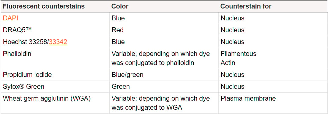
7. Ensure your fluorophore staining is spectrally differentiable from your counterstain, which provides background contrast and puts the observed staining into perspective (e.g. by visualizing nuclei). For example, DRAQ5™ should be used as a nuclear counterstain rather than DAPI when using antibodies conjugated to blue emitting fluorophores such as Alexa Fluor® 405 or DyLight® Fluor 405.
For your reference the most commonly used fluorescent counterstains and their respective emission colors are shown below (adapted from Paul 2013).
8. Use fluorophores with narrow emission spectra in multi-color IF experiments. We make this recommendation because narrow emission spectra minimize the risk of spectral overlap. This process is also known as bleed-through and describes the detection of one fluorophore in another fluorophore's filter set. Bleed-through makes it difficult to observe discrete fluorescence signals and complicates the evaluation of co-localization experiments. Therefore when selecting fluorophores the emission and excitation spectra should be checked for spectral overlap with the help of a spectrum viewer. Ideally, there should be no spectral overlap between the fluorophores. Quantum dot conjugates, which have broad absorption and very narrow emission spectra, are therefore perfectly suited for multi-color experiments. Indeed multiple antibodies conjugated to quantum dots can simultaneously be detected with negligible spectral overlap (Olympus 2015).
9. After having selected the most suitable fluorophores for your experiment you have to decide what antigen to detect with which fluorophore in your multi-color IF experiment. The brightest fluorophore should be reserved for the detection of the antigen with the least abundant expression level. The dimmest fluorophore should be used for detecting the most abundant antigen.
10. Include appropriate controls to verify that the observed fluorescence is a result of fluorophore staining rather than an unspecific artifact. We recommend you consider conducting these three controls:
- Autofluorescence / cell background staining control. As cells (especially those that have been damaged or have started to undergo apoptosis (Hibbs 2004)) can display natural fluorescence it is crucial to observe IF samples microscopically before every staining experiment. Additionally, a label/fluorophore control should be included which basically entails performing the complete staining protocol without the addition of fluorophore conjugated antibodies.
- Positive and negative controls. Include cell lines in which your protein of interest is either over-expressed or absent (e.g. a knock-out cell line). If you do not see staining in the positive control something has gone wrong with the staining protocol. Alternatively, if you see staining in the negative control you know that the staining/observed fluorescence is unspecific.
- If secondary antibodies rather than directly fluorophore conjugated primary antibodies are to be used a secondary antibody only control should be performed (also called no primary antibody control; follow the same staining protocol without the addition of a primary antibody). This type of control is used to verify that the secondary antibody does not unspecifically bind to certain cellular compartments. For multi-color immunofluorescence experiments we recommend the use of cross-adsorbed/pre-adsorbed secondary antibodies as those minimize the risk of the secondary antibody reacting with endogenous immunoglobulins or an undesired primary antibody.
References
Cancer Institute, The University of Mississippi Medical Centre (2015). Selecting Fluorochromes for Analysis and Sorting.
(Accessed: May 13, 2015).
Paul C (2013). Counterstaining for Immunohistochemistry: Choices, Choices.
Available here (Accessed: December 02, 2014).
Olympus. Theory of Confocal Microscopy, Fluorophores for Confocal Microscopy.
Available here (Accessed: May 13, 2015).
Hibbs AR (2004). Confocal Microscopy for Biologists (New York: Kluwer Academic/Plenum Publishers).
You may also be interested in...

This blog discusses some best practices for storing and using your antibodies to get the most out of them in your experiments....

Treat Them Right! – Best Practices for Storing and Working with Antibodies
Fluorescent proteins have expanded the capabilities of scientists, thus allowing observations that would not have been possible without them....
Illuminating biomedical research — the story of fluorescent proteins
View more Applications or Immunofluorescence blogs